IRF5 Promotes the Progression of Hepatocellular Carcinoma and is Regulated by...
CKRBSACI 2005 SM
1. PO 1021
ABSTRACT
Rationale: Chemokine receptors are involved in the recruitment of T
lymphocytes to sites of infection and inflammation. CCR4 and
CXCR3 expression has been associated with Th2 and Th1 immune
responses respectively. We examined the expression of the
chemokine receptors CCR4 and CXCR3 on CD3+ cells after allergen
and diluent challenge in the skin.
Methods: Skin biopsies were obtained from atopic allergic and non-
atopic individuals after allergen PPD and diluent challenge.
Biopsies were examined for CD3+CCR4+ cells and CD3+CXCR3+
cells using dual immunofluorescence and IL-4 and IFN-γ mRNA
expressing cells using in situ hybridisation.
Results: Following allergen challenge there was a significant
increase in CD3+/CCR4+ cell numbers within the atopic group
(p<0.001) and not in the non-atopics (p=0.28). Both atopics and non
atopics showed an increase in CD3+/CXCR3+ cells at 48 hours after
PPD challenge (p=0.01 and p=0.06 respectively). Furthermore IL-4
mRNA+ cells peaked at 8 hours after allergen challenge while IFNg
mRNA+ cells were elevated after PPD challenge at 48 hours, with no
change in the nonatopics.
Conclusion: These data suggest that, in vivo, in man CCR4 recruits
Th2 cells to the skin in the late response to specific allergen,
whereas CXCR3 recruits Th1 cells to the skin in response to PPD
challenge.
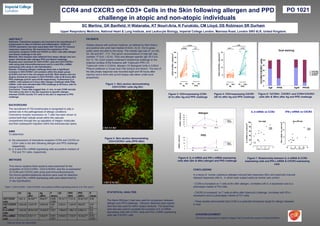
Figure 6: IL-4 mRNA and IFN-γ mRNA expressing
cells after (8hr & 48hr) allergen and PPD challenge
BACKGROUND
The recruitment of Th2 lymphocytes is recognised to play a
central role in the pathogenesis of allergic conditions.
Chemokine receptor expression on T cells has been shown to
control both their cellular arrest within the vascular
compartment through the up regulation of integrin molecules
and their subsequent migration within the extravascular space.
METHODS
Figure 3: CD3+expressing CCR4
(8 hr) after Ag and PPD challenge
CONCLUSIONS
Figure 7: Relationship between IL-4 mRNA & CCR4
expressing cells and IFN-γ mRNA & CXCR3 expressing
cells
PATIENTS
Sixteen atopics with summer hayfever, as defined by their history
and positive skin prick test (median 8.0mm, IQ (5, 10)) to grass
pollen were recruited to this study. The median age was 27 yrs (IQ,
24, 29) and M:F, 11:5. The serum concentrations of total IgE was
(median 171IU/L ( IQ 42, 720)) and allergen specific IgE 24.0 IU/L
(IQ 13, 79). Each subject underwent intradermal challenge to the
extensor surface of the forearms with Tuberculin PPD (10
Tuberculin Units in 0.02ml), allergen (10 Biological Units in 0.02ml
Phleum pratense or house dust mite extract) and diluent. The size of
the late phase response was recorded eight and 48 hours after
injection and a 4mm skin punch biopsy was taken under local
anaesthetic.
AIMS
To determine:
a) the expression of chemokine receptors CCR4 and CXCR3 on
CD3+ cells in the skin following allergen and PPD challenge
respectively
b) IL-4 and IFN-γ mRNA expressing cells as putative markers of
Th2 and Th1 cells, respectively
CCR4 and CXCR3 on CD3+ Cells in the Skin following allergen and PPD
challenge in atopic and non-atopic individuals
SC Martins, GK Banfield, H Watanabe, KT Nouri-Aria, K Furukido, CM Lloyd, DS Robinson SR Durham
Upper Respiratory Medicine, National Heart & Lung Institute, and Leukocyte Biology, Imperial College London, Manresa Road, London SW3 6LR, United Kingdom.
Allergen
Dil. 8 hrs 48 hrs
0
10
20
30
40
50
mRNA+cells/mm2
IL-4
IFN-γ
PPD
0
5
10
15
20
Dil. 8 hrs 48 hrs
mRNA+cells/mm2
IL-4
IFN-γ
CD3+
/CXCR3+
CD3+
/CCR4+
Dual staining
0
20
40
60
80
100
Ag8 PPD8 Ag48 PPD48
%CCR4+
CXCR3+
cells
%double
%CXCR3
%CCR4
-20 0 20 40 60 80
CXCR3 +ve cells/mm2
0
10
20
30
IFN-γmRNA+vecells/mm2
r=0.61
p=0.03
Skin (PPD 48)
0 20 40 60 80 100
CCR4 +ve cells/mm
2
0
20
40
60
80
100
IL-4mRNA+vecells/mm2
r=0.64
p=0.01
Skin (Ag8)
IL-4 mRNA vs CCR4 IFN-γ mRNA vs CXCR3
In a study of human cutaneous allergen-induced late responses (8hr) and tuberculin-induced
delayed responses (48) hr, in which each subject acted as his/her own control:
- CCR4 is increased on T cells at 8hr after allergen, correlates with IL-4 expression and is a
phenotypic marker of Th2 cells
- CXCR3 is increased on T cells at 48hrs after tuberculin challenge, correlates with IFN-γ
expression and is a phenotypic marker of Th1 cells
- These studies demonstrate that CCR4 is a potential therapeutic target for allergic diseases
in man
Table 1: CD3+/CCR4+, CD3+/CXCR3+ and cytokine mRNA expressing cells (IL-4 & IFN-γ)/mm2
Five micron acetone fixed sections were examined for the
proportion of CD3+CCR4+, CD3+CXCR3+ and the co-expression
of CCR4 and CXCR3 cells using dual immunofluorescence.
Six micron paraformaldehyde sections were used for detection
of IL-4 and IFN-γ mRNA expressing cells were determined by
In situ hybridisation.
STATISTICAL ANALYSIS
Figure 5: %CCR4+, CXCR3+ and CCR4+CXCR3+
cells (8hr & 48hr) after Ag and PPD challenge
The Mann-Whitney U test was used for comparison between
allergen and PPD challenge. Wilcoxon Matched-pairs signed-
rank test was used for within subject analysis. The Spearman
rank test was used to correlate the numbers of IL-4 mRNA
expressing cells with CCR4+ cells and IFN-γ mRNA expressing
cells with CXCR3+ cells ACKNOWLEDGMENT
This work is sponsored by Imperial College Trust Fund with the support of GlaxoSmithKline
Figure 1: Skin section demonstrating
CD3+CCR4+ cells (Ag 8hr)
Figure 2: Skin section demonstrating
CD3+CXCR3+ cells (PPD 48hr)
Figure 4: CD3+expressing CXCR3
(48 hr) after Ag and PPD challenge
Dil. Ag
(8hr)
Ag
(48hr)
P
values
Dil. PPD
(8hr)
PPD
(48hr)
P
values
CD3+
/CCR4+
cells
7.6±1.4 40.5±6*** 35±9.5** 0.000
0.009
18.7±7.7 7.7±1.6 53.8±13.6* 0.02
CD3+
/CXCR3+
cell
0.5±0.26 2.5±0.75* 0.8±0.45 0.04
0.7
3.9±3.4 1.3±0.47 20±5.6* 0.01
0.4
IL-4 mRNA+
cells
1.0±0.53 36.9±10.8* 19.9±8.5* 0.001 1.65±0.8 1.88±1.2 2.68±1.4 0.9
0.6
IFN-γ mRNA+
cells
0.57±0.2 6.5±1.1* 5.5±0.9* 0.001
0.001
0.44±0.2 1.7±0.5* 10.3±2.3** 0.03
0.003
Data are shown as mean±SEM
Dil 48hr
Ag
0
10
20
30
CD3+/CXCR3+cells/mm
2
Dil 48hr
PPD
p=0.7 p=0.03
p=002
(74)
(57)
(57)
(55)
Dil 8hr
Ag
0
20
40
60
80
100
CD3+/CCR4+cells/mm
2
Dil 8hr
PPD
p=0.0000 p=0.1
p=0.000
(126)