Immunology Pathway of During Autoimmune Disease: A Review Article
C3C8AAAAI SM
1. No.1088
ABSTRACT
RATIONALE: Chemokine receptors are involved in the recruitment of
T lymphocytes to sites of infection and inflammation. We examined
the expression of three chemokine receptors CCR4, CCR3 and CCR8
on CD3+ cells after cutaneous allergen, PPD and diluent challenge in
the skin
METHODS: 4mm punch skin biopsies were obtained from 16 atopic
allergic individuals at 8 hours after 10 BU intradermal allergen
(Phleum Pratense or house dust mite extract) and following 10 TU
intradermal Purified Protein Derivative (PPD). Biopsies were
analysed for CD3+CCR4+, CD3+CCR3+ and CD3+CCR8+ cells using
dual immunofluorescence and IL-4 and IFN-γ mRNA expressing cells
using in situ hybridisation.
RESULTS: Following allergen challenge there was a significant
increase in CD3+CCR4+ cells which was not observed after PPD
challenge (p<0.001 and p=0.15 respectively; n=16). There was no
significant difference in CD3+CCR3+ cell numbers following either
allergen or PPD challenge (p=0.62 and p=0.83 respectively; n=8).
Similarly, there was no significant difference in CD3+CCR8+ cell
numbers following either allergen or PPD challenge (p=0.86 and
p=0.20 respectively; n=8). The percentage increase in CD3+CCR4+
cells was significantly greater than CD3+CCR3+ (p=0.002) and
CD3+CCR8+ (p=0.001) cells after allergen. IL-4+/IFN-γ+ ratio was
greater after allergen compared to PPD (3 and 0.2 respectively;
p=0.0004). Conversely, IFN-γ+/IL-4+ ratio was greater after PPD
compared to allergen (3.5 and 0.38 respectively; p=0.0002).
CONCLUSIONS: CCR4 but not CCR3 or CCR8 may contribute to Th2
T lymphocyte recruitment during allergen-induced late skin
responses. These data support CCR4 as a selective target for therapy
of allergic disease.
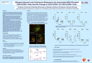
Figure 4: IL-4 mRNA and IFN-γ mRNA expressing
cells after (8hr & 48hr) allergen and PPD challenge
BACKGROUND
The recruitment of Th2 lymphocytes is recognised to play a
central role in the pathogenesis of allergic conditions.
Chemokine receptor expression on T cells has been shown to
control both their cellular arrest within the vascular
compartment through the up regulation of integrin molecules
and their subsequent migration within the extravascular space.
METHODS
Figure 2: CD3+ CCR4+, CD3+ CCR3+ and CD3+ CCR8+ cells following allergen and PPD
challenge in atopic individuals
CONCLUSIONS
PATIENTS
Sixteen atopics with summer hayfever, as defined by their history
and positive skin prick test (median 8.0mm, IQ (5, 10)) to grass
pollen were recruited to this study. The median age was 27 yrs (IQ,
24, 29) and M:F, 11:5. The serum concentrations of total IgE was
(median 171IU/L ( IQ 42, 720)) and allergen specific IgE 24.0 IU/L
(IQ 13, 79). Each subject underwent intradermal challenge to the
extensor surface of the forearms with Tuberculin PPD (10
Tuberculin Units in 0.02ml), allergen (10 Biological Units in 0.02ml
Phleum pratense or house dust mite extract) and diluent. The size
of the late phase response was recorded eight and 48 hours after
injection and a 4mm skin punch biopsy was taken under local
anaesthetic.
AIMS
To determine:
a) the expression of chemokine receptors CCR4, CCR3 and CCR8
on CD3+ cells in the skin following allergen and PPD challenge
respectively in atopic individuals
b) IL-4 and IFN-γ mRNA expressing cells as putative markers of
Th2 and Th1 cells, respectively
Allergen-Induced Late Cutaneous Responses Are Associated With Elevated
CD3+CCR4+ Cells And No Change In CD3+CCR3+ Or CD3+CCR8+ Cells
SC Martins, KT Nouri-Aria, GK Banfield, MR Jacobson, H Watanabe, S Radulovic, DS Robinson, CM Lloyd, SR Durham
Upper Respiratory Medicine, National Heart & Lung Institute, and Leukocyte Biology, Imperial College London, Manresa Road, London SW3 6LR, United Kingdom.
Allergen
Dil. 8 hrs 48 hrs
0
10
20
30
40
50
mRNA+cells/mm2
IL-4
IFN-γ
PPD
0
5
10
15
20
Dil. 8 hrs 48 hrs
mRNA+cells/mm2
IL-4
IFN-γ
CD3+
CCR4+
In a study of human cutaneous allergen-induced late responses (8hr) and tuberculin-
induced delayed responses:
- CCR4 is increased on T cells at 8hr after allergen, correlates with IL-4 expression and is a
phenotypic marker of Th2 cells in atopic individuals
- CCR3 and CCR8 are not associated with allergic responses in atopic skin
These studies demonstrate that CCR3 and CCR8 are not associated with allergic diseases.
CCR4 is a potential therapeutic target for allergic diseases in man.
Five micron acetone fixed sections were examined for the
proportion of CD3+CCR4+, CD3+CCR3+ and CD3+CCR8+ using
dual immunofluorescence. Six micron paraformaldehyde sections
were used for detection of IL-4 and IFN-γ mRNA expressing cells
were determined by In situ hybridisation.
STATISTICAL ANALYSIS
Wilcoxon Matched-pairs signed-rank test was used for within
subject analysis. The Spearman rank test was used to correlate
the numbers of IL-4 mRNA expressing cells with CCR4+ cells
ACKNOWLEDGMENT
This work is sponsored by Imperial College Trust Fund with the support of GlaxoSmithKline
Travel grant awarded by INDOOR Biotechnologies to attend the AAAAI annual meeting in Miami, FL.
Figure 1: Skin sections demonstrating
CD3+CCR4+, CD3+CCR3+ and CD3+CCR8+ cells (Ag 8hr)
CD3+
CCR3+
Dil Ag
CD3+CCR4+
0
100
200
300
Cells/mm2
Dil PPD
p= p=0.001 0.15
Dil Ag
CD3+CCR3+
0
100
200
300
Cells/mm2
Dil PPD
p= p=0.62 0.83
Dil Ag
CD3+CCR8+
0
100
200
300
Cells/mm2
Dil PPD
p= p=0.86 0.20
Figure 3: CCR4+, CCR3+ and CCR8+ cells following allergen and PPD challenge in atopic individuals
CD3+
CCR8+
Dil Ag
CCR4+
0
200
400
600
800
1000
Cells/mm2
Dil PPD
p= p=0.000 0.121
Dil Ag
CCR3+
0
200
400
600
800
1000
Cells/mm2
Dil PPD
p= p=0.944 0.208
Dil Ag
CCR8+
0
200
400
600
800
1000
Cells/mm2
Dil PPD
p= p= 0.5540.675