
Recommended
More Related Content
What's hot
What's hot (20)
Similar to Supports of Uterus
Similar to Supports of Uterus (20)
Recently uploaded
Recently uploaded (20)
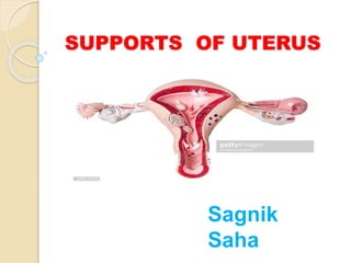
Supports of Uterus
- 2. Introduction Uterus is inverted pear shaped, highly mobile ,thick walled ,muscular organ in the female body. It is situated obliquely in the lesser pelvis between urinary bladder anteriorly and the rectum & sigmoid colon posteriorly.
- 3. Importance of Support The Supporter of the Uterus is needed to kept it in it’s actual position & prevented from sagging down . The uterus is supported and prevented from prolapse(downward Displacement) by a number of structures providing support to it.
- 4. Supports Primary support Muscular/ active Pelvic diaphragm Perineal body Urogenital diaphragm Visceral Urinary bladder vagina Uterine axis Fibro-muscular Mackenrodt’s Ligament Pubocervical ligaments Uterosacral ligaments Round ligaments Secondary support Peritoneal folds Broad Ligaments Utero –vesical fold Recto-uterine fold Pelvic cellular tissue
- 5. PRIMARY SUPPORT 1. Muscular / Active Support
- 6. A. Pelvic Diaphragm It is a gutter shaped musculo-fascial partition between the pelvic cavity & perinium . Pubococcygeus part of levetor ani plays the most significant supportive role of uterus. Pubococcygeus has 4 components: Pubococccygeus proper, Puborectalis, puboanalis and pubovaginalis/ pubovesicalis Pubovaginalis narrows the hiatus urogenitalis & constricts the vagina from side to side & prevents the discent of Uterus.
- 8. B. Perineal Body It is a pyramidal fibromuscular node intervening between the anal canal & urogenital apparaters. It is known as gynaecological perineum which receives the attachment of following muscle : longitudinal muscle coat of anal canal ,sphincter ani exturnus, superficial transverse perini (2) , deep transverse perini (2) Bulbospongious, pubococcygeus part of levetor ani The perineal body acts as an anchor for the pelvic diaphragm & maintains the integrity of the pelvic floor. If it is torn during delivery & not properly repaired the ,urogenital hiatus becomes enlarged, the vagina dilates & uterus descent.
- 10. C. Urogenital Diaphragm It is a musculofascial partition between the pelvic cavity & the anterior part of pelvic outlet. It helps in closure of urogenital hiatus from bellow & constricts the vagina.
- 12. Uterine Axis Angle of Anteversion It is forward angle formed by long axis of vagina & that of cervix at the level of external os measuring approx. 90° provided Urinary Bladder and rectum are empty. Angle of Anteflexion It is forward angle formed by long axis of uterus & that of cervix at the level of internal os measuring approx. 125° -170° provided Urinary Bladder and rectum are empty
- 13. 3. Fibromuscular support Mackenrodt’s Ligaments : These are fan shaped fibro- muscular band derived from condensation of parametric tissue .They form a hammock that keep the cervix in position & prevent downward displacement of uterus. It is also known as lateral /transverse cervical ligament/ cardinal ligament. Pubocervical ligaments : these are a pair of thin fibrous band which extend from cervix to the posterior surface of pubis. Uterosacral ligaments : Each ligaments extends from cervix to the 3rd sacral vertebra .They pull the cervix backwards & help in maintenance of anteversion & anteflexion position of uterus.
- 15. Round Ligaments of Uterus It is a remnant of distal part of gubernaculum of ovary . Attached proximally to the cornu of uterus and distally to the labia majora. It pull the fundus forwards & maintains the Anteversion & anteflexion.
- 16. SECONDARY SUPPORT 1. Peritoneal folds / Ligaments
- 17. Broad Ligaments Each Ligament is Broad , quadrilateral fold of peritoneum that extends from lateral borders of Uterus to the lateral pelvic wall.
- 18. Peritoneal Folds
- 19. Utero vesical fold : It is formed by reflection of peritoneum that extends from anterior border of the body of uterus to the upper surface of urinary bladder. Rectovaginal / Rectouterine fold : It is formed by peritoneal reflection from posterior fornix of vagina to the rectum. It forms the floor of pouch of Doglus .
- 20. 2. Pelvic cellular Tissue/ Loose packing meterial The parametric tissue made up of fibro- areolar & fibro-muscular tissue fills up the dead space between pelvic peritoneum & pelvic floor & act as a packing structure . Ovarian hormones control the growth of parametric tissue