
Recommended
More Related Content
Similar to 876.ppt
Similar to 876.ppt (20)
Recently uploaded
Recently uploaded (20)
876.ppt
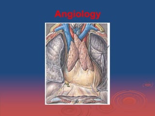
- 1. Angiology
- 2. The heart and lungs are situated in the thorax, the walls of which afford them protection. The heart lies between the two lungs, and is enclosed within a fibrous bag, the pericardium, The capacity of the cavity of the thorax does not correspond with its apparent size externally, because
- 3. - the space enclosed by the lower ribs is occupied by some of the abdominal viscera; and - the cavity extends above the anterior parts of the 1st ribs into the neck. The size of the thoracic cavity is constantly varying during life with the movements of the ribs and diaphragm, and with the degree of distention of the abdominal viscera.
- 4. From the collapsed state of the lungs as seen when the thorax is opened in the dead body, it would appear as if the viscera only partly filled the cavity, but during life there is no vacant space, that which is seen after death being filled up by the expanded lungs.
- 5. The Upper Opening of the Thorax. The parts which pass through the upper opening of the thorax are, from before backward, in or near the middle line, the Sternohyoideus and Sternothyreoideus muscles, the remains of the thymus, the inferior thyroid veins, the trachea, esophagus thoracic duct, and the Longus colli muscles;
- 6. The Lower Opening of the Thorax. The lower opening of the thorax is wider transversely than from before backward. It slopes obliquely downward and backward, so that the thoracic cavity is much deeper behind than in front. The diaphragm closes the opening and forms the floor of the thorax.
- 7. The floor is flatter at the center than at the sides, and higher on the right side than on the left; in the dead body the right side reaches the level of the upper border of the 5th costal cartilage, while the left extends only to the corresponding part of the 6th costal cartilage.
- 8. The pericardium is a conical fibro-serous sac, in which the heart and the roots of the great vessels are contained. It is placed behind the sternum and the cartilages of the 3rd, 4th , 5th , 6th , and 7th ribs of the left side, in the mediastinal cavity.
- 9. In front, it is separated from the anterior wall of the thorax, in the greater part of its extent, by the lungs and pleuræ; but a small area, somewhat variable in size, and usually corresponding with the left half of the lower portion of the body of the sternum and the medial ends of the cartilages of the 4th and 5th ribs of the left side, comes into direct relationship with the chest wall.
- 10. The lower extremity of the thymus, in the child, is in contact with the front of the upper part of the pericardium. Behind, it rests upon the bronchi, the esophagus, the descending thoracic aorta, and the posterior part of the mediastinal surface of each lung.
- 11. Laterally, it is covered by the pleuræ, and is in relation with the mediastinal surfaces of the lungs; the phrenic nerve, with its accompanying vessels, descends between the pericadium and pleura on either side. Posterior wall of the pericardial sac, showing the lines of reflection of the serous pericardium on the great vessels.
- 12. Pericardium. Although the pericardium is usually described as a single sac, an examination of its structure shows that it consists essentially of two sacs intimately connected with one another, but totally different in structure. The outer sac, known as the fibrous pericardium, consists of fibrous tissue.
- 13. The inner sac, or serous pericardium, is a delicate membrane which lies within the fibrous sac and lines its walls; it is composed of a single layer of flattened cells resting on loose connective tissue.The heart invaginates the wall of the serous sac from above and behind, and practically obliterates its cavity, the space being merely a potential one.
- 14. The fibrous pericardium forms a flask- shaped bag, the neck of which is closed by its fusion with the external coats of the great vessels, while its base is attached to the central tendon and to the muscular fibers of the left side of the diaphragm.
- 15. Over a small area the central tendon of the diaphragm and the pericardium are completely fused. Above, the fibrous pericardium not only blends with the external coats of the great vessels, but is continuous with the pretracheal layer of the deep cervical fascia.
- 16. By means of these upper and lower connections it is securely anchored within the thoracic cavity. It is also attached to the posterior surface of the sternum by the superior and inferior sterno pericardiac ligaments; The upper passing to the manubrium, and the lower to the xiphoid process.
- 17. The vessels receiving fibrous prolongations from this membrane are: the aorta, the superior vena cava, the right and left pulmonary arteries, and the four pulmonary veins. The inferior vena cava enters the pericardium through the central tendon of the diaphragm, and receives no covering from the fibrous layer.
- 18. The serous pericardium is, as already stated, a closed sac which lines the fibrous pericarp- dium and is invaginated by the heart; it therefore consists of a visceral and a parietal portion. The visceral portion, or epicardium, covers the heart and the great vessels, from the latter is continuous with the parietal layer which lines the fibrous pericardium.
- 19. The portion which covers the vessels is arranged in the form of two tubes. The aorta and pulmonary artery are enclosed in one tube, the arterial mesocardium. The superior and inferior venæ cavæ and the four pulmonary veins are enclosed in a second tube, the venous mesocardium,
- 20. between the aorta and pulmonary artery in front and the atria behind — is the transverse sinus.
- 21. Cor-The heart is a hollow muscular organ of a somewhat conical form; it lies between the lungs in the middle mediastinum and is enclosed in the pericardium. It is placed obliquely in the chest behind the body of the sternum and adjoining parts of the rib cartilages
- 22. The heart projects farther into the left than into the right half of the thoracic cavity, so that about one-third of it is situated on the right and two-thirds on the left of the median plane.
- 23. The heart, in the adult, measures about 12 cm. in length, 8 to 9 cm. in breadth at the broadest part, and 6 cm. in thickness. Its weight, in the male, varies from 280 to 340 grams; In the female, from 230 to 280 grams. The heart continues to increase in weight and size up to an advanced period of life; this increase is more marked in men than in women.
- 24. Component Parts. As has already been stated, the heart is subdivided by septa into right and left halves, and a constriction subdivides each half of the organ into two cavities, the upper cavity being called the atrium, the lower the ventricle. The heart therefore consists of four chambers, viz., right and left atria, and right and left ventricles.
- 25. The division of the heart into four cavities is indicated on its surface by grooves. The atria are separated from the ventricles by the coronary sulcus (auriculoventricular groove); this contains the trunks of the nutrient vessels of the heart, and is deficient in front, where it is crossed by the root of the pulmonary artery.
- 26. The interatrial groove, separating the two atria, is scarcely marked on the posterior surface, while anteriorly it is hidden by the pulmonary artery and aorta. The ventricles are separated by two grooves, one of which, the anterior longitudinal sulcus, the other posterior longitudinal sulcus,
- 27. The anterior longitudinal sulcus, is situated on the sternocostal surface of the heart, close to its left margin, the other posterior longitudinal sulcus, on the diaphragmatic surface near the right margin;
- 28. these grooves extend from the base of the ventricular portion to a notch, the incisura apicis cordis, on the acute margin of the heart just to the right of the apex.
- 29. Base (basis cordis), directed upward, backward, and to the right, is separated from the 5th, 6th, 7th , and 8th thoracic vertebræ by the esophagus, aorta, and thoracic duct. It is formed mainly by the left atrium, and, to a small extent, by the back part of the right atrium.
- 30. Somewhat quadrilateral in form, it is in relation above with the bifurcation of the pulmonary artery, and is bounded below by the posterior part of the coronary sulcus, containing the coronary sinus.
- 31. The four pulmonary veins, two on either side, open into the left atrium, while the superior vena cava opens into the upper, and the inferior vena cava into the lower, part of the right.
- 32. Apex cordis is directed downward, forward, and to the left, and is overlapped by the left lung and pleura:
- 33. Apex lies behind the 5th left intercostal space, 8 to 9 cm. from the mid-sternal line, or about 4 cm. below and 2 mm. to the medial side of the left mammary papilla.
- 34. The superior vena cava returns the blood from the upper half of the body, and opens into the upper and back part of the right atrium, the direction of its orifice being downward and forward. Its opening has no valve.
- 35. The inferior vena cava, larger than the superior, returns the blood from the lower half of the body, and opens into the lowest part of the atrium, near the atrial septum, its orifice being directed upward and backward, and guarded by a rudiment- tary valve, the valve of the inferior vena cava (Eustachian valve).
- 36. The coronary sinus opens into the atrium, between the orifice of the inferior vena cava and the atrioventricular opening. It returns blood from the substance of the heart and is protected by a semicircular valve, the valve of the coronary sinus. The foramina venarum minimarum are the orifices of minute veins, which return blood directly from the muscular substance of the heart.
- 37. The right atrioventricular opening (tricuspid orifice) is the large oval aperture of communication between the atrium and the ventricle; it will be described with the right ventricle.
- 38. The valve of the inferior vena cava, Eustachian valve is situated in front of the orifice of the inferior vena cava. It is semilunar in form, its convex margin being attached to the anterior margin of the orifice; its concave margin, which is free, ends in two cornua, of which the left is continuous with the anterior edge of the limbus fossæ ovalis while the right is lost on the wall of the atrium.
- 39. The valve of the coronary sinus (valvula sinus coronarii ,The besian valve) is a semicircular fold of the lining membrane of the atrium, at the orifice of the coronary sinus. It prevents the regurgitation of blood into the sinus during the contraction of the atrium. This valve may be double or it may be cribriform.
- 40. The fossa ovalis is an oval depression on the septal wall of the atrium, and corresponds to the situation of the foramen ovale in the fetus. It is situated at the lower part of the septum, above and to the left of the orifice of the inferior vena cava.
- 41. The limbus fossæ ovalis (annulus ovalis) is the prominent oval margin of the fossa ovalis. It is most distinct above and at the sides of the fossa; below, it is deficient. A small slit-like valvular opening is occasionally found, at the upper margin of the fossa, leading upward beneath the limbus, into the left atrium; it is the remains of the fetal aperture between the two atria.
- 42. The intervenous tubercle (tuberculum intervenosum; tubercle of Lower) is a small projection on the posterior wall of the atrium, above the fossa ovalis. It is in man is scarcely visible. It was supposed by Lower to direct the blood from the superior vena cava toward the atrioventricular opening.
- 43. Right Ventricle - ventriculus dexter is triangular in form, and extends from the right atrium to near the apex of the heart. Its anterosuperior surface is rounded and convex, and forms the larger part of the sternocostal surface of the heart. Its under surface is flattened, rests upon the diaphragm, and forms a small part of the diaphragmatic surface of the heart.
- 44. Its posterior wall is formed by the ventricular septum, which bulges into the right ventricle, so that a transverse section of the cavity presents a semilunar outline. Right ventricle Left ventricle
- 45. Its upper and left angle forms a conical pouch, the conus arteriosus, from which the pulmonary artery arises. A tendinous band, which may be named the tendon of the conus arteriosus, extends upward from the right atrioventricular fibrous ring and connects the poste- rior surface of the conus arteriosus to the aorta.
- 46. The right atrioventricular orifice is the large oval aperture of communication between the right atrium and ventricle. Situated at the base of the ventricle, it measures about 4 cm. in diameter and is surrounded by a fibrous ring, covered by the lining membrane of the Heart.
- 47. It is considerably larger than the corresponding aperture on the left side, being sufficient to admit the ends of four fingers. The right atrioventricular orifice is guarded by the tricuspid valve.
- 48. The opening of the pulmonary artery is circular in form, and situated at the summit of the conus arteriosus, close to the ventricular septum. It is placed above and to the left of the atrioventricular opening, and is guarded by the pulmonary semilunar valves.
- 49. The tricuspid valve (valvula tricuspidalis) consists of three somewhat triangular cusps or segments. The largest cusp is the anterior cusp. A second, the posterior cusp, and a third, the medial or septal cusp.
- 50. They are formed by duplicatures of the lining membrane of the heart, strengthened by intervening layers of fibrous tissue: their central parts are thick and strong, their marginal portions thin and translucent, and in the angles between the latter small intermediate segments are sometimes seen.
- 51. Their bases are attached to a fibrous ring surrounding the atrioventricular orifice and are also joined to each other so as to form a continuous annular membrane, while their apices project into the ventricular cavity.
- 53. The pulmonary semilunar valves are three in number, two in front and one behind, formed by duplicatures of the lining membrane, strengthened by fibrous tissue. They are attached, by their convex margins, to the wall of the artery, at its junction with the ventricle, their free borders being directed upward into the lumen of the vessel.
- 54. The free and attached margins of each are strengthened by tendinous fibers, and the former presents, at its middle, a thickened nodule (corpus Arantii). From this nodule tendinous fibers radiate through the segment to its attached margin, but are absent from two narrow crescentic portions, the lunulæ, placed one on either side of the nodule immediately adjoining the free margin.
- 55. Left Atrium, atrium sinistum - is rather smaller than the right, but its walls are thicker, measuring about 3 mm.; it consists, like the right, of two parts, a principal cavity and an auricula.
- 56. The principal cavity is cuboidal in form, and concealed, in front, by the pulmonary artery and aorta; in front and to the right it is separated from the right atrium by the atrial septum; opening into it on either side are the two pulmonary veins.
- 57. Auricula (auricula sinistra) is somewhat constricted at its junction with the principal cavity; it is longer, narrower, and more curved than that of the right side, and its margins are more deeply indented. It is directed forward and toward the right and overlaps the root of the pulmonary artery.
- 58. The interior of the left atrium presents the following parts for examination: - Openings of the four pulmonary veins. - Left atrioventricular opening. - Musculi pectinati.
- 59. The pulmonary veins, four in number, open into the upper part of the posterior surface of the left atrium —two on either side of its middle line: they are not provided with valves. The two left veins frequently end by a common opening.
- 60. The left atrioventricular opening is the aperture between the left atrium and ventricle, and is rather smaller than the corresponding opening on the right side. The musculi pectinati, fewer and smaller than in the right auricula, are confined to the inner surface of the auricula.
- 61. Left Ventricle, ventriculus sinister- is longer and more conical in shape than the right, and on transverse section its concavity presents an oval or nearly circular outline.
- 62. The left atrioventricular opening (mitral orifice) is placed below and to the left of the aortic orifice. It is a little smaller than the corresponding aperture of the opposite side, admitting only two fingers. It is guarded by the bicuspid or mitral valve.
- 63. Aorta laid open to show the semilunar valves. The aortic opening is a circular aperture, in front and to the right of the atrioventricular, from which it is separated by the anterior cusp of the bicuspid valve. Its orifice is guarded by the aortic semilunar.
- 64. The bicuspid or mitral valve is (valvula bicuspidalis) attached to the circumference of the left atrioventricular orifice in the same way that the tricuspid valve is on the opposite side.
- 65. The cusps are of unequal size, and are larger, thicker, and stronger than those of the tricuspid valve. The larger cusp is placed in front and to the right between the atrioventricular and aortic orifices, and is known as the anterior or aortic cusp; the smaller or posterior cusp is placed behind and to the left of the opening.
- 66. The aortic semilunar valves are three in number, and surround the orifice of the aorta; two are anterior (right and left) and one posterior. They are similar in structure, and in their mode of attachment, to the pulmonary semilunar valves, but are larger, thicker, and stronger.
- 67. Opposite the valves the aorta presents slight dilatations, the aortic sinuses (sinuses of Valsalva), which are larger than those at the origin of the pulmonary artery.
- 68. The musculi papillares are two in number, one being connected to the anterior, the other to the posterior wall; they are of large size, and end in rounded extremities from which the chordæ tendineæ arise. The chordæ tendineæ from each papillary muscle are connected to both cusps of the bicuspid valve.
- 69. Ventricular Septum (interventricular septum) is directed obliquely backward and to the right, and is curved with the convexity toward the right ventricle: its margins correspond with the anterior and posterior longitudinal sulci. The greater portion of it is thick and muscular and constitutes the muscular ventricular septum.
- 70. its upper and posterior part, which separates the aortic vestibule from the lower part of the right atrium and upper part of the right ventricle, is thin and fibrous, and is termed the membranous ventricular septum. An abnormal communication may exist between the ventricles at this part owing to defective development of the membranous septum.
- 71. Strucutre. The heart consists of muscular fibers, and of fibrous rings which serve for their attachment. It is covered by the visceral layer of the serous pericardium-epicardium, and lined by the endocardium. Between these two membranes is the muscular wall or - myocardium. Epicard ium Myocardium Endocardium Parietal pericardium
- 72. endocardium is a thin, smooth membrane which lines and gives the glistening appearance to the inner surface of the heart; it assists in forming the valves by its reduplications, and is continuous with the lining membrane of the large bloodvessels. It consists of connective tissue and elastic fibers, and is attached to the muscular structure by loose elastic tissue which contains bloodvessels and nerves; its free surface is covered by endothelial cells.
- 73. The fibrous rings surround the atrioventricular and arterial orifices, and are stronger upon the left than on the right side of the heart. The atrioventricular rings serve for the attachment of the muscular fibers of the atria and ventricles, and for the attachment of the bicuspid and tricuspid valves.
- 74. The fibers of the heart differ very remarkably from those of other striped muscles. They are smaller by one-third, and their transverse striæ are by no means so well- marked. They show faint longitudinal striation. The fibers are made up of distinct quadrangular cells, joined end to end so as to form a syncytium.
- 75. Each cell contains a clear oval nucleus, situated near its center. The extremities of the cells have a tendency to branch or divide, the subdivisions uniting with offsets from other cells, and thus producing an anastomosis of the fibers.
- 76. The connective tissue between the bundles of fibers is much less than in ordinary striped muscle, and no sarcolemma has been proved to exist.
- 77. Purkinje Fibers Between the endocardium and the ordinary cardiac muscle are found, imbedded in a small amount of connective tissue, peculiar fibers known as Purkinje fibers. The fibers are very much larger in size than the cardiac cells and differ from them in several ways. In longitudinal section they are quadrilateral in shape, being about twice as long as they are broad.
- 78. The muscular structure of the heart consists of bands of fibers, which present an exceedingly intricate interlacement. They comprise the fibers - of the atria, - of the ventricles, and - the atrioventricular bundle of His.
- 79. The fibers of the atria are arranged in two layers: a superficial, common to both cavities, and a deep, proper to each. The superficial fibers are most distinct on the front of the atria, across the bases of which they run in a transverse direction, forming a thin and incomplete layer. Some of these fibers run into the atrial septum. The deep fibers consist of looped and annular fibers.
- 80. The fibers of the ventricles are arranged in a complex manner, and various accounts have been given of their course and connections. They consist of superficial and deep layers, all of which, with the exception of two, are inserted into the papillary muscles of the ventricles.
- 81. The superficial layers consist of the following: (a) Fibers which spring from the tendon of the conus arteriosus and sweep downward and toward the left across the anterior longitudinal sulcus and around the apex of the heart, where they pass upward and inward to terminate in the papillary muscles of the left ventricle;
- 82. The deep layers are three in number; they arise in the papillary muscles of one ventricle and turn in at the longitudinal sulcus and end in the papillary muscles of the other ventricle. The layer which is most superficial in the right ventricle lies next the lumen of the left.
- 83. Those of the first layer almost encircle the right ventricle and, crossing in the septum to the left, unite with the superficial fibers from the right atrioventricular ring to form the posterior papillary muscle.
- 84. Those of the second layer have a less extensive course in the wall of the right ventricle, and a correspondingly greater course in the left, where they join with the superficial fibers from the anterior half of the tendon of the conus arteriosus to form the papillary muscles of the septum.
- 85. The atrioventricular bundle of His, is the only direct muscular connection known to exist between the atria and the ventricles. Its cells differ from ordinary cardiac muscle cells in being more spindle-shaped. They are, moreover, more loosely arranged and have a richer vascular supply than the rest of the heart muscle.
- 87. It arises in connection with two small collections of spindle-shaped cells, the sinoatrial and atrioventricular nodes. The sinoatrial node is situated on the anterior border of the opening of the superior vena cava; from its strands of fusiform fibers run under the endocardium of the wall of the atrium to the atrioventricular node.
- 88. The atrioventricular node lies near the orifice of the coronary sinus in the annular and septal fibers of the right atrium; from it the atrioventricular bundle passes forward in the lower part of the membranous septum, and divides into right and left fasciculi. These run down in the right and left ventricles, one on either side of the ventricular septum, covered by endocardium.
- 89. In the lower parts of the ventricles they break up into numerous strands which end in the papillary muscles and in the ventricular muscle generally. The greater portion of the atrioventricular bundle consists of narrow, somewhat fusiform fibers, but its terminal strands are composed of Purkinje fibers.
- 90. Vessels and Nerves. The arteries supplying the heart are the right and left coronary from the aorta; the veins end in the right atrium. The lymphatics end in the thoracic and right lymphatic ducts.The nerves are derived from the cardiac plexus, which are formed partly from the vagi, and partly from the sympathetic trunks. They are freely distributed both on the surface and in the substance of the heart, the separate nerve filaments being furnished with small ganglia.
- 91. The Cardiac Cycle and the Actions of the Valves. By the contractions of the heart the blood is pumped through the arteries to all parts of the body. These contractions occur regularly and at the rate of about seventy per minute. Each wave of contraction or period of activity is followed by a period of rest, the two periods constituting what is known as a cardiac cycle.
- 92. Each cardiac cycle consists of three phases, which succeed each other as follows: (1) a short simultaneous contraction of both atria, termed the atrial systole, followed, after a slight pause, by (2) a simultaneous, but more prolonged, contraction of both ventricles, named the ventricular systole, and (3) a period of rest, during which the whole heart is relaxed.
- 93. The atrial contraction commences around the venous openings, and sweeping over the atria forces their contents through the atrioventricular openings into the ventricles, regurgitation into the veins being prevented by the contraction of their muscular coats.
- 95. Peculiarities in the Vascular System in the Fetus The chief peculiarities of the fetal heart are the direct communication between the atria through the foramen ovale, and the large size of the valve of the inferior vena cava. Among other peculiarities the following may be noted. In early fetal life the heart is relatively large in size. As development proceeds it is gradually drawn within the thorax, but at first it lies in the middle line; toward the end of pregnancy it gradually becomes oblique in direction.
- 96. (2) For a time the atrial portion exceeds the ventricular in size, and the walls of the ventricles are of equal thickness: toward the end of fetal life the ventricular portion becomes the larger and the wall of the left ventricle exceeds that of the right in thickness. (3) Its size is large as compared with that of the rest of the body, the proportion at the second month being 1 to 50, and at birth, 1 to 120, while in the adult the average is about 1 to 160.
- 97. The foramen ovale, situated at the lower part of the atrial septum, forms a free communication between the atria until the end of fetal life. After birth the foramen ovale is obliterated. The valve of the inferior vena cava serves to direct the blood from that vessel through the foramen ovale into the left atrium.
- 98. The peculiarities in the arterial system of the fetus are the communication between the pulmonary artery and the aorta by means of the ductus arteriosus, and the continuation of the hypogastric arteries as the umbilical arteries to the placenta.
- 99. The ductus arteriosus is a short tube, about 1.25 cm. in length at birth, and of the diameter of a goose-quill. In the early condition it forms the continuation of the pulmonary artery, and opens into the aorta, just beyond the origin of the left subclavian artery; and so conducts the greater amount of the blood from the right ventricle into the aorta. When the branches of the pulmonary artery have become larger relatively to the ductus arteriosus, the latter is chiefly connected to the left pulmonary artery.
- 100. The hypogastric arteries run along the sides of the bladder and thence upward on the back of the anterior abdominal wall to the umbilicus; here they pass out of the abdomen and are continued as the umbilical arteries in the umbilical cord to the placenta. They convey the fetal blood to the placenta.
- 101. Fetal Circulation The fetal blood is returned from the placenta to the fetus by the umbilical vein. This vein enters the abdomen at the umbilicus, and passes upward along the free margin of the falciform ligament of the liver to the under surface of that organ, where it gives off two or three branches, one of large size to the left lobe, and others to the lobus quadratus and lobus caudatus.
- 102. At the porta hepatis (transverse fissure of the liver) it divides into two branches: of these, the larger is joined by the portal vein, and enters the right lobe; the smaller is continued upward, under the name of the ductus venosus, and joins the inferior vena cava. The blood, therefore, which traverses the umbilical vein, passes to the inferior vena cava in three different ways.
- 103. The lungs of the fetus being inactive, only a small quantity of the blood of the pulmonary artery is distributed to them by the right and left pulmonary arteries, and returned by the pulmonary veins to the left atrium: the greater part passes through the ductus arteriosus into the aorta, where it mixes with a small quantity of the blood transmitted by the left ventricle into the aorta.
- 104. Through this vessel it descends, and is in part distributed to the lower extremities and the viscera of the abdomen and pelvis, but the greater amount is conveyed by the umbilical arteries to the placenta.
- 105. The placenta serves the purposes of nutrition and excretion, receiving the impure blood from the fetus, and returning it purified and charged with additional nutritive material. Nearly the whole of the blood of the umbilical vein traverses the liver before entering the inferior vena cava; hence the large size of the liver, especially at an early period of fetal life.
- 106. The right atrium is the point of meeting of a double current, the blood in the inferior vena cava being guided by the valve of this vessel into the left atrium, while that in the superior vena cava descends into the right ventricle. At an early period of fetal life it is highly probable that the two streams are quite distinct; for the inferior vena cava opens almost directly into the left atrium, and the valve of the inferior vena cava would exclude the current from the right ventricle.
- 107. At a later period, as the separation between the two atria becomes more distinct, it seems probable that some mixture of the two streams must take place. The pure blood carried from the placenta to the fetus by the umbilical vein, mixed with the blood from the portal vein and inferior vena cava, passes almost directly to the arch of the aorta, and is distributed by the branches of that vessel to the head and upper extremities.
- 108. The blood contained in the descending aorta, chiefly derived from that which has already circulated through the head and limbs, together with a small quantity from the left ventricle, is distributed to the abdomen and lower extremities.
- 109. Changes in the Vascular System at Birth. At birth, when respiration is established, an increased amount of blood from the pulmonary artery passes through the lungs, and the placental circulation is cut off. The foramen ovale is closed by about the 10th day after birth: the valvular fold above mentioned adheres to the margin of the foramen for the greater part of its circumference, but a slit-like opening is left between the two atria above, and this sometimes persists.
- 110. The ductus arteriosus begins to contract immediately after respiration is established, and is completely closed from the 4th to the 10th day; it ultimately degenerates into an impervious cord, the ligamentum arteriosum, which connects the left pulmonary artery to the arch of the aorta.
- 111. Of the hypogastric arteries, the parts extending from the sides of the bladder to the umbilicus become obliterated between the second and fifth days after birth, and project as fibrous cords, the lateral umbilical ligaments, toward the abdominal cavity, carrying on them folds of peritoneum.
- 112. The umbilical vein and ductus venosus are completely obliterated between the second and fifth days after birth; the former becomes the ligamentum teres, the latter the ligamentum venosum, of the liver.
- 113. The blood entering the atrium through the superior vena cava is directed downward and forward, i.e., toward the atrioventricular orifice, while that entering through the inferior vena cava is directed upward and back- ward, toward the atrial septum. This is the normal direction of the two currents in fetal life.
- 114. The pulmonary veins are not provided with valves. The superior vena cava, Its opening has no valve.