1. METHODS/ RESULTS
INTRODUCTION
ABSTRACT
Overview
PERIPHERAL NEUROPATHY & NERVEPERIPHERAL NEUROPATHY & NERVE
REGENERATIONREGENERATION
Robert Sutherland
Course: Pharmacology (Bio 560-60), Instructor: Prof. Emmanuel Onaivi
t
CONCLUSION
RECOMMENDATION
REFERENCES
Figurative representation of the role of
neurotrophic factors in promoting axon outgrowth
from the proximal nerve stump of axotomized
neurons. Electrical stimulation that elevates
intracellular cAMP levels is mimicked by blocking
phosphodiesterase with rolipram. The cAMP
initiates gene transcription and translation of
neurotrophins and their trk receptors via PKA. The
trk receptors act downstream on adenyl cyclase to
sustain elevated intracellular cAMP levels and, in
turn, promote expression of growth-associated
genes for axon outgrowth.
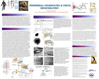
Transmitting electron micrographs of Schwann cells
and their axons showing the effect of the
neurotrophic factors BDNF and GDNF in promoting
regenerative sprouting of axons after nerve injury and
surgical repair. A: A single axon is visible in
association with a myelinating Schwann cell in the
intact nerve. B: One month after nerve transection of
the tibial nerve and surgical cross-union of tibial
(proximal nerve stump) and common peroneal (distal
nerve stump) nerves in Sprague-Dawley rats, typical
regenerating units, consisting of a Schwann cell and
several (4) daughter axons, are seen after daily saline
administration via an implanted mini-osmotic pump.
C: The number of axon sprouts/Schwann cells is
increased following exogenous GDNF
administration. D: The number of axon
sprouts/Schwann cells is increased further following
exogenous daily administration of both GDNF and
BDNF. The arrows point to axon profiles.
Peripheral neuropathy describes damages to the peripheral nervous system, the vast communications
network responsible for conducting information into and out of the CNS. This information is
deciphered by the brain as being pain, pleasure, pressure and temperature. These sensations can be
can be interfered with by damage to the PNS as described in the pictures. Distortion of this kind can
be caused by the degeneration of the myelin sheath around peripheral nerves and or possibly a
complete severing of the axon (nerve fiber). The purpose of the presentation is outline the factors
contributing to peripheral neuropathy provide information in its treatment and prevention and to take
more detailed look at a study done on rodents that regenerated nerve cells and deciphered some
related chemical signal transduction mechanisms. Genetically inherited peripheral neuropathy has no
treatment. Understanding the biological mechanisms in repairing this vital tissue is an important and
necessary step forward for science simply for its countless future applications in the medical field.
This review considers the two sources of neurotrophic factors in the peripheral nervous system
(PNS), the neurons and the nonneuronal cells in the denervated distal nerve stumps, and their role in
axon regeneration. Morphological assessment of regenerative success in response to administration
of exogenous growth factors after nerve injury and repair has indicated a role of the endogenous
neurotrophic factors from Schwann cells in the distal nerve stump. However, the increased number
of axons may reflect more neurons regenerating their axons and/or increased numbers of axon
sprouts from the same number of neurons. Using fluorescent dyes to count neurons that regenerated
their axons across a suture site and into distal nerve stumps, brain-derived neurotrophic factor
(BDNF) and glial cell–derived neurotrophic factor (GDNF) were found not to increase the number
of neurons that regenerated their axons after immediate nerve repair. Nevertheless, the factors did
reverse the deleterious effect of delayed nerve repair, indicating that the axons that regenerate into
the distal nerve stump normally have access to sufficient levels of endogenous neurotrophic factors
to sustain their regeneration, while neurons that do not have access to these factors require
exogenous factors to sustain axon regeneration. Neurons upregulate neurotrophic factors after
axotomy. The upregulation is normally slow, beginning after 7 days and occurring in association
with a protracted period of axonal regeneration in which axons grow out from the proximal nerve
stump across a suture site over a period of 1 month in rodents. This staggered axon regeneration
across the suture site is accelerated by a 1-hour period of low-frequency electrical stimulation that
simultaneously accelerates the expression of BDNF and its trkB receptor in the neurons. Elevation
of the level of BDNF after 2 days to > 3 times that found in unstimulated neurons was accompanied
by elevation of the level of cAMP and followed by accelerated upregulation of growth-associated
genes, tubulin, actin, and GAP-43 and downregulation of neurofilament protein. Elevation of
cAMP levels via rolipram inhibition of phosphodiesterase 4 mimicked the effect of the low-
frequency electrical stimulation. In conclusion, the enhanced upregulation of neurotrophic factors in
the electrically stimulated axotomized neurons accelerates axon outgrowth into the distal nerve
stumps where endogenous sources of growth factors in the Schwann cells support the regeneration
of the axons toward the denervated targets. The findings provide strong support for endogenous
neurotrophic factors of axotomized neurons and of denervated Schwann cells playing a critical role
in supporting axon regeneration in the PNS. A visual synopsis of the anatomy of the PNS and some
types of peripheral neuropathy are provided. Following these pictures are research related pictures
accompanied by text to illustrate the methods and results used in this multifaceted experiment.
Some portions of the research had to be omitted due to constraints on space.
Treatment is dependent on the symptoms and causes. There has been research on
animals that has shown that neurotrophin-3 can curb the loss of myelin. Drugs
originally intended as antidepressants and antiepileptic that act on the central nervous
system, have been found to be useful against neuropathic pain. Use of tricyclic
antidepressants, such as gabapentin or sodium valproate, has been known to be
beneficial in some cases, and are relatively low cost. Recent studies have shown some
natural therapies to be safe. Alpha-lipoic acid, an anti-oxidant found in many foods, is
an effective remedy for relieving pain in peripheral neuropathy. Patients have shown
appreciable improvement in health, by following techniques like yoga, meditation and
deep-breathing.