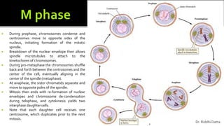
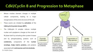
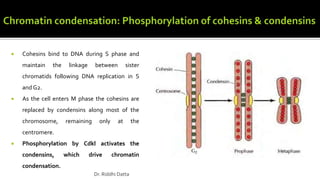
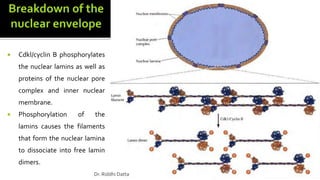
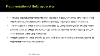
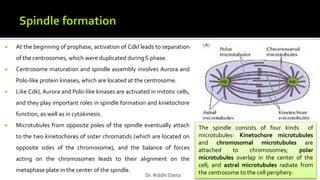
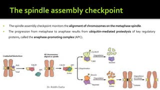
SlideShare a Scribd company logo
1 of 50
WBSU
Semester I
Botany Core Course II (Group B)
Dr. Riddhi Datta
 A typical eukaryotic cell cycle is illustrated by human cells in culture, which divide
approximately every 24 hours.
 The cell cycle is divided into two basic parts:
 mitosis (or meiosis)
 interphase.
 Mitosis and cytokinesis last only about an hour, so approximately 95% of the cell
cycle is spent in interphase-the period between mitoses.
 However, interphase is the time during which both cell growth and DNA
replication occur in an orderly manner in preparation for cell division.
Dr. Riddhi Datta
 The timing of DNA synthesis thus divides the cycle of eukaryotic
cells into four discrete phases:
 The M phase of the cycle corresponds to mitosis, which is
usually followed by cytokinesis.
 This phase is followed by the G1 phase (gap 1), which
corresponds to the interval (gap) between mitosis and
initiation of DNA replication. During G1 the cell is
metabolically active.
 G1 is followed by S phase (synthesis) during which DNA
replication takes place.
 The completion of DNA synthesis is followed by the G2 phase
(gap 2) during which cell growth continues and proteins are
synthesized in preparation for mitosis.
Dr. Riddhi Datta
 Cells at different stages of cell cycle can be differentiated by their
DNA content.
 Animal cells in G1 are diploid (2n). During S phase, replication
increase the DNA content from 2n to 4n. It remains in the 4n stage in
G2 phase as well and is then decreased to 2n stage after M phase.
 Some cells in adult animals cease division altogether (e.g., nerve
cells) and many other cells divide only occasionally (e.g. skin
fibroblasts).
 These cells exit G1 to enter a quiescent stage of the cycle called G0
phase, where they remain metabolically active but no longer
proliferate unless called on to do so by appropriate intracellular
signals.
Dr. Riddhi Datta
 The progression of cells through the division cycle is regulated by extracellular signals from
the environment, as well as by internal signals that monitor and coordinate the various
processes that take place during different cell cycle phases.
 This is accomplished by some control points that regulate progression through the cell
cycle:
 START
 Restriction point
 Control of G2 to M transition
Dr. Riddhi Datta
 A major cell cycle regulatory point in many types of cells occurs late in G1 and controls
progression from G1 to S.
 This regulatory point was first defined by studies of budding yeast (Saccharomyces
cerevisiae), where it is known as START.
 Once cells have passed START, they are committed to entering S phase and undergoing one
cell division cycle.
 The importance of this regulation is particularly evident in budding yeasts in which cell
division produces progeny cells of very different sizes: a large mother cell and a small
daughter cell.
Dr. Riddhi Datta
 Some of the activities monitored at this check point are:
 Optimum cell size: In order for yeast cells to maintain a constant size, the small daughter cell, after one
division, must grow more than the large mother cell does before they divide again. This regulation is
accomplished by a control mechanism that requires each cell to reach a minimum size before it can pass
START.
 Nutrient status: If yeasts are faced with a shortage of
nutrients, they arrest their cell cycle at START and enter
a resting state rather than proceeding to S phase. Thus
START represents a decision point at which the cell
determines whether sufficient nutrients are available to
support progression through the rest of the division
cycle.
 Polypeptide factors that signal yeast mating also arrest
the cell cycle at START, allowing haploid yeast cells to
fuse with one another instead of progressing to S phase.
Dr. Riddhi Datta
 Such arrested cells then enter a quiescent stage, called G0, in which they can
remain for long periods of time without proliferating. G0 cells are metabolically
active, although they cease growth and have reduced rates of protein synthesis.
 For example, skin fibroblasts are arrested in G0 until they are stimulated to
divide as required to repair damage resulting from a wound. The proliferation of
these cells is triggered by platelet-derived growth factor, which is released from
blood platelets during clotting and signals the proliferation of fibroblasts in the
vicinity of the injured tissue.
 In animals, a decision point in late G1, called restriction point, functions analogously to START in yeasts.
 In contrast to yeasts, however, the passage of animal cells through the cell cycle is regulated primarily by the
extracellular growth factors that signal cell proliferation, rather than by the availability of nutrients.
 In the presence of the appropriate growth factors, cells pass the restriction point and enter S phase. Once it has
passed through the restriction point, the cell is committed to proceed through S phase and the rest of the cell
cycle. On the other hand, if appropriate growth factors are not available in G1 progression through the cell cycle
stops at the restriction point.
Dr. Riddhi Datta
 Although the proliferation of most cells is regulated primarily in G1 some cell cycles are
instead controlled principally in G2.
 In fission yeast Schizosaccharomyces pombe, in contrast to Saccharomyces cerevisiae, the
cell cycle is regulated primarily by control of the transition from G2 to M, which is the
principal point at which cell size and nutrient availability are monitored.
 Vertebrate oocytes can remain arrested in G 2 for long periods of time (several decades in
humans) until their progression to M phase is triggered by hormonal stimulation.
Extracellular signals can thus control cell proliferation by regulating progression from the
G2 to M phase of the cell cycle.
 In addition, some haploid mosses use G2 control point.
Dr. Riddhi Datta
 In addition to cell size and extracellular signals, the events that take place during
different stages of the cell cycle must be coordinated with one another so that they occur
in the appropriate order.
 This coordination between different phases of the cell cycle is dependent on a series of
cell cycle checkpoints that prevent entry into the next phase of the cell cycle until the
events of the preceding phase have been completed.
Stage of cell cycle
progression arrest
Response
G1 DNA Damage
S DNA Damage/incomplete DNA replication
G2 DNA Damage/incomplete DNA replication
M Chromosome misalignment
Dr. Riddhi Datta
 Several cell cycle checkpoints function to ensure that incomplete or damaged
chromosomes are not replicated and passed on to daughter cells.
 Cell cycle arrest at the G1, S, and G2 checkpoints is mediated by two related protein
kinases, designated ATM and ATR, that recognize damaged or unreplicated DNA and
are activated in response to DNA damage.
 ATM and ATR then activate a signaling pathway that leads not only to cell cycle
arrest, but also to the activation of DNA repair and, in some cases, programmed cell
death.
Dr. Riddhi Datta
 Another important cell cycle checkpoint that maintains the integrity of the genome occurs
toward the end of mitosis .
 This check point, called the spindle assembly checkpoint, monitors the alignment of
chromosomes on the mitotic spindle, thus ensuring that a complete set of chromosomes is
distributed accurately to the daughter cells.
 The failure of one or more chromosomes to align properly on the spindle causes mitosis to
arrest at metaphase, prior to the segregation of the newly replicated chromosomes to
daughter nuclei.
 As a result of the spindle assembly checkpoint, the chromosomes do not separate until a
complete complement of chromosomes has been organized for distribution to each
daughter cell.
Dr. Riddhi Datta
 DNA replication is restricted to once per cell cycle by
the MCM helicase proteins that bind to origins of
replication together with ORC (origin recognition
complex) proteins and are required for the initiation
of DNA replication.
 MCM proteins are only able to bind to DNA in G1,
allowing DNA replication to initiate in S phase.
 Once initiation has occurred, the MCM proteins are
displaced so that replication cannot initiate again
until after mitosis.
Dr. Riddhi Datta
• Our current understanding of cell cycle regulation has emerged from a
convergence of results obtained through experiments on organisms as
diverse as yeasts, sea urchins, frogs, and mammals.
• Three initially distinct experimental approaches contributed to identifica-
tion of the key molecules responsible for cell cycle regulation.
Dr. Riddhi Datta
1st approach:
 In 1971, two independent teams of researchers (Yoshio Masui and Clement Markert, as
well as Dennis Smith and Robert Ecker) found that oocytes arrested in G2 could be
induced to enter M phase by microinjection of cytoplasm from oocytes that had been
hormonally stimulated.
 Thus a cytoplasmic factor present in hormone treated oocytes was sufficient to
trigger transition from G2 to M.
 Because the entry of oocytes into meiosis is frequently referred to as oocyte
maturation, this cytoplasmic factor was called maturation promoting factor (MPF).
 However, MPF also induces entry into M phase of the mitotic cycle. MPF thus
appeared to act as a general regulator of the transition from G2 to M. Dr. Riddhi Datta
2nd approach:
 Studying the budding yeast Saccharomyces cerevisiae, Hartwell and his colleagues in the early 1970s identified
temperature-sensitive mutants that were defective in cell cycle progression.
 The key characteristic of these mutants (called
cdc for cell division cycle mutants) was that
they underwent growth arrest at specific points
in the cell cycle.
 A particularly important mutant designated
cdc28 caused the cell cycle to arrest at START,
indicating that the Cdc28 protein is required for
passage through this critical regulatory point in
G1. Dr. Riddhi Datta
 A similar collection of cell cycle mutants was isolated in the fission yeast Schizosaccharomyces
pombe by Paul Nurse and his collaborators.
 These mutants included cdc2, which arrests the S. pombe cell cycle both in G1 and at the G2
to M transition (the major regulatory point in fission yeast).
 Further studies showed that S. cerevisiae Cdc28 and S. pombe Cdc2 are functionally
homologous genes, which are required for passage through START as well as for entry into
mitosis in both species of yeasts.
Dr. Riddhi Datta
 Further studies of cdc2 yielded two important insights:
 Molecular cloning and nucleotide sequencing revealed that Cdc2 encodes a
protein kinase—the first indication of the prominent role of protein
phosphorylation in regulating the cell cycle.
 A human gene related to Cdc2 was identified and shown to function in yeasts,
providing a dramatic demonstration of the conserved activity of this cell cycle
regulator.
Dr. Riddhi Datta
3rd approach:
 In 1983, Tim Hunt and his colleagues identified two proteins that display a periodic pattern of accumulation and
degradation in sea urchin and clam embryos.
 These proteins accumulate throughout interphase and are then rapidly degraded toward the end of each
mitosis.
 Hunt called these proteins cyclins (the two proteins were designated cyclin A and cyclin B) and suggested that
they might function to induce mitosis, with their periodic accumulation and destruction controlling entry and exit
from M phase.
 Direct support for such a role was provided in 1986,
when Joan Ruderman and her colleagues showed that
microinjection of cyclin A into frog oocytes is
sufficient to trigger the G2 to M transition.
Dr. Riddhi Datta
 These initially independent approaches converged dramatically in 1988 when MPF was purified
from frog eggs in the laboratory of James Maller.
 MPF, a conserved regulator of the cell cycle is composed of two key subunits:
 Cdkl
 cyclin B
 Cyclin B is a regulatory subunit required for catalytic activity of the Cdkl protein kinase.
 MPF activity is controlled by periodic accumulation and degradation of cyclin B during cell cycle
progression.
Dr. Riddhi Datta
 Cdkl forms complexes with cyclin B duringG2.
 CdkI is then phosphorylated on threonine-161 by
MO15, which is required for Cdkl activity.
 It is then phosphorylated on tyrosine-15 (and
threonine-14 in vertebrate cells) by a protein kinase
called Wee1, which inhibits Cdkl activity and leads to
the accumulation of inactive Cdkl/cyclin B complexes
throughout G2 phase.
 Dephosphorylation of Thr14 and Tyr15 by a protein
phosphatase called Cdc25C activates MPF at the G2
to M transition.
 Activated Cdkl protein kinase
phosphorylates a variety of target
proteins that initiate the M phase.
 MPF activity is terminated at the end of
mitosis by degradation of cyclin B. Dr. Riddhi Datta
The activity of Cdk's during cell cycle progression is regulated by four molecular mechanisms:
Activities of CdkI/cyclin B
complexes can also be
regulated by binding of
inhibitory proteins called CKI’s
(Cdk inhibitors). In mammals
2 families of CKI’s are present:
 Ink4 family: binds to Cdk4
& Cdk6; inhibits G1 to S
progression
 Kip/Cip family: binds to
Cdk1 & Cdk2; inhibits
various phases of cell
cycle progression
Dr. Riddhi Datta
 In yeast, passage through START is
controlled by Cdkl in association with G1
cyclins (Cln1 , Cln2, and Cln3).
 Complexes of Cdkl with distinct B-type
cyclins (Clb’s) then regulate progression
through S phase and entry into mitosis.
 In animal cells, progression through the G1 restriction point is
controlled by complexes of Cdk4 and Cdk6 with D-type cyclins.
 Cdk2/cyclin E complexes function later in G1 and are required for
the G1 to S transition.
 Cdk2/ cyclin A complexes are then required for progression
through S phase.
 CdkI/cyclin A regulates progression to G2 Cdkl/cyclin B complexes
drive the G2 to M transition.
Dr. Riddhi Datta
 Growth factors regulate cell cycle progression through the G1 restriction point by inducing
synthesis of D-type cyclins via the Ras/Raf/MEK/ERK signaling pathway.
 Cyclin D1 continues to be synthesized as long as growth factors are present. However, cyclin D1 is
also rapidly degraded.
 As long as growth factors are present through G1,
complexes of Cdk4, 6/ cyclin D1 drive cells through the
restriction point.
 If growth factors are removed prior to this key
regulatory point, the levels of cyclin D1 rapidly fall and
cells are unable to progress through G1 to S, instead
becoming quiescent and entering G0. Dr. Riddhi Datta
 While cyclin D and oncogene proteins like Ras drive cell proliferation, proteins encoded by
many tumor suppressor genes (like Rb and Ink4 Cdk inhibitors ) act as brakes that slows
down cell cycle progression.
 Mutations resulting in continual unregulated expression of cyclin D1 contribute to the
development of a variety of human cancers, including lymphomas and breast cancers.
 Similarly, mutations that inactivate the Ink4 Cdk inhibitors that bind to Cdk4 and Cdk6 are
commonly found in human cancer cells.
Dr. Riddhi Datta
 Connection between cyclin D and cancer is further fortified by the fact that a key substrate protein of Cdk4, 6/
cyclin D complexes, Rb, is itself frequently mutated in a wide array of human tumors.
 Rb is the prototype of a tumor suppressor gene whose inactivation leads to tumor development.
 In its under-phosphorylated form, Rb binds to members
of the E2F family (transcription factors that initiates
transcription of genes required for initiation of S phase).
This represses transcription of E2F-regulated genes.
 Phosphorylation of Rb by Cdk4,6/cyclin D complexes
results in its dissociation from E2F in late G1.
 E2F then stimulates expression of its target genes,
which encode proteins required for cell cycle
progression. Dr. Riddhi Datta
 Progression through the restriction point and
entry into S phase is mediated by the activation of
Cdk2/cyclin E complexes.
 ln early G1, Cdk2/cyclin E complexes are inhibited
by the Cdk inhibitor p27. Passage through the
restriction point induces the synthesis of cyclin E
via activation of E2F.
 In addition, growth factor signaling reduces the
levels of p27 by inhibiting its transcription and
translation. The resulting activation of Cdk2/ cyclin
E leads to activation of the MCM helicase and
initiation of DNA replication (i.e. S phase).
Dr. Riddhi Datta
 DNA damage check points arrests cell cycle progression in
response to damaged or incompletely replicated DNA.
 Cell cycle arrest at the DNA damage checkpoints is initiated
by the ATM or ATR protein kinases, which are components
of protein complexes that recognize damaged or un-
replicated DNA.
 ATM is activated principally by double-strand breaks, while
ATR is activated by single stranded or un-replicated DNA.
 ATM and ATR then phosphorylate and activate the CHK2
and CHKI protein kinases, respectively. CHKI and CHK2
phosphorylate and inhibit the Cdc25A and Cdc25C protein
phosphatases.
 Cdc25A and Cdc25C are required to activate Cdk2 and Cdkl,
respectively, so their inhibition leads to arrest at the DNA
damage checkpoints in G1, S, and G2. Dr. Riddhi Datta
 In mammalian cells, arrest at the G1 checkpoint is also mediated by the action of
an additional protein known as p53, which is phosphorylated by both ATM and
CHK2.
 Phosphorylation by ATM and CHK2 stabilize p53, resulting in rapid increases in
p53 levels in response to DNA damage.
 The protein p53 then activates transcription of the gene encoding the Cdk
inhibitor p21, leading to inhibition of Cdk2/cyclin E complexes and cell cycle
arrest.
Loss of p53 function as a result of mutations prevents G1 arrest in response to DNA damage,
so the damaged DNA is replicated and passed on to daughter cells instead of being repaired.
This inheritance of damaged DNA results in an increased frequency of mutations and general
instability of the cellular genome, which contributes to cancer development.
Dr. Riddhi Datta
 During prophase, chromosomes condense and
centrosomes move to opposite sides of the
nucleus, initiating formation of the mitotic
spindle.
 Breakdown of the nuclear envelope then allows
spindle microtubules to attach to the
kinetochores of chromosomes.
 During pro-metaphase the chromosomes shuffle
back and forth between the centrosomes and the
center of the cell, eventually aligning in the
center of the spindle (metaphase).
 At anaphase, the sister chromatids separate and
move to opposite poles of the spindle.
 Mitosis then ends with re-formation of nuclear
envelopes and chromosome de-condensation
during telophase, and cytokinesis yields two
interphase daughter cells.
 Note that each daughter cell receives one
centrosome, which duplicates prior to the next
mitosis.
Dr. Riddhi Datta
Dr. Riddhi Datta
 Mitosis involves dramatic changes in multiple
cellular components, leading to a major
reorganization of the entire structure of the cell.
 These events are initiated by activation of the
Cdkl/cyclin B protein kinase (MPF).
 The Cdkl/cydin B complex induces multiple
nuclear and cytoplasmic changes at the onset of
M phase both by activating other protein kinases
and by phosphorylating proteins such as
condensins, components of the nuclear
envelope, Golgi matrix proteins, and proteins
associated with centrosomes and microtubules.
Dr. Riddhi Datta
 Cohesins bind to DNA during S phase and
maintain the linkage between sister
chromatids following DNA replication in S
and G2.
 As the cell enters M phase the cohesins are
replaced by condensins along most of the
chromosome, remaining only at the
centromere.
 Phosphorylation by Cdkl activates the
condensins, which drive chromatin
condensation.
Dr. Riddhi Datta
 Cdkl/cyclin B phosphorylates
the nuclear lamins as well as
proteins of the nuclear pore
complex and inner nuclear
membrane.
 Phosphorylation of the
lamins causes the filaments
that form the nuclear lamina
to dissociate into free lamin
dimers.
Dr. Riddhi Datta
 The Golgi apparatus fragments into small vesicles at mitosis, which may either be absorbed
into the endoplasmic reticulum or distributed directly to daughter cells at cytokinesis.
 The breakdown of these membranes is mediated by Cdkl phosphorylation of Golgi matrix
proteins (such as GM130 and GRASP-65), which are required for the docking of COPI-
coated vesicles to the Golgi membrane.
 Phosphorylation of these proteins by Cdkl inhibits vesicle docking and fusion, leading to
fragmentation of the Golgi apparatus.
Dr. Riddhi Datta
 At the beginning of prophase, activation of Cdkl leads to separation
of the centrosomes, which were duplicated during S phase.
 Centrosome maturation and spindle assembly involves Aurora and
Polo-like protein kinases, which are located at the centrosome.
 Like Cdkl, Aurora and Polo-like kinases are activated in mitotic cells,
and they play important roles in spindle formation and kinetochore
function, as well as in cytokinesis.
 Microtubules from opposite poles of the spindle eventually attach
to the two kinetochores of sister chromatids (which are located on
opposite sides of the chromosome), and the balance of forces
acting on the chromosomes leads to their alignment on the
metaphase plate in the center of the spindle.
The spindle consists of four kinds of
microtubules: Kinetochore microtubules
and chromosomal microtubules are
attached to chromosomes; polar
microtubules overlap in the center of the
cell; and astral microtubules radiate from
the centrosome to the cell periphery.Dr. Riddhi Datta
 The spindle assembly checkpoint monitors the alignment of chromosomes on the metaphase spindle.
 The progression from metaphase to anaphase results from ubiquitin-mediated proteolysis of key regulatory
proteins, called the anaphase-promoting complex (APC).
Dr. Riddhi Datta
 Activation of APC is induced at the beginning of mitosis, so the activation of Cdkl/cyclin B ultimately triggers its
own destruction.
 The APC remains inhibited until the cell passes the spindle assembly checkpoint, after which activation of ubiquitin
degradation system brings about the transition from metaphase to anaphase and progression through the rest of
mitosis.
Dr. Riddhi Datta
 Unattached kinetochores lead to assembly of Mad/Bub protein complex that bind to Cdc20-a required
component of the APC. Mad proteins are activated in this complex, and then released in active form that inhibits
Cdc20, maintaining the APC in inactive state.
 Once all chromosomes are aligned on the spindle, the Mad/Bub complex dissociates, relieving inhibition of
Cdc20 and leading to APC activation.
Dr. Riddhi Datta
 Activation of APC results in ubiquitination and degradation of two key target proteins.
 APC ubiquitinates cyclin B, leading to its degradation and inactivation of Cdkl.
 APC also ubiquitinates securin, leading to activation of separase.
 Separase degrades a subunit of cohesin, breaking the link between sister chromatids and initiating anaphase.
Dr. Riddhi Datta
Dr. Riddhi Datta
Dr. Riddhi Datta
Growth factors
Ras/Raf/ ERK/MEK
Synthesis of Cyclin D
Cdk4,6/CycD
Rb
E2F
Rb
E2F
P
Transcriptional activation
Cyclin E
Cyclin E
Cdk2
p27
Growth factorsp27
Cyclin E
Cdk2
MCM
helicase
ATMATR
Double strand breakSingle strand break
CHK1
Cdc25C Cdc25A
Cdk2
Cdk1
PP
p53
p21
CHK2
Unattached kinetochore
Mad
Bub
Mad Cdc20
APC
All kinetochores attached
Bub
Bub
Mad
Cdc20
APC
Cyclin B degraded
Cdk1 inactive
Securin degraded
Separase active
Anaphase
Dr. Riddhi Datta
 Vertebrate oocytes (developing eggs) have been particularly useful
models for research on the cell cycle.
 The discovery and subsequent purification of MPF (Cdkl/cyclin B) was
made from frog oocytes.
 The first regulatory point in oocyte meiosis is in the diplotene stage
of the first meiotic division.
 Meiosis is arrested at the diplotene stage, during which oocytes grow
to a large size.
 Oocytes then resume meiosis in response to hormonal stimulation
and complete the first meiotic division, with asymmetric cytokinesis
giving rise to a small polar body.
 Most vertebrate oocytes are then arrested again at metaphase II.
Dr. Riddhi Datta
 Like the M phase of somatic cells, the meiosis of
oocytes is controlled by the activity of Cdkl/cyclinB
complexes.
 The regulation of Cdkl during oocyte meiosis,
however, displays unique features that are
responsible for progression from meiosis l to
meiosis II and for metaphase II arrest.
 Hormonal stimulation of diplotene-arrested
oocytes initially triggers the resumption of meiosis
by activating Cdkl, as at the G2 to M transition of
somatic cells.
Dr. Riddhi Datta
 Cdkl then induces chromosome condensation,
nuclear envelope breakdown, and formation of
the spindle.
 Activation of the anaphase-promoting complex
then leads to the metaphase to anaphase
transition of meiosis I, accompanied by a decrease
in the activity of Cdkl.
 However, in contrast to mitosis, Cdkl activity is
only partially decreased, so the oocyte remains
in M phase, chromatin remains condensed, and
nuclear envelopes do not re-form.
 Following cytokinesis, Cdkl activity again rises
and remains high while the egg is arrested at
metaphase II.
Dr. Riddhi Datta
 Cytoplasm from a metaphase II egg is microinjected into
one cell of a two-cell embryo.
 The injected embryo cell arrests at metaphase, while the
uninjected cell continues to divide.
 A factor in metaphase II in egg cytoplasm (cytostatic
factor) therefore has induced metaphase arrest of the
injected embryo cell.
Dr. Riddhi Datta
 More recent experiments have identified a protein-
serine/threonine kinase known as Mos as an essential component
of CSF.
 Mos is specifically synthesized in oocytes around the time of
completion of meiosis I.
 The Mos protein kinase maintains Cdkl I cyclin B activity both by
stimulating the synthesis of cyclin B and by inhibiting the
degradation of cyclin B by the anaphase-promoting complex.
 The action of Mos is mediated by MEK, ERK, and Rsk protein
kinases, and inhibition of the anaphase-promoting complex is
mediated by Mad/Bub proteins.
 Mos thus maintains Cdkl/cyclin B activity during oocyte meiosis,
leading to the arrest of oocytes at metaphase II.
 Oocytes can remain arrested at this point in the meiotic cell cycle
for several days, awaiting fertilization.
Dr. Riddhi Datta
 At fertilization, the sperm binds to a receptor on the surface of the egg and fuses with the egg plasma membrane,
initiating the development of a new diploid organism containing genetic information derived from both parents.
 A sperm binds to a plasma membrane receptor of the egg and this induces an increase in Ca2+ level in egg cytoplasm,
via hydrolysis of PIP2 (phosphatidylinositol4,5-isphosphate).
 The Ca2+ induces exocytosis of secretory vesicles that are present in large numbers beneath the egg plasma membrane.
 This, in turn, induces surface alterations that prevent additional sperm from entering the egg.
 The increase in cytosolic Ca2+ following fertilization also signals the completion of meiosis.
 Following completion of oocyte meiosis, the fertilized egg (zygote) contains two haploid nuclei (called pronuclei), one
derived from each parent.
 The two pronuclei then enter S phase and replicate their DNA as they migrate toward each other.
 As they meet, the zygote enters M phase of its first mitotic division.
Dr. Riddhi Datta
Dr. Riddhi Datta

More Related Content

What's hot

Cell cycle regulation 17BCB0016
Cell cycle regulation 17BCB0016Cell cycle regulation 17BCB0016
Cell cycle regulation 17BCB0016AkanchaAgarwal1
 
Cyclin dependent kinases
Cyclin dependent kinasesCyclin dependent kinases
Cyclin dependent kinasesMandira bhosale
 
Cell cycle and cell death
Cell cycle and cell deathCell cycle and cell death
Cell cycle and cell deathVamsi kumar
 
Cell cycle and regulation
Cell cycle and regulation Cell cycle and regulation
Cell cycle and regulation mohamed abusalih
 
Prokaryotic Replication presentation
Prokaryotic Replication presentationProkaryotic Replication presentation
Prokaryotic Replication presentationUsama Aamir
 
Cel cycle and its regulation
Cel cycle and its regulationCel cycle and its regulation
Cel cycle and its regulationarti yadav
 
Transcription in prokaryotes
Transcription in prokaryotesTranscription in prokaryotes
Transcription in prokaryotesKaayathri Devi
 
Polyadenylation
PolyadenylationPolyadenylation
PolyadenylationEmaSushan
 
cell cycle and its check points and regulation
cell cycle and its check points and regulationcell cycle and its check points and regulation
cell cycle and its check points and regulationSayanti Sau
 
Cell cycle regulation and checkpoints
Cell cycle regulation and checkpointsCell cycle regulation and checkpoints
Cell cycle regulation and checkpointsDhaval Bhatt
 
CELL CYCLE , MITOSIS ,MEIOSIS AND CELL REGULATION
CELL CYCLE , MITOSIS ,MEIOSIS AND CELL  REGULATIONCELL CYCLE , MITOSIS ,MEIOSIS AND CELL  REGULATION
CELL CYCLE , MITOSIS ,MEIOSIS AND CELL REGULATIONLIFE SCIENCES
 
Regulation of gene expression in eukaryotes
Regulation of gene expression in eukaryotesRegulation of gene expression in eukaryotes
Regulation of gene expression in eukaryotesKristu Jayanti College
 
Transcription in Eukaryotes
Transcription in EukaryotesTranscription in Eukaryotes
Transcription in EukaryotesRuchiRawal1
 
Assignment on Cell signaling
Assignment on Cell signalingAssignment on Cell signaling
Assignment on Cell signalingDeepak Kumar
 
Mitochondrial genome and its manipulation
Mitochondrial genome and its manipulationMitochondrial genome and its manipulation
Mitochondrial genome and its manipulationAvinash Gowda H
 
Transcription in eukaryotes
Transcription in eukaryotesTranscription in eukaryotes
Transcription in eukaryotesHemantkrdu
 

What's hot (20)

Cell cycle regulation 17BCB0016
Cell cycle regulation 17BCB0016Cell cycle regulation 17BCB0016
Cell cycle regulation 17BCB0016
 
Transcription in eukaryotes
Transcription in eukaryotesTranscription in eukaryotes
Transcription in eukaryotes
 
Cyclin dependent kinases
Cyclin dependent kinasesCyclin dependent kinases
Cyclin dependent kinases
 
Cell cycle and cell death
Cell cycle and cell deathCell cycle and cell death
Cell cycle and cell death
 
Cell cycle and regulation
Cell cycle and regulation Cell cycle and regulation
Cell cycle and regulation
 
Prokaryotic Replication presentation
Prokaryotic Replication presentationProkaryotic Replication presentation
Prokaryotic Replication presentation
 
Cel cycle and its regulation
Cel cycle and its regulationCel cycle and its regulation
Cel cycle and its regulation
 
Transcription in prokaryotes
Transcription in prokaryotesTranscription in prokaryotes
Transcription in prokaryotes
 
Transcription regulatory elements
Transcription regulatory elementsTranscription regulatory elements
Transcription regulatory elements
 
Cell Cycle Regulation
Cell Cycle RegulationCell Cycle Regulation
Cell Cycle Regulation
 
Polyadenylation
PolyadenylationPolyadenylation
Polyadenylation
 
cell cycle and its check points and regulation
cell cycle and its check points and regulationcell cycle and its check points and regulation
cell cycle and its check points and regulation
 
Cell cycle regulation and checkpoints
Cell cycle regulation and checkpointsCell cycle regulation and checkpoints
Cell cycle regulation and checkpoints
 
CELL CYCLE , MITOSIS ,MEIOSIS AND CELL REGULATION
CELL CYCLE , MITOSIS ,MEIOSIS AND CELL  REGULATIONCELL CYCLE , MITOSIS ,MEIOSIS AND CELL  REGULATION
CELL CYCLE , MITOSIS ,MEIOSIS AND CELL REGULATION
 
Regulation of gene expression in eukaryotes
Regulation of gene expression in eukaryotesRegulation of gene expression in eukaryotes
Regulation of gene expression in eukaryotes
 
Transcription in Eukaryotes
Transcription in EukaryotesTranscription in Eukaryotes
Transcription in Eukaryotes
 
Assignment on Cell signaling
Assignment on Cell signalingAssignment on Cell signaling
Assignment on Cell signaling
 
Prokaryotic transcription
Prokaryotic transcriptionProkaryotic transcription
Prokaryotic transcription
 
Mitochondrial genome and its manipulation
Mitochondrial genome and its manipulationMitochondrial genome and its manipulation
Mitochondrial genome and its manipulation
 
Transcription in eukaryotes
Transcription in eukaryotesTranscription in eukaryotes
Transcription in eukaryotes
 

Similar to Cell cycle regulation

Similar to Cell cycle regulation (20)

Eukaryotic cell division cycle
Eukaryotic cell division cycleEukaryotic cell division cycle
Eukaryotic cell division cycle
 
Cell cycle and cell division
Cell cycle and cell divisionCell cycle and cell division
Cell cycle and cell division
 
Lecture 18 cell cycle and its regulation converted
Lecture 18 cell cycle and its regulation convertedLecture 18 cell cycle and its regulation converted
Lecture 18 cell cycle and its regulation converted
 
cellcycleanditsregulation11.pptx
cellcycleanditsregulation11.pptxcellcycleanditsregulation11.pptx
cellcycleanditsregulation11.pptx
 
Cell cycle
Cell cycleCell cycle
Cell cycle
 
18042020100230Cell cycle.pdf
18042020100230Cell cycle.pdf18042020100230Cell cycle.pdf
18042020100230Cell cycle.pdf
 
Cell cycle
Cell cycleCell cycle
Cell cycle
 
Cell cycle and signaling
Cell cycle and signalingCell cycle and signaling
Cell cycle and signaling
 
Cell Cycle- an essential part of cell growth
Cell Cycle- an essential part of cell growthCell Cycle- an essential part of cell growth
Cell Cycle- an essential part of cell growth
 
Cell cycle By KK Sahu Sir
Cell cycle By KK Sahu SirCell cycle By KK Sahu Sir
Cell cycle By KK Sahu Sir
 
Cell cycle
Cell cycleCell cycle
Cell cycle
 
Cell cycle and signaling
Cell cycle and signalingCell cycle and signaling
Cell cycle and signaling
 
Cell cycle, check point,
Cell cycle, check point, Cell cycle, check point,
Cell cycle, check point,
 
Cell cycle and mitosis
Cell cycle and mitosisCell cycle and mitosis
Cell cycle and mitosis
 
cell cycle.pdf
cell cycle.pdfcell cycle.pdf
cell cycle.pdf
 
Cell cycle
Cell cycleCell cycle
Cell cycle
 
Cell division
Cell divisionCell division
Cell division
 
Concept 12.3
Concept 12.3Concept 12.3
Concept 12.3
 
Cell cycle
Cell cycleCell cycle
Cell cycle
 
Cell cycle
Cell cycleCell cycle
Cell cycle
 

More from Riddhi Datta

Regulation of Floral Development
Regulation of Floral DevelopmentRegulation of Floral Development
Regulation of Floral DevelopmentRiddhi Datta
 
Introduction to Chromosomal Aberration
Introduction to Chromosomal AberrationIntroduction to Chromosomal Aberration
Introduction to Chromosomal AberrationRiddhi Datta
 
A practical approach to Southern, Northern and Western Blot analyses
A practical approach to Southern, Northern and Western Blot analysesA practical approach to Southern, Northern and Western Blot analyses
A practical approach to Southern, Northern and Western Blot analysesRiddhi Datta
 
Introduction to the world of Fungi
Introduction to the world of FungiIntroduction to the world of Fungi
Introduction to the world of FungiRiddhi Datta
 
DNA as genetic material
DNA as genetic materialDNA as genetic material
DNA as genetic materialRiddhi Datta
 
Basics of Protein Translation
Basics of Protein TranslationBasics of Protein Translation
Basics of Protein TranslationRiddhi Datta
 
Plant Diseases (Part I)
Plant Diseases (Part I)Plant Diseases (Part I)
Plant Diseases (Part I)Riddhi Datta
 
Basics of DNA Structure
Basics of DNA StructureBasics of DNA Structure
Basics of DNA StructureRiddhi Datta
 
Basics of DNA Replication
Basics of DNA ReplicationBasics of DNA Replication
Basics of DNA ReplicationRiddhi Datta
 
Basics of Gene cloning
Basics of Gene cloningBasics of Gene cloning
Basics of Gene cloningRiddhi Datta
 
Types of Ecosystems
Types of EcosystemsTypes of Ecosystems
Types of EcosystemsRiddhi Datta
 
Exploring the nucleus
Exploring the nucleusExploring the nucleus
Exploring the nucleusRiddhi Datta
 
Basics of Lipid Biochemistry
Basics of Lipid BiochemistryBasics of Lipid Biochemistry
Basics of Lipid BiochemistryRiddhi Datta
 
Basics of Carbohydrate Biochemistry
Basics of Carbohydrate BiochemistryBasics of Carbohydrate Biochemistry
Basics of Carbohydrate BiochemistryRiddhi Datta
 

More from Riddhi Datta (14)

Regulation of Floral Development
Regulation of Floral DevelopmentRegulation of Floral Development
Regulation of Floral Development
 
Introduction to Chromosomal Aberration
Introduction to Chromosomal AberrationIntroduction to Chromosomal Aberration
Introduction to Chromosomal Aberration
 
A practical approach to Southern, Northern and Western Blot analyses
A practical approach to Southern, Northern and Western Blot analysesA practical approach to Southern, Northern and Western Blot analyses
A practical approach to Southern, Northern and Western Blot analyses
 
Introduction to the world of Fungi
Introduction to the world of FungiIntroduction to the world of Fungi
Introduction to the world of Fungi
 
DNA as genetic material
DNA as genetic materialDNA as genetic material
DNA as genetic material
 
Basics of Protein Translation
Basics of Protein TranslationBasics of Protein Translation
Basics of Protein Translation
 
Plant Diseases (Part I)
Plant Diseases (Part I)Plant Diseases (Part I)
Plant Diseases (Part I)
 
Basics of DNA Structure
Basics of DNA StructureBasics of DNA Structure
Basics of DNA Structure
 
Basics of DNA Replication
Basics of DNA ReplicationBasics of DNA Replication
Basics of DNA Replication
 
Basics of Gene cloning
Basics of Gene cloningBasics of Gene cloning
Basics of Gene cloning
 
Types of Ecosystems
Types of EcosystemsTypes of Ecosystems
Types of Ecosystems
 
Exploring the nucleus
Exploring the nucleusExploring the nucleus
Exploring the nucleus
 
Basics of Lipid Biochemistry
Basics of Lipid BiochemistryBasics of Lipid Biochemistry
Basics of Lipid Biochemistry
 
Basics of Carbohydrate Biochemistry
Basics of Carbohydrate BiochemistryBasics of Carbohydrate Biochemistry
Basics of Carbohydrate Biochemistry
 

Recently uploaded

How to Give a Domain for a Field in Odoo 17
How to Give a Domain for a Field in Odoo 17How to Give a Domain for a Field in Odoo 17
How to Give a Domain for a Field in Odoo 17Celine George
 
Beyond_Borders_Understanding_Anime_and_Manga_Fandom_A_Comprehensive_Audience_...
Beyond_Borders_Understanding_Anime_and_Manga_Fandom_A_Comprehensive_Audience_...Beyond_Borders_Understanding_Anime_and_Manga_Fandom_A_Comprehensive_Audience_...
Beyond_Borders_Understanding_Anime_and_Manga_Fandom_A_Comprehensive_Audience_...Pooja Bhuva
 
Food safety_Challenges food safety laboratories_.pdf
Food safety_Challenges food safety laboratories_.pdfFood safety_Challenges food safety laboratories_.pdf
Food safety_Challenges food safety laboratories_.pdfSherif Taha
 
TỔNG ÔN TẬP THI VÀO LỚP 10 MÔN TIẾNG ANH NĂM HỌC 2023 - 2024 CÓ ĐÁP ÁN (NGỮ Â...
TỔNG ÔN TẬP THI VÀO LỚP 10 MÔN TIẾNG ANH NĂM HỌC 2023 - 2024 CÓ ĐÁP ÁN (NGỮ Â...TỔNG ÔN TẬP THI VÀO LỚP 10 MÔN TIẾNG ANH NĂM HỌC 2023 - 2024 CÓ ĐÁP ÁN (NGỮ Â...
TỔNG ÔN TẬP THI VÀO LỚP 10 MÔN TIẾNG ANH NĂM HỌC 2023 - 2024 CÓ ĐÁP ÁN (NGỮ Â...Nguyen Thanh Tu Collection
 
Interdisciplinary_Insights_Data_Collection_Methods.pptx
Interdisciplinary_Insights_Data_Collection_Methods.pptxInterdisciplinary_Insights_Data_Collection_Methods.pptx
Interdisciplinary_Insights_Data_Collection_Methods.pptxPooja Bhuva
 
Key note speaker Neum_Admir Softic_ENG.pdf
Key note speaker Neum_Admir Softic_ENG.pdfKey note speaker Neum_Admir Softic_ENG.pdf
Key note speaker Neum_Admir Softic_ENG.pdfAdmir Softic
 
Python Notes for mca i year students osmania university.docx
Python Notes for mca i year students osmania university.docxPython Notes for mca i year students osmania university.docx
Python Notes for mca i year students osmania university.docxRamakrishna Reddy Bijjam
 
Fostering Friendships - Enhancing Social Bonds in the Classroom
Fostering Friendships - Enhancing Social Bonds  in the ClassroomFostering Friendships - Enhancing Social Bonds  in the Classroom
Fostering Friendships - Enhancing Social Bonds in the ClassroomPooky Knightsmith
 
Micro-Scholarship, What it is, How can it help me.pdf
Micro-Scholarship, What it is, How can it help me.pdfMicro-Scholarship, What it is, How can it help me.pdf
Micro-Scholarship, What it is, How can it help me.pdfPoh-Sun Goh
 
REMIFENTANIL: An Ultra short acting opioid.pptx
REMIFENTANIL: An Ultra short acting opioid.pptxREMIFENTANIL: An Ultra short acting opioid.pptx
REMIFENTANIL: An Ultra short acting opioid.pptxDr. Ravikiran H M Gowda
 
Unit 3 Emotional Intelligence and Spiritual Intelligence.pdf
Unit 3 Emotional Intelligence and Spiritual Intelligence.pdfUnit 3 Emotional Intelligence and Spiritual Intelligence.pdf
Unit 3 Emotional Intelligence and Spiritual Intelligence.pdfDr Vijay Vishwakarma
 
2024-NATIONAL-LEARNING-CAMP-AND-OTHER.pptx
2024-NATIONAL-LEARNING-CAMP-AND-OTHER.pptx2024-NATIONAL-LEARNING-CAMP-AND-OTHER.pptx
2024-NATIONAL-LEARNING-CAMP-AND-OTHER.pptxMaritesTamaniVerdade
 
Sociology 101 Demonstration of Learning Exhibit
Sociology 101 Demonstration of Learning ExhibitSociology 101 Demonstration of Learning Exhibit
Sociology 101 Demonstration of Learning Exhibitjbellavia9
 
ICT role in 21st century education and it's challenges.
ICT role in 21st century education and it's challenges.ICT role in 21st century education and it's challenges.
ICT role in 21st century education and it's challenges.MaryamAhmad92
 
Unit-V; Pricing (Pharma Marketing Management).pptx
Unit-V; Pricing (Pharma Marketing Management).pptxUnit-V; Pricing (Pharma Marketing Management).pptx
Unit-V; Pricing (Pharma Marketing Management).pptxVishalSingh1417
 
FSB Advising Checklist - Orientation 2024
FSB Advising Checklist - Orientation 2024FSB Advising Checklist - Orientation 2024
FSB Advising Checklist - Orientation 2024Elizabeth Walsh
 
How to setup Pycharm environment for Odoo 17.pptx
How to setup Pycharm environment for Odoo 17.pptxHow to setup Pycharm environment for Odoo 17.pptx
How to setup Pycharm environment for Odoo 17.pptxCeline George
 
SKILL OF INTRODUCING THE LESSON MICRO SKILLS.pptx
SKILL OF INTRODUCING THE LESSON MICRO SKILLS.pptxSKILL OF INTRODUCING THE LESSON MICRO SKILLS.pptx
SKILL OF INTRODUCING THE LESSON MICRO SKILLS.pptxAmanpreet Kaur
 
On National Teacher Day, meet the 2024-25 Kenan Fellows
On National Teacher Day, meet the 2024-25 Kenan FellowsOn National Teacher Day, meet the 2024-25 Kenan Fellows
On National Teacher Day, meet the 2024-25 Kenan FellowsMebane Rash
 
Graduate Outcomes Presentation Slides - English
Graduate Outcomes Presentation Slides - EnglishGraduate Outcomes Presentation Slides - English
Graduate Outcomes Presentation Slides - Englishneillewis46
 

Recently uploaded (20)

How to Give a Domain for a Field in Odoo 17
How to Give a Domain for a Field in Odoo 17How to Give a Domain for a Field in Odoo 17
How to Give a Domain for a Field in Odoo 17
 
Beyond_Borders_Understanding_Anime_and_Manga_Fandom_A_Comprehensive_Audience_...
Beyond_Borders_Understanding_Anime_and_Manga_Fandom_A_Comprehensive_Audience_...Beyond_Borders_Understanding_Anime_and_Manga_Fandom_A_Comprehensive_Audience_...
Beyond_Borders_Understanding_Anime_and_Manga_Fandom_A_Comprehensive_Audience_...
 
Food safety_Challenges food safety laboratories_.pdf
Food safety_Challenges food safety laboratories_.pdfFood safety_Challenges food safety laboratories_.pdf
Food safety_Challenges food safety laboratories_.pdf
 
TỔNG ÔN TẬP THI VÀO LỚP 10 MÔN TIẾNG ANH NĂM HỌC 2023 - 2024 CÓ ĐÁP ÁN (NGỮ Â...
TỔNG ÔN TẬP THI VÀO LỚP 10 MÔN TIẾNG ANH NĂM HỌC 2023 - 2024 CÓ ĐÁP ÁN (NGỮ Â...TỔNG ÔN TẬP THI VÀO LỚP 10 MÔN TIẾNG ANH NĂM HỌC 2023 - 2024 CÓ ĐÁP ÁN (NGỮ Â...
TỔNG ÔN TẬP THI VÀO LỚP 10 MÔN TIẾNG ANH NĂM HỌC 2023 - 2024 CÓ ĐÁP ÁN (NGỮ Â...
 
Interdisciplinary_Insights_Data_Collection_Methods.pptx
Interdisciplinary_Insights_Data_Collection_Methods.pptxInterdisciplinary_Insights_Data_Collection_Methods.pptx
Interdisciplinary_Insights_Data_Collection_Methods.pptx
 
Key note speaker Neum_Admir Softic_ENG.pdf
Key note speaker Neum_Admir Softic_ENG.pdfKey note speaker Neum_Admir Softic_ENG.pdf
Key note speaker Neum_Admir Softic_ENG.pdf
 
Python Notes for mca i year students osmania university.docx
Python Notes for mca i year students osmania university.docxPython Notes for mca i year students osmania university.docx
Python Notes for mca i year students osmania university.docx
 
Fostering Friendships - Enhancing Social Bonds in the Classroom
Fostering Friendships - Enhancing Social Bonds  in the ClassroomFostering Friendships - Enhancing Social Bonds  in the Classroom
Fostering Friendships - Enhancing Social Bonds in the Classroom
 
Micro-Scholarship, What it is, How can it help me.pdf
Micro-Scholarship, What it is, How can it help me.pdfMicro-Scholarship, What it is, How can it help me.pdf
Micro-Scholarship, What it is, How can it help me.pdf
 
REMIFENTANIL: An Ultra short acting opioid.pptx
REMIFENTANIL: An Ultra short acting opioid.pptxREMIFENTANIL: An Ultra short acting opioid.pptx
REMIFENTANIL: An Ultra short acting opioid.pptx
 
Unit 3 Emotional Intelligence and Spiritual Intelligence.pdf
Unit 3 Emotional Intelligence and Spiritual Intelligence.pdfUnit 3 Emotional Intelligence and Spiritual Intelligence.pdf
Unit 3 Emotional Intelligence and Spiritual Intelligence.pdf
 
2024-NATIONAL-LEARNING-CAMP-AND-OTHER.pptx
2024-NATIONAL-LEARNING-CAMP-AND-OTHER.pptx2024-NATIONAL-LEARNING-CAMP-AND-OTHER.pptx
2024-NATIONAL-LEARNING-CAMP-AND-OTHER.pptx
 
Sociology 101 Demonstration of Learning Exhibit
Sociology 101 Demonstration of Learning ExhibitSociology 101 Demonstration of Learning Exhibit
Sociology 101 Demonstration of Learning Exhibit
 
ICT role in 21st century education and it's challenges.
ICT role in 21st century education and it's challenges.ICT role in 21st century education and it's challenges.
ICT role in 21st century education and it's challenges.
 
Unit-V; Pricing (Pharma Marketing Management).pptx
Unit-V; Pricing (Pharma Marketing Management).pptxUnit-V; Pricing (Pharma Marketing Management).pptx
Unit-V; Pricing (Pharma Marketing Management).pptx
 
FSB Advising Checklist - Orientation 2024
FSB Advising Checklist - Orientation 2024FSB Advising Checklist - Orientation 2024
FSB Advising Checklist - Orientation 2024
 
How to setup Pycharm environment for Odoo 17.pptx
How to setup Pycharm environment for Odoo 17.pptxHow to setup Pycharm environment for Odoo 17.pptx
How to setup Pycharm environment for Odoo 17.pptx
 
SKILL OF INTRODUCING THE LESSON MICRO SKILLS.pptx
SKILL OF INTRODUCING THE LESSON MICRO SKILLS.pptxSKILL OF INTRODUCING THE LESSON MICRO SKILLS.pptx
SKILL OF INTRODUCING THE LESSON MICRO SKILLS.pptx
 
On National Teacher Day, meet the 2024-25 Kenan Fellows
On National Teacher Day, meet the 2024-25 Kenan FellowsOn National Teacher Day, meet the 2024-25 Kenan Fellows
On National Teacher Day, meet the 2024-25 Kenan Fellows
 
Graduate Outcomes Presentation Slides - English
Graduate Outcomes Presentation Slides - EnglishGraduate Outcomes Presentation Slides - English
Graduate Outcomes Presentation Slides - English
 

Cell cycle regulation

  • 1. WBSU Semester I Botany Core Course II (Group B) Dr. Riddhi Datta
  • 2.  A typical eukaryotic cell cycle is illustrated by human cells in culture, which divide approximately every 24 hours.  The cell cycle is divided into two basic parts:  mitosis (or meiosis)  interphase.  Mitosis and cytokinesis last only about an hour, so approximately 95% of the cell cycle is spent in interphase-the period between mitoses.  However, interphase is the time during which both cell growth and DNA replication occur in an orderly manner in preparation for cell division. Dr. Riddhi Datta
  • 3.  The timing of DNA synthesis thus divides the cycle of eukaryotic cells into four discrete phases:  The M phase of the cycle corresponds to mitosis, which is usually followed by cytokinesis.  This phase is followed by the G1 phase (gap 1), which corresponds to the interval (gap) between mitosis and initiation of DNA replication. During G1 the cell is metabolically active.  G1 is followed by S phase (synthesis) during which DNA replication takes place.  The completion of DNA synthesis is followed by the G2 phase (gap 2) during which cell growth continues and proteins are synthesized in preparation for mitosis. Dr. Riddhi Datta
  • 4.  Cells at different stages of cell cycle can be differentiated by their DNA content.  Animal cells in G1 are diploid (2n). During S phase, replication increase the DNA content from 2n to 4n. It remains in the 4n stage in G2 phase as well and is then decreased to 2n stage after M phase.  Some cells in adult animals cease division altogether (e.g., nerve cells) and many other cells divide only occasionally (e.g. skin fibroblasts).  These cells exit G1 to enter a quiescent stage of the cycle called G0 phase, where they remain metabolically active but no longer proliferate unless called on to do so by appropriate intracellular signals. Dr. Riddhi Datta
  • 5.  The progression of cells through the division cycle is regulated by extracellular signals from the environment, as well as by internal signals that monitor and coordinate the various processes that take place during different cell cycle phases.  This is accomplished by some control points that regulate progression through the cell cycle:  START  Restriction point  Control of G2 to M transition Dr. Riddhi Datta
  • 6.  A major cell cycle regulatory point in many types of cells occurs late in G1 and controls progression from G1 to S.  This regulatory point was first defined by studies of budding yeast (Saccharomyces cerevisiae), where it is known as START.  Once cells have passed START, they are committed to entering S phase and undergoing one cell division cycle.  The importance of this regulation is particularly evident in budding yeasts in which cell division produces progeny cells of very different sizes: a large mother cell and a small daughter cell. Dr. Riddhi Datta
  • 7.  Some of the activities monitored at this check point are:  Optimum cell size: In order for yeast cells to maintain a constant size, the small daughter cell, after one division, must grow more than the large mother cell does before they divide again. This regulation is accomplished by a control mechanism that requires each cell to reach a minimum size before it can pass START.  Nutrient status: If yeasts are faced with a shortage of nutrients, they arrest their cell cycle at START and enter a resting state rather than proceeding to S phase. Thus START represents a decision point at which the cell determines whether sufficient nutrients are available to support progression through the rest of the division cycle.  Polypeptide factors that signal yeast mating also arrest the cell cycle at START, allowing haploid yeast cells to fuse with one another instead of progressing to S phase. Dr. Riddhi Datta
  • 8.  Such arrested cells then enter a quiescent stage, called G0, in which they can remain for long periods of time without proliferating. G0 cells are metabolically active, although they cease growth and have reduced rates of protein synthesis.  For example, skin fibroblasts are arrested in G0 until they are stimulated to divide as required to repair damage resulting from a wound. The proliferation of these cells is triggered by platelet-derived growth factor, which is released from blood platelets during clotting and signals the proliferation of fibroblasts in the vicinity of the injured tissue.  In animals, a decision point in late G1, called restriction point, functions analogously to START in yeasts.  In contrast to yeasts, however, the passage of animal cells through the cell cycle is regulated primarily by the extracellular growth factors that signal cell proliferation, rather than by the availability of nutrients.  In the presence of the appropriate growth factors, cells pass the restriction point and enter S phase. Once it has passed through the restriction point, the cell is committed to proceed through S phase and the rest of the cell cycle. On the other hand, if appropriate growth factors are not available in G1 progression through the cell cycle stops at the restriction point. Dr. Riddhi Datta
  • 9.  Although the proliferation of most cells is regulated primarily in G1 some cell cycles are instead controlled principally in G2.  In fission yeast Schizosaccharomyces pombe, in contrast to Saccharomyces cerevisiae, the cell cycle is regulated primarily by control of the transition from G2 to M, which is the principal point at which cell size and nutrient availability are monitored.  Vertebrate oocytes can remain arrested in G 2 for long periods of time (several decades in humans) until their progression to M phase is triggered by hormonal stimulation. Extracellular signals can thus control cell proliferation by regulating progression from the G2 to M phase of the cell cycle.  In addition, some haploid mosses use G2 control point. Dr. Riddhi Datta
  • 10.  In addition to cell size and extracellular signals, the events that take place during different stages of the cell cycle must be coordinated with one another so that they occur in the appropriate order.  This coordination between different phases of the cell cycle is dependent on a series of cell cycle checkpoints that prevent entry into the next phase of the cell cycle until the events of the preceding phase have been completed. Stage of cell cycle progression arrest Response G1 DNA Damage S DNA Damage/incomplete DNA replication G2 DNA Damage/incomplete DNA replication M Chromosome misalignment Dr. Riddhi Datta
  • 11.  Several cell cycle checkpoints function to ensure that incomplete or damaged chromosomes are not replicated and passed on to daughter cells.  Cell cycle arrest at the G1, S, and G2 checkpoints is mediated by two related protein kinases, designated ATM and ATR, that recognize damaged or unreplicated DNA and are activated in response to DNA damage.  ATM and ATR then activate a signaling pathway that leads not only to cell cycle arrest, but also to the activation of DNA repair and, in some cases, programmed cell death. Dr. Riddhi Datta
  • 12.  Another important cell cycle checkpoint that maintains the integrity of the genome occurs toward the end of mitosis .  This check point, called the spindle assembly checkpoint, monitors the alignment of chromosomes on the mitotic spindle, thus ensuring that a complete set of chromosomes is distributed accurately to the daughter cells.  The failure of one or more chromosomes to align properly on the spindle causes mitosis to arrest at metaphase, prior to the segregation of the newly replicated chromosomes to daughter nuclei.  As a result of the spindle assembly checkpoint, the chromosomes do not separate until a complete complement of chromosomes has been organized for distribution to each daughter cell. Dr. Riddhi Datta
  • 13.  DNA replication is restricted to once per cell cycle by the MCM helicase proteins that bind to origins of replication together with ORC (origin recognition complex) proteins and are required for the initiation of DNA replication.  MCM proteins are only able to bind to DNA in G1, allowing DNA replication to initiate in S phase.  Once initiation has occurred, the MCM proteins are displaced so that replication cannot initiate again until after mitosis. Dr. Riddhi Datta
  • 14. • Our current understanding of cell cycle regulation has emerged from a convergence of results obtained through experiments on organisms as diverse as yeasts, sea urchins, frogs, and mammals. • Three initially distinct experimental approaches contributed to identifica- tion of the key molecules responsible for cell cycle regulation. Dr. Riddhi Datta
  • 15. 1st approach:  In 1971, two independent teams of researchers (Yoshio Masui and Clement Markert, as well as Dennis Smith and Robert Ecker) found that oocytes arrested in G2 could be induced to enter M phase by microinjection of cytoplasm from oocytes that had been hormonally stimulated.  Thus a cytoplasmic factor present in hormone treated oocytes was sufficient to trigger transition from G2 to M.  Because the entry of oocytes into meiosis is frequently referred to as oocyte maturation, this cytoplasmic factor was called maturation promoting factor (MPF).  However, MPF also induces entry into M phase of the mitotic cycle. MPF thus appeared to act as a general regulator of the transition from G2 to M. Dr. Riddhi Datta
  • 16. 2nd approach:  Studying the budding yeast Saccharomyces cerevisiae, Hartwell and his colleagues in the early 1970s identified temperature-sensitive mutants that were defective in cell cycle progression.  The key characteristic of these mutants (called cdc for cell division cycle mutants) was that they underwent growth arrest at specific points in the cell cycle.  A particularly important mutant designated cdc28 caused the cell cycle to arrest at START, indicating that the Cdc28 protein is required for passage through this critical regulatory point in G1. Dr. Riddhi Datta
  • 17.  A similar collection of cell cycle mutants was isolated in the fission yeast Schizosaccharomyces pombe by Paul Nurse and his collaborators.  These mutants included cdc2, which arrests the S. pombe cell cycle both in G1 and at the G2 to M transition (the major regulatory point in fission yeast).  Further studies showed that S. cerevisiae Cdc28 and S. pombe Cdc2 are functionally homologous genes, which are required for passage through START as well as for entry into mitosis in both species of yeasts. Dr. Riddhi Datta
  • 18.  Further studies of cdc2 yielded two important insights:  Molecular cloning and nucleotide sequencing revealed that Cdc2 encodes a protein kinase—the first indication of the prominent role of protein phosphorylation in regulating the cell cycle.  A human gene related to Cdc2 was identified and shown to function in yeasts, providing a dramatic demonstration of the conserved activity of this cell cycle regulator. Dr. Riddhi Datta
  • 19. 3rd approach:  In 1983, Tim Hunt and his colleagues identified two proteins that display a periodic pattern of accumulation and degradation in sea urchin and clam embryos.  These proteins accumulate throughout interphase and are then rapidly degraded toward the end of each mitosis.  Hunt called these proteins cyclins (the two proteins were designated cyclin A and cyclin B) and suggested that they might function to induce mitosis, with their periodic accumulation and destruction controlling entry and exit from M phase.  Direct support for such a role was provided in 1986, when Joan Ruderman and her colleagues showed that microinjection of cyclin A into frog oocytes is sufficient to trigger the G2 to M transition. Dr. Riddhi Datta
  • 20.  These initially independent approaches converged dramatically in 1988 when MPF was purified from frog eggs in the laboratory of James Maller.  MPF, a conserved regulator of the cell cycle is composed of two key subunits:  Cdkl  cyclin B  Cyclin B is a regulatory subunit required for catalytic activity of the Cdkl protein kinase.  MPF activity is controlled by periodic accumulation and degradation of cyclin B during cell cycle progression. Dr. Riddhi Datta
  • 21.  Cdkl forms complexes with cyclin B duringG2.  CdkI is then phosphorylated on threonine-161 by MO15, which is required for Cdkl activity.  It is then phosphorylated on tyrosine-15 (and threonine-14 in vertebrate cells) by a protein kinase called Wee1, which inhibits Cdkl activity and leads to the accumulation of inactive Cdkl/cyclin B complexes throughout G2 phase.  Dephosphorylation of Thr14 and Tyr15 by a protein phosphatase called Cdc25C activates MPF at the G2 to M transition.  Activated Cdkl protein kinase phosphorylates a variety of target proteins that initiate the M phase.  MPF activity is terminated at the end of mitosis by degradation of cyclin B. Dr. Riddhi Datta
  • 22. The activity of Cdk's during cell cycle progression is regulated by four molecular mechanisms: Activities of CdkI/cyclin B complexes can also be regulated by binding of inhibitory proteins called CKI’s (Cdk inhibitors). In mammals 2 families of CKI’s are present:  Ink4 family: binds to Cdk4 & Cdk6; inhibits G1 to S progression  Kip/Cip family: binds to Cdk1 & Cdk2; inhibits various phases of cell cycle progression Dr. Riddhi Datta
  • 23.  In yeast, passage through START is controlled by Cdkl in association with G1 cyclins (Cln1 , Cln2, and Cln3).  Complexes of Cdkl with distinct B-type cyclins (Clb’s) then regulate progression through S phase and entry into mitosis.  In animal cells, progression through the G1 restriction point is controlled by complexes of Cdk4 and Cdk6 with D-type cyclins.  Cdk2/cyclin E complexes function later in G1 and are required for the G1 to S transition.  Cdk2/ cyclin A complexes are then required for progression through S phase.  CdkI/cyclin A regulates progression to G2 Cdkl/cyclin B complexes drive the G2 to M transition. Dr. Riddhi Datta
  • 24.  Growth factors regulate cell cycle progression through the G1 restriction point by inducing synthesis of D-type cyclins via the Ras/Raf/MEK/ERK signaling pathway.  Cyclin D1 continues to be synthesized as long as growth factors are present. However, cyclin D1 is also rapidly degraded.  As long as growth factors are present through G1, complexes of Cdk4, 6/ cyclin D1 drive cells through the restriction point.  If growth factors are removed prior to this key regulatory point, the levels of cyclin D1 rapidly fall and cells are unable to progress through G1 to S, instead becoming quiescent and entering G0. Dr. Riddhi Datta
  • 25.  While cyclin D and oncogene proteins like Ras drive cell proliferation, proteins encoded by many tumor suppressor genes (like Rb and Ink4 Cdk inhibitors ) act as brakes that slows down cell cycle progression.  Mutations resulting in continual unregulated expression of cyclin D1 contribute to the development of a variety of human cancers, including lymphomas and breast cancers.  Similarly, mutations that inactivate the Ink4 Cdk inhibitors that bind to Cdk4 and Cdk6 are commonly found in human cancer cells. Dr. Riddhi Datta
  • 26.  Connection between cyclin D and cancer is further fortified by the fact that a key substrate protein of Cdk4, 6/ cyclin D complexes, Rb, is itself frequently mutated in a wide array of human tumors.  Rb is the prototype of a tumor suppressor gene whose inactivation leads to tumor development.  In its under-phosphorylated form, Rb binds to members of the E2F family (transcription factors that initiates transcription of genes required for initiation of S phase). This represses transcription of E2F-regulated genes.  Phosphorylation of Rb by Cdk4,6/cyclin D complexes results in its dissociation from E2F in late G1.  E2F then stimulates expression of its target genes, which encode proteins required for cell cycle progression. Dr. Riddhi Datta
  • 27.  Progression through the restriction point and entry into S phase is mediated by the activation of Cdk2/cyclin E complexes.  ln early G1, Cdk2/cyclin E complexes are inhibited by the Cdk inhibitor p27. Passage through the restriction point induces the synthesis of cyclin E via activation of E2F.  In addition, growth factor signaling reduces the levels of p27 by inhibiting its transcription and translation. The resulting activation of Cdk2/ cyclin E leads to activation of the MCM helicase and initiation of DNA replication (i.e. S phase). Dr. Riddhi Datta
  • 28.  DNA damage check points arrests cell cycle progression in response to damaged or incompletely replicated DNA.  Cell cycle arrest at the DNA damage checkpoints is initiated by the ATM or ATR protein kinases, which are components of protein complexes that recognize damaged or un- replicated DNA.  ATM is activated principally by double-strand breaks, while ATR is activated by single stranded or un-replicated DNA.  ATM and ATR then phosphorylate and activate the CHK2 and CHKI protein kinases, respectively. CHKI and CHK2 phosphorylate and inhibit the Cdc25A and Cdc25C protein phosphatases.  Cdc25A and Cdc25C are required to activate Cdk2 and Cdkl, respectively, so their inhibition leads to arrest at the DNA damage checkpoints in G1, S, and G2. Dr. Riddhi Datta
  • 29.  In mammalian cells, arrest at the G1 checkpoint is also mediated by the action of an additional protein known as p53, which is phosphorylated by both ATM and CHK2.  Phosphorylation by ATM and CHK2 stabilize p53, resulting in rapid increases in p53 levels in response to DNA damage.  The protein p53 then activates transcription of the gene encoding the Cdk inhibitor p21, leading to inhibition of Cdk2/cyclin E complexes and cell cycle arrest. Loss of p53 function as a result of mutations prevents G1 arrest in response to DNA damage, so the damaged DNA is replicated and passed on to daughter cells instead of being repaired. This inheritance of damaged DNA results in an increased frequency of mutations and general instability of the cellular genome, which contributes to cancer development. Dr. Riddhi Datta
  • 30.  During prophase, chromosomes condense and centrosomes move to opposite sides of the nucleus, initiating formation of the mitotic spindle.  Breakdown of the nuclear envelope then allows spindle microtubules to attach to the kinetochores of chromosomes.  During pro-metaphase the chromosomes shuffle back and forth between the centrosomes and the center of the cell, eventually aligning in the center of the spindle (metaphase).  At anaphase, the sister chromatids separate and move to opposite poles of the spindle.  Mitosis then ends with re-formation of nuclear envelopes and chromosome de-condensation during telophase, and cytokinesis yields two interphase daughter cells.  Note that each daughter cell receives one centrosome, which duplicates prior to the next mitosis. Dr. Riddhi Datta
  • 32.  Mitosis involves dramatic changes in multiple cellular components, leading to a major reorganization of the entire structure of the cell.  These events are initiated by activation of the Cdkl/cyclin B protein kinase (MPF).  The Cdkl/cydin B complex induces multiple nuclear and cytoplasmic changes at the onset of M phase both by activating other protein kinases and by phosphorylating proteins such as condensins, components of the nuclear envelope, Golgi matrix proteins, and proteins associated with centrosomes and microtubules. Dr. Riddhi Datta
  • 33.  Cohesins bind to DNA during S phase and maintain the linkage between sister chromatids following DNA replication in S and G2.  As the cell enters M phase the cohesins are replaced by condensins along most of the chromosome, remaining only at the centromere.  Phosphorylation by Cdkl activates the condensins, which drive chromatin condensation. Dr. Riddhi Datta
  • 34.  Cdkl/cyclin B phosphorylates the nuclear lamins as well as proteins of the nuclear pore complex and inner nuclear membrane.  Phosphorylation of the lamins causes the filaments that form the nuclear lamina to dissociate into free lamin dimers. Dr. Riddhi Datta
  • 35.  The Golgi apparatus fragments into small vesicles at mitosis, which may either be absorbed into the endoplasmic reticulum or distributed directly to daughter cells at cytokinesis.  The breakdown of these membranes is mediated by Cdkl phosphorylation of Golgi matrix proteins (such as GM130 and GRASP-65), which are required for the docking of COPI- coated vesicles to the Golgi membrane.  Phosphorylation of these proteins by Cdkl inhibits vesicle docking and fusion, leading to fragmentation of the Golgi apparatus. Dr. Riddhi Datta
  • 36.  At the beginning of prophase, activation of Cdkl leads to separation of the centrosomes, which were duplicated during S phase.  Centrosome maturation and spindle assembly involves Aurora and Polo-like protein kinases, which are located at the centrosome.  Like Cdkl, Aurora and Polo-like kinases are activated in mitotic cells, and they play important roles in spindle formation and kinetochore function, as well as in cytokinesis.  Microtubules from opposite poles of the spindle eventually attach to the two kinetochores of sister chromatids (which are located on opposite sides of the chromosome), and the balance of forces acting on the chromosomes leads to their alignment on the metaphase plate in the center of the spindle. The spindle consists of four kinds of microtubules: Kinetochore microtubules and chromosomal microtubules are attached to chromosomes; polar microtubules overlap in the center of the cell; and astral microtubules radiate from the centrosome to the cell periphery.Dr. Riddhi Datta
  • 37.  The spindle assembly checkpoint monitors the alignment of chromosomes on the metaphase spindle.  The progression from metaphase to anaphase results from ubiquitin-mediated proteolysis of key regulatory proteins, called the anaphase-promoting complex (APC). Dr. Riddhi Datta
  • 38.  Activation of APC is induced at the beginning of mitosis, so the activation of Cdkl/cyclin B ultimately triggers its own destruction.  The APC remains inhibited until the cell passes the spindle assembly checkpoint, after which activation of ubiquitin degradation system brings about the transition from metaphase to anaphase and progression through the rest of mitosis. Dr. Riddhi Datta
  • 39.  Unattached kinetochores lead to assembly of Mad/Bub protein complex that bind to Cdc20-a required component of the APC. Mad proteins are activated in this complex, and then released in active form that inhibits Cdc20, maintaining the APC in inactive state.  Once all chromosomes are aligned on the spindle, the Mad/Bub complex dissociates, relieving inhibition of Cdc20 and leading to APC activation. Dr. Riddhi Datta
  • 40.  Activation of APC results in ubiquitination and degradation of two key target proteins.  APC ubiquitinates cyclin B, leading to its degradation and inactivation of Cdkl.  APC also ubiquitinates securin, leading to activation of separase.  Separase degrades a subunit of cohesin, breaking the link between sister chromatids and initiating anaphase. Dr. Riddhi Datta
  • 43. Growth factors Ras/Raf/ ERK/MEK Synthesis of Cyclin D Cdk4,6/CycD Rb E2F Rb E2F P Transcriptional activation Cyclin E Cyclin E Cdk2 p27 Growth factorsp27 Cyclin E Cdk2 MCM helicase ATMATR Double strand breakSingle strand break CHK1 Cdc25C Cdc25A Cdk2 Cdk1 PP p53 p21 CHK2 Unattached kinetochore Mad Bub Mad Cdc20 APC All kinetochores attached Bub Bub Mad Cdc20 APC Cyclin B degraded Cdk1 inactive Securin degraded Separase active Anaphase Dr. Riddhi Datta
  • 44.  Vertebrate oocytes (developing eggs) have been particularly useful models for research on the cell cycle.  The discovery and subsequent purification of MPF (Cdkl/cyclin B) was made from frog oocytes.  The first regulatory point in oocyte meiosis is in the diplotene stage of the first meiotic division.  Meiosis is arrested at the diplotene stage, during which oocytes grow to a large size.  Oocytes then resume meiosis in response to hormonal stimulation and complete the first meiotic division, with asymmetric cytokinesis giving rise to a small polar body.  Most vertebrate oocytes are then arrested again at metaphase II. Dr. Riddhi Datta
  • 45.  Like the M phase of somatic cells, the meiosis of oocytes is controlled by the activity of Cdkl/cyclinB complexes.  The regulation of Cdkl during oocyte meiosis, however, displays unique features that are responsible for progression from meiosis l to meiosis II and for metaphase II arrest.  Hormonal stimulation of diplotene-arrested oocytes initially triggers the resumption of meiosis by activating Cdkl, as at the G2 to M transition of somatic cells. Dr. Riddhi Datta
  • 46.  Cdkl then induces chromosome condensation, nuclear envelope breakdown, and formation of the spindle.  Activation of the anaphase-promoting complex then leads to the metaphase to anaphase transition of meiosis I, accompanied by a decrease in the activity of Cdkl.  However, in contrast to mitosis, Cdkl activity is only partially decreased, so the oocyte remains in M phase, chromatin remains condensed, and nuclear envelopes do not re-form.  Following cytokinesis, Cdkl activity again rises and remains high while the egg is arrested at metaphase II. Dr. Riddhi Datta
  • 47.  Cytoplasm from a metaphase II egg is microinjected into one cell of a two-cell embryo.  The injected embryo cell arrests at metaphase, while the uninjected cell continues to divide.  A factor in metaphase II in egg cytoplasm (cytostatic factor) therefore has induced metaphase arrest of the injected embryo cell. Dr. Riddhi Datta
  • 48.  More recent experiments have identified a protein- serine/threonine kinase known as Mos as an essential component of CSF.  Mos is specifically synthesized in oocytes around the time of completion of meiosis I.  The Mos protein kinase maintains Cdkl I cyclin B activity both by stimulating the synthesis of cyclin B and by inhibiting the degradation of cyclin B by the anaphase-promoting complex.  The action of Mos is mediated by MEK, ERK, and Rsk protein kinases, and inhibition of the anaphase-promoting complex is mediated by Mad/Bub proteins.  Mos thus maintains Cdkl/cyclin B activity during oocyte meiosis, leading to the arrest of oocytes at metaphase II.  Oocytes can remain arrested at this point in the meiotic cell cycle for several days, awaiting fertilization. Dr. Riddhi Datta
  • 49.  At fertilization, the sperm binds to a receptor on the surface of the egg and fuses with the egg plasma membrane, initiating the development of a new diploid organism containing genetic information derived from both parents.  A sperm binds to a plasma membrane receptor of the egg and this induces an increase in Ca2+ level in egg cytoplasm, via hydrolysis of PIP2 (phosphatidylinositol4,5-isphosphate).  The Ca2+ induces exocytosis of secretory vesicles that are present in large numbers beneath the egg plasma membrane.  This, in turn, induces surface alterations that prevent additional sperm from entering the egg.  The increase in cytosolic Ca2+ following fertilization also signals the completion of meiosis.  Following completion of oocyte meiosis, the fertilized egg (zygote) contains two haploid nuclei (called pronuclei), one derived from each parent.  The two pronuclei then enter S phase and replicate their DNA as they migrate toward each other.  As they meet, the zygote enters M phase of its first mitotic division. Dr. Riddhi Datta