Magnetic Resonance Cholangiopancreatography (MRCP)
•Download as PPTX, PDF•
7 likes•3,898 views

Magnetic Resonance Cholangiopancreatography

Recommended
More Related Content
What's hot
What's hot (20)
Similar to Magnetic Resonance Cholangiopancreatography (MRCP)
Similar to Magnetic Resonance Cholangiopancreatography (MRCP) (20)
More from Rahman Ud Din
More from Rahman Ud Din (12)
Recently uploaded
Recently uploaded (20)
Magnetic Resonance Cholangiopancreatography (MRCP)
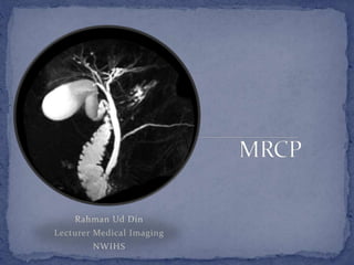
- 1. Rahman Ud Din Lecturer Medical Imaging NWIHS
- 2. Wide spread clinical acceptance Replaced ERCP To visualise biliary and pancreatic tree Non-invasive No contrast injection No radiation
- 3. Heavily T2-w images To visualise static fluid or bile in the PB-tree Longer TE 600-1200 ms Long TE, only fluid or tissues with high T2 relaxation time will retain signal Background tissues with shorter TE do not retain sufficient signal at longer TEs and are suppressed
- 4. Seqs; used are 3D FSE and single-shot FSE Other seqs; include balanced SSFP and contrast enhanced T1-w GRE seq 3D FSE sequence High TE Respiratory triggering by tying bellows over abdomen MIP technique is used for 3D data Takes 4-5 minutes Limitations include respiration
- 5. Single-shot FSE (SSFE/HASTE) Slab of 2-5 cm thickness Radial coronal slabs Acquired with breath hold No need for MIP Suppressed background tissues shows ducts Balanced SSFP (TrueFISP/FIESTA/bTFE) Breath hold Motion insensitive imaging Good quality imaging Shows ducts without motion artifacts
- 6. Contrast-enhanced T1-w GRE sequences (THRIVE/VBE/LAVA) IV injection Specific contrast gadobenate (Multihance), gadoxetate (Eovist/primovist) and mangafodifir trisodium (Mn- DPDP, Teslascan) Excreted through bile opacifying the bile ducts on T1-w image Contrast-enhanced MRCP Detection of bile leak Visualisation of small ducts
- 7. Secretin MRCP Secretin a hormone secreted by duodenal mucosa in response to acid stimulation It increases secretion of water and bicarbonate by pancreas IV (1 unit/kg) and T2-w images are acquired every 30 seconds for 10 minutes Distends pancreatic duct up to 3mm diameter Peak response occur at about 3-5 minutes after injection and response completely vanish after 10 minutes S-MRCP improves visualisation of branches of pancreas to diagnose chronic pancreatitis Main limitation is high cost of secretin
- 8. Patient preparation NPO 8-12 hours No food in upper GIT Blue-berry juice and barium can empty GIT if any food NPO also dilate GB and bile ducts Examination Routine T2-w axial seq for planning MRCP 3D FSE applied later (takes 4-5 minutes) Single-shot seq; applied Thin contagious section (3-4 mm) single-shot seq; in coronal and axial plane (TE=200-300 ms) Coronal balanced-SSFP and axial T1-w fat sat seq;
- 9. Cystic disease of bile duct Choledochal cyst Choledochocele Caroli’s disease
- 10. Congenital anomalies Pancreas divisum Cystic duct insertion Medial cystic duct insertion Parallel course of the cystic and hepatic duct Aberrant right hepatic duct These variations are important to know in order to avoid any complications during cholecystectomy especially the laproscopic Pancreas divisum
- 11. Choledocholithiasis Accurate diagnosis of stone in CBD More accurate modality Compared to USG and CT Primary Sclerosing Cholangistis Characterized by multiple irregular strictures and saccular dilatation of intraheptic and extrahepatic bile ducts producing beaded appearance Good in diagnosis and in follow-up in such conditions ERCP may result in progression of cholestasis and may not show ducts proximal to severe stenosis
- 12. Postsurgical complications Benign strictures, retained stones, biliary leaks and fistula Patency of biliary-enteric anastomosis can be seen by MRCP
- 13. Chronic pancreatitis It is characterised by pancreatic duct dilatation, narrowing or stricture and irregularity Alcoholic chronic pancreatitis is usually heterogeneous and characterised by side-branch dilatation and ductal calcifications Whereas obstructive pancreatitis is more homogenous, lack calcifications and is associated more often with main duct dilatation MRCP is useful in such detection Identification of a surgically or endoscopically correctable lesion
- 14. Neoplasmic Lesions MRCP can show duct proximal to the obstruction Cause by i.e. Cholangiocarcinoma Pancreatic head carcinoma Fat saturated postcontrast T1-w images for the evaluation of extent and spread of the lesion.