
Recommended
Recommended
More Related Content
What's hot
What's hot (20)
Similar to ruchika ppt.pptx
Similar to ruchika ppt.pptx (20)
Recently uploaded
Recently uploaded (20)
ruchika ppt.pptx
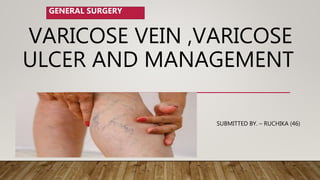
- 1. VARICOSE VEIN ,VARICOSE ULCER AND MANAGEMENT SUBMITTED BY. – RUCHIKA (46) GENERAL SURGERY
- 2. DEFINITION > VARICOSE VEINS ARE DEFINED AS DILATED, ELONGATED, TORTUOUS AND PALPABLE SUPERFICIAL VEINS AS A RESULT OF VENOUS HYPERTENSION.
- 3. VARICOSE VEIN • More common in males in India • Left lower limb more commonly involved • Long saphenous system affected in 2/3 rd of cases veins • It is common – may be present in up to 30% of the population – 40,000 operations/year in England &
- 4. VENOUS SYSTEM OF LOWER LIMBS Consists of •Deep system of veins which lies below the deep fascia. •Superficial system of veins which lies outside the deep fascia (carry 10% blood) •Perforating veins which pass through the deep fascia joining the superficial to the deep system of veins.
- 5. FACTORS HELPING IN VENOUS . RETURN 1. Negative pressure in thorax during inspiration to -6 mm. 2. Calf muscle pump. 3. Vis a tergo. 4. Competent valves 5. Venae commitants.
- 6. TYPES 1. Primary ( idiopathic) • More common in women • Lower extremities • Strong family history 2.SECONDARY PREVIOUS DVT other identifiable obstruction Also occur in esophagus • Haemorrhoids • arterivenous malformation
- 7. ETIOLOGY 1. Long hours of standingwhich increase the hydrostatic pressure of gravity, 2. • Family history 3. • Pregnancy 4. • Ageing 5. • Deep vein thrombosis 6. • Oral contraceptives 7. • obesity
- 10. VARICOSE ULCERS VENOUS ULCERS, STASIS ULCERS, ULCUS CRURIS . . A VENOUS LEG ULCER IS THE MOST COMMON TYPE OF LEG ULCER, ACCOUNTING FOR 80- 85% OF ALL CASES. VENOUS LEG ULCERS DEVELOP WHEN PERSISTENTLY HIGH BLOOD PRESSURE IN THE VEINS OF THE LEGS (VENOUS HYPERTENSION) CAUSES DAMAGE TO THE SKIN, WHICH EVENTUALLY BREAKS DOWN AND FORMS AN ULCER.
- 12. Symptoms a chronic non-healing wound with broken skin and exposed tissue. -usually found on the inside of the leg, just above the ankle . painful, particularly when infected . pitting oedema . lipodermatosclerosis . atrophie blanche .
- 13. DIAGNOSIS Gp : diagnosis based on symptoms and a physical examination. Look for symptoms of a venous leg ulcer and feel your pulse at ankles (check arteries) . Doppler study. Colour duplex ultrasound Test.
- 14. TREATMENT 70% of small ulcers will heal within 12 weeks. -Larger ulcers may take longer to heal. Treatment involves cleaning and dressing the wound and using compression bandages to control blood pressure inside the legs. 4E’s: education, elevation, elastic compression and evaluation. Active movement -Leg elevation -Emollient use -Treating the underlying condition -Treatment of any infection
- 15. COMPLICATIONS 1. Unless underlying risk factors such as immobility, obesity and varicose veins are addressed, there is a high risk of a venous leg ulcer reoccurring. 1. > Loss of mobility - infection (rarely infection could lead to more serious conditions such as osteomyelitis or sepsis)
- 16. Conservative management Avoiding prolonged standing Crepe bandaging and elastic stockings from toe to thigh, which causes decreased edema, venous volume and reflux and increases venous return. Limb elevation above the level of heart while lying down
- 17. Sclerotherapy A chemical is injected into the vein, irritating the venous endothelium and producing localized phlebitis and fibrosis, thereby obliterating the lumen of the vein • Under Ultrasound guidance. hypertonic sodium chloride solution • Sodium morrhuate • Ethanolamine oleate • Polidocanol. Spread of foam monitored under USG guidance as it spreads. • Apex of saphenous opening compressed by probe to prevent foam entering deep veins. • Leg also elevated • After leg is
- 18. SURGICAL MANAGEMENT Surgical management High end ligation and stripping • Ligation of entire vein and dissection and removal of its tributaries Laser fiber produce endoluminal heat that destroy the vascular endothelium
- 19. NURSING MANAGEMENT Bed rest is maintained for 24 hours, after which the patient begins walking every 2 hours for 5 to 10 minutes. • Elastic compression stockings are used to maintaincompression of the leg.They are worn continuously for about 1 week after vein stripping . •The foot of the bed should be elevated, Standing still and sitting are discouraged • Usually, the patient may shower after the first 24 hours. The patient is instructed to dry the incisions well with a clean towel using a patting technique rather than rubbing.
- 20. Thankyou