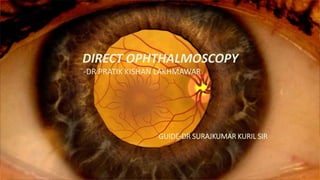
normal fundus , the retina how it works & how it is visualised is described here.
the procedure of direct ophthalmoscopy how the image is seen.
Abnormalities of retina how are they seen.
2. Contents
• Introduction
• History
• Direct ophthalmoscope
• Optics
• Distant direct ophthalmoscopy
• Procedure of direct ophthalmoscopy
• Normal fundus
3. What are the different methods used for
fundus examination?
Fundus examination
1 Direct ophthalmoscopy
2 Indirect ophthalmoscopy
3 Slit lamp biomicroscopy
4 Fundus camera
4. Introduction
• It is handheld instrument used to examine the fundus of eye.
• It is the most commonly practiced method for routine fundus
examination.
• It is used to examine central 7 to 10 degree of retina
• “The physician using a direct ophthalmoscope is like a one-eyed
Eskimo peering into an igloo from the entryway with a flashlight.”
6. Optics
• Convergent beam of light is reflected on to patients retina.
• The emergent rays from patients retina reach the observer through
the viewing hole present in the ophthalmoscope.
• If the observer is not emmetropic then the correcting lenses must be
interposed which are present in the ophthalmoscope.
7.
8. How is the image formed?
• The image formed on observers retina is-
• Erect
• Virtual
• 15 times magnified (in emmetropes)
9. Procedure
• It should be performed in semi dark room.
• Ideally the pupils should be dilated with mydriatic agent.
• Patient should be looking straight.
10. Position of examination
• The subject should be examined in sitting or lying down position.
• While examining the left eye of the subject the examiner should be
on left side of the subject and should hold the instrument in the left
hand and use left eye.
• And while examining right eye stand on right side ,hold the
instrument in right eye and use right eye.
11.
12. What is distant direct ophthalmoscopy?
• The ophthalmoscope should be kept at a distance of 20-25 cm from
the patient’s eye.
• Normally a red reflex is seen at the pupillary area.
• It is used to examine the condition of different media of the eye.
13. Uses
Opacities in the ocular media are seen as dark spots in the red glow at
the pupillary area.
The plane of the opacities can be assessed by asking the patient to
move the eye from side to side while the examiner is observing the
pupillary glow (based on parallax principle).
Opacities in front of the pupil move in the direction of eye movement.
Opacities in the pupillary plane do not move.
Ospacities behind the pupillary plane move opposite the direction of
eye movement.
14. • Once the red reflex is seen on distant direct ophthalmoscopy
observer should move as close to the patients eye as possible(it
should be at the anterior focal palne of the patients eye i.e. 15.4 mm
from the cornea.
• Once the retina is focused details should be examined like disc, blood
vessels, macula etc.
15. Parts of direct ophthalmoscope
• Body-
To hold the battery.
The body has switch with a rheostat that can control illumination.
• Head-
Eyepiece
Lens rack
Power dial
Filter selector
22. Half-circle diaphragm
• Used to reduce reflections by limiting the illumination beam.
• It is also used to observe some fine retinal details.
• It is used to avoid fundus reflections while examining.
23. Red-free filter
• Lack of red light makes the red elements very dark so that vessels &
pin point haemorrhages stand out.
• It is also used to differentiate between retinal & choroidal lesions.
24. Cobalt blue filter
• Used to enhance visibility of flourescein angiography and it is also
used as hand held light source for flourescein staining of cornea.
26. Normal fundus
• The fundus of the eye is the interior surface of the eye opposite the
lens and includes the retina, optic disc, macula, fovea, and posterior
pole.
• The term fundus may also be inclusive of Bruch's membrane and the
choroid.
28. What is RETINA?
• Retina is the innermost tunic of the eye.
• It is thin,transparent, delicate membrane.
• It is the most developed tissue of the eye.
29.
30. Fundus examination is used for
• Routinely used to assess and diagnose vitreo-retinal diseases such
as diabetic retinopathy, hypertensive retinopathy, retinal tear and
detachment, macular hole retinal haemorrhages, retinal artery
and vein occlusion, Choroidal tumour, macular oedema.
• Examine the extent of the defects, abnormalities to plan a proper
treatment.
• Evaluate the Success of the treatment.
31. Appearance of normal fundus
• Appearance-
• r- Orange to Vermilion
Factors Responsible- 1)Amount of pigment in the Choroid
2)hexagonal epithelium of Retina
3)Choroidal vasculature.
33. Optic disc
• Optic disc- It’s a Ophthalmoscopic view of nerve head , while Papilla
is the term for Microscopical sections.
• Its an insensitive area in Retina where Ganglion cell axons exit to
form optic nerve.
• Location- Nasal to Geometrical axis
• Diameter- 1.5 mm(1 Disc Diopter)
34. • Colour- Pale red or Yellowish red tint
• Normal colour of the disc is due to
1. Axonal bundles – transparent
2. Transmit the light
3. Reflected by disc capillaries
4. Disc appears pale red to yellowish tint
35. • Shape- Round or vertically Oval
• Edges- Regular
• Physiological Cup- It’s a depression seen in the disc.
• The size of Physiological Cup not affected by Age Or
Hypermetropia.
• Vessels- Central Retinal artery and vein emerge through its
centre.
• Normal C/D Ratio—0.3-0.5
36. Physiological cup
• At the centre of the optic disc, there is a short funnel shaped
depression from which retinal vessels appear to emerge.
• 15% optic discs do not show physiological cupping.
• Nasal border is steeper than temporal border.
• C/D ratio- 0.3-0.5
• Depth of cup depends upon size, shape, obliguity and vascularity of
disc.
• Size of physiological cup is not affected by age or hypermetropia.
37. Macula
• Diameter-5.5 mm
• Location- 2 DD temporal to disc
• Colour-Yellow
• Function- Photopic vision and Colour vision
• Macular region is supplied by twigs of superior and inferior
temporal arteries, also by cilioretinal arteries.
40. Fovea
• It is the centre of macula.
• Fully developed after 4 years.
• Thick basement membrane.
• Prone for macular holes.
41. Foveola
• Centre of Fovea-Foveola
• Foveola- It is small ,circular, area of deeper red than surrounding
fundus.
• Foveola has only Cones, No Rods.
• Centre of foveola- Umbo , which is seen as bright spot of light on
ophthalmoscopy called as Foveal reflex.
• Vision is most acute at foveola.
• Foveal avascular zone- is devoid of retinal blood vessels. It is 0.4-
0.6mm in diameter.
42. •At fovea- 1 lacs to 3 lacs per mm2
•3mm away from fovea- 6000 per
mm2
•10 mm away from fovea- 4000
per mm2
Cone
density in
and
around
fovea-
43. Other uses of direct ophthalmoscope
Pupil-
Pupil can be evaluated by comparing size & shape.
Anterior segment-
With +13 to +15D lens in ophthalmoscope cornea, lids, sclera, lashes &
iris can also be examined.
Vitreous-
With +6 to +7D lens vitreous floaters & opacities can be examined
44. Advantages
Easy procedure
It can be used in non dilating pupils as well
It can be carried everywhere
Refractive power can be adjusted