
Recommended
More Related Content
What's hot
What's hot (20)
Similar to Tmj ankylosis-oral and maxillofacial surgery
Similar to Tmj ankylosis-oral and maxillofacial surgery (20)
Recently uploaded
Recently uploaded (20)
Tmj ankylosis-oral and maxillofacial surgery
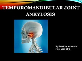
- 2. • Greek terminology meaning ‘stiff joint’. • Fusion between cranium and condyle. • Jaw function is affected. • Hypomobility or immobility of joint can lead to inability to open mouth from partial to complete. 2
- 3. ETIOPATHOLOGY OF ANKYLOSIS OF TMJ • • • • congenital At birth (forceps delivery ) hemarthrosis condylar # - intra / extra capsulra Trauma • • • • • Parotitis tonsilitis Abscess around the joint osteomyelitis of the jaw actinomycosis Infections 3
- 4. Inflammation • • • Rheumatoid arthritis osteoarthritis Septic arthritis Systemic diseases • • • • • small pox scarlet fever Scleroderma beriberi Ankylosing spondylitis Other causes • • • • bifid condyle prolonged trismus prolonged immobilization Burns 4
- 5. Pathophysiology Trauma ↓ Extravasation of blood into the joint space ↓ Heamarthrosis ↓ Period of restricted mobility due to pain ↓ Fibrosis leading to further restriction ↓ Gradual bone formation 5
- 6. CLASSIFICATION 1. Based on the location: - - Intra articular or true ankylosis Extra articular or false ankylosis 2. - - - Based on the Bony. Fibrous. Mixed. type of tissue involved: 3. Based on the extent of fusion/severity of ankylosis: - - Complete. Incomplete. 4. Based on the side involved: - - Unilateral. Bilateral. 6
- 7. SAWHNEY CLASSIFICATION 1. Type I: Head of the condyle is flattened or deformed with close approximation to the upper make movement possible. articular surface. Dense fibrous adhesions 2. Type II: Head misshapen or flattened but is distinguishable. Bony fusion of head to outer edge of articular surface. 7
- 8. 3. Type III: Bony block seems to bridge across ramus and zygomatic arch. Displaced condylar head. Elongation of coronoid process seen. 4. Type IV: Bony block is wide and deep and extends between ramus and upper articular surface thereby completely replacing joint architecture. 8
- 9. CLINICAL MANIFESTATIONS → Unilateral Ankylosis: • Facial asymmetry. • Deviation of mandible and chin on affected side. • Roundness and fullness of face on affected side. • Cross bite maybe seen. • Lower border of mandible has a concavity on affected side. 9
- 10. → Bilateral Ankylosis: • Inability to open mouth progresses to decreased interincisal opening. • Typical ‘bird face’ deformity with receding chin. • Neck chin angle reduced or completely absent. • Class II malocclusion. • Protrusive upper incisors with anterior open bite. • Multiple carious teeth with bad periodontal health. 10
- 11. DIAGNOSIS Diagnosis is based on the following: 1. 2. 3. a. History of trauma, infection etc. Clinical findings. Radiographic findings: OPG: Shows both joints picture which can be compared in unilateral cases. Lateral oblique view: Gives anteroposterior dimension of condylarb. mass. Elongation of coronoid process seen. c. Cephalometric radiograph: Taken to evaluate associated skeletal deformities. 11
- 12. d. CT scan: • Very helpful guide for surgery. • Relation to middle cranial fossa, anteroposterior width can be assessed. • Any presence of fractured condylar head can be located. 12
- 13. MANAGEMENT OF TMJ ANKYLOSIS Aims and Objectives of Surgery: 1. Release the ankylosed mass and creation of a gap to mobilize joint. Creation of a functional joint. the 2. 3. 4. 5. 6. To To To To reconstruct the joint and restore vertical height prevent recurrence. restore normal facial growth pattern. of ramus. improve esthetics and rehabilitate the patient. Surgical Techniques: I: Condylectomy. II: Gap Arthroplasty. III: Interpositional Arthroplasty. 13
- 14. Management: Adult • Cause: • Cause: • Trauma • Aim: • Restoration of satisfactory movement • Trauma •Infection • Aim: •Restoring function and movement • Bony replacement with CCG • Correction of occlusal and cosmetic deformity Childhood 14
- 15. SURGICALAPPROACHES Blair inverted hockey stick vertical incision Dingman question markAl-Kayat & Bramley in 1979- modified preauricular approach and Ivy in 1936- Thoma in 1958- angulated Preauricular incision- Popowich and Crane in 1982- 15
- 16. Submandibular approach Postramal incision (Hinds) Endaural incision Coronal approach Postauricular approach 16
- 17. Condylectomy: Advocated in cases of fibrous ankylosis, where joint space is obilterated with deposition of fibrous bands but there is not much deformity of condylar head. • Preauricular approach used commonly, others include Al Kayat Bramley, inverted hockey stick. 17
- 18. Gap Arthroplasty: • Section consists of two horizontal osteotomy cuts and removal of bony wedge for creation of a gap. • No substance is interposed between the two cut bony surfaces. • Minimum gap of 1 cm to prevent reankylosis. 18
- 19. Interpositional Arthroplasty: • Involves creation of a gap, but in addition a barrier is inserted between the cut bony surfaces to minimize risk of recurrence and to maintain vertical height of ramus. 18 19
- 20. 20
- 21. Materials used in Interpositional Arthroplasty: 21
- 22. KABAN’S PROTOCOL FOR MANAGEMENT OF TMJ ANKYLOSIS 1. Early surgical intervention. 2. - Aggressive resection: Gap of at least 1 – 1.5 cm should be created. 3. - Ipsilateral coronoidectomy and temporalis myotomy: After gap arthroplasty, coronoidectomy on the same side carried out. should be - Temporalis muscle attachments are severed by carrying out temporalis myotomy. 4. Contralateral coronoidectomy and temporalis myotomy. 22
- 23. 5. Lining of glenoid fossa region with temporalis fascia. 6. Reconstruction of ramus with costochondral graft. 7. Early mobilization and aggressive physiotherapy for months postoperatively. at least six 8. Regular long term follow up. 9. To carry out cosmetic surgery at later date, when growth of patient is completed. 23
- 24. COMPLICATIONS DURING SURGERY During Anesthesia: a. As the patient cannot open the mouth, awake blind intubation has to be done where co – operation is required which is difficult to achieve sometimes. b. Because of small mandible and altered position of larynx, intubation poses a problem. c. Aspiration of blood clot, tooth or foreign body during extubation. d. Danger of falling back of tongue and obstructing airway is always there after extubation. 24
- 25. During Surgery: a. b. c. d. e. f. Hemorrhage. Damage Damage Damage Damage Damage to to to to to external auditory meatus. zygomatic and temporal auriculotemporal nerve. parotid gland. glenoid fossa. branch of facial nerve. During Postoperative Followup: a. b. c. Infection. Open bite. Recurrence of ankylosis. 25
- 26. FREY SYNDROME: 1st described by frey. It is localised gustatory sweating in the area supplied by auriculotemporal nerve. Cause: Congenital or acquired Surgery of parotid gland, TMJ , parotid abscess, facial wound. Clinical feature: Pain in area supplied by ATN Gustatory sweating Erythema & flushing Positive iodine starch test 1. 2. 3. 4. 26
- 27. 1. 2. 3. 4. 5. i. ii. iii. Treatment: Antiperspirants Anticholinergic prepn: glycopyrolate Botulinum toxin A inj. Radiation therapy: 50 Gy Surgical: Skin excision: for localise & small area ATN section: not permanent Tympanic neurectomy: safe procedure 27
- 28. RECURRENCE OFANKYLOSIS Several factors said to be responsible: 1. 2. 3. Inadequate gap created between fragments. Fracture of costochondral graft. Loosening of costochondral graft due to inadequate ramus. Inadequate postoperative physiotherapy. Inadequate coverage of glenoid fossa surface. fixation to 4. 5. 6. Higher osteogenic potential and periosteal osteogenic responsible for high rate of recurrence in children. power maybe 28
- 29. THANK YOU! 29