
Recommended
More Related Content
What's hot
What's hot (20)
Similar to IMPLANT SITE PREPARATION.pptx
Similar to IMPLANT SITE PREPARATION.pptx (20)
More from PrasanthThalur
More from PrasanthThalur (20)
Recently uploaded
Recently uploaded (20)
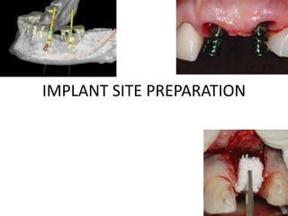
IMPLANT SITE PREPARATION.pptx
- 2. Contents • Introduction. • Implant site assessment. • Alveolar morphology. • Alveolar ridge defects and classification. • Hard tissue and soft tissue augmentation. • Grafts and membranes. • Guided bone regeneration. • Soft tissue augmentation. • Ridge preservation. • Advanced procedures. • Systematic reviews and meta analysis. • Summary and Conclusion. • References.
- 5. Bone at Implant site Bone quality Bone quantity
- 7. Bone Quality assessment Bone density Classification: Misch
- 9. Radiographic assessment BONE QUALITY DENSITY HOUNSFIELD UNITS D 1 1250 D 2 850-1250 D 3 350-850 D 4 150-350 D 5 < 150
- 10. Soft tissue assessment Thin scalloped Periodontium Thick flat periodontium
- 12. Reverse Engineering Bone based implant placement Prosthesis based implant placement
- 13. Alveolar morphology “A normal ridge is defined as one that retains the general shape of the alveolar process following uneventful extraction socket healing”.
- 14. Reasons for loss of alveolar morphology Periodontal disease Peri-apical lesions Improper tooth extraction Trauma Tumor Implant failures
- 15. Classifications of alveolar ridge defects Seibert JS, 1983: Seibert ClassI • Buccolingual loss of tissue • Normal ridge height in apico-coronal direction Seibert Class II • Apico-coronal loss of tissue . • Normal ridge width in bucco lingual direction Seibert Class III • Bucco-lingual and apico-coronal loss of tissue • Loss of height and width
- 16. Classifications of alveolar ridge defects Modification by Allen et al 1983 Mild : < 3mm. Moderate : 3-6 mm. Severe: > 6 mm. A: Apico-coronal loss of bone. B: Bucco-longual loss of bone C: Combination of both
- 17. HVC Ridge deficiency classification (Hom Lay Wang et al 2002) • Combination of Seibert and Allen classification. • Therapeutically oriented. • H: corelates with Seibert I, V- co-relates with Seibert II, C- co-relates with Seibert III. • In addition there is a sub-classification in H, V and C as follows: – Small (< 3mm). – Medium (4-6 mm). – Large ( > 7 mm). – H (s,m,l), V (s,m,l), C (s, m, l)
- 18. Risk assessment for edentulous sites (Martin et al ) Risk factors Low risk Medium risk High risk Medical status Healthy patient and intact immune system --------------- Reduced immune system Smoking habit Non smoker Light smoker ( < 10 cigarettes per day) Heavy smoker (>10 cigarettes per day) Patient’s esthetic demands Low Medium High Lip line Low Medium High Gingival biotype Low scalloped, thick Medium scalloped, medium thick High scalloped , thin Shape of tooth crowns Regular ----------------- Triangular Bone level at adjacent teeth < 5 mm to contact point = 5 mm to contact point > 5 mm to contact point Periodontal health status Healthy Moderately compromised Severely compromised Restorative status of neighboring teeth Untouched --------------- Restored Width of edentulous span 1 tooth (>7 mm) 1 tooth (> 5.5 mm) 1 tooth (< 7 mm) 1 tooth (< 5.5 mm) 2 Teeth or more Soft tissue anatomy Intact soft tissue ------------------ Soft tissue defects Bone anatomy of alveolus No bone deficiency Horizontal bpne deficiency Vertical bone deficiency
- 19. Implant site development: Sequence, Timing, Selection, Duration Treatment Period Treatment timing Procedures performed Treatment Duration Initial Site Development Prior to implant placement Extraction site management Hard tissue grafting Soft tissue grafting Prosthetic soft tissue management Upto 12 months Intermediate site development Implant placement through osseointegration period Extraction site management Hard tissue grafting Soft tissue grafting Prosthetic soft tissue management 6 weeks to 9 months Final site development Transmucosal emergence of implant to final restoration Abutment connection. Prosthesis guided soft tissue healing. Esthetic soft tissue re-surfacing Upto 4 months Recall phase Recall period Soft tissue grafting. Esthetic soft tissue re-surfacing Variable
- 20. Hard tissue augmentation Soft tissue augmentation Implant site Development
- 22. Hard tissue Augmentation Guided bone regeneration concept: • Advocates that regeneration of osseous defects is predictably attainable via the application of occlusive membranes, which mechanically exclude non-osteogenic cell populations from the surrounding soft tissues, thereby allowing osteogenic cell populations originating from the parent bone to inhabit the osseous wound. • Based on pioneer experiments investigating healing of periodontal tissues following surgical therapy, a principle of tissue healing was discovered by Nyman & Karring in the early 1980’s.
- 23. • Based on osteo promotion principle. • Osteopromotion refers to the use of physical means to seal off an anatomical site the site where bone is intended to be (re) formed – in order to prevent other tissues, notably connective tissue, to interfere with osteogenesis as well as to direct bone formation. How do Guided Bone Regeneration works ?
- 25. • A viable source of bone cells must be available from the surrounding bone. • New bone formation requires an adequate blood supply. • The wound must remain mechanically stable during healing. • Adequate space must be created and maintained between the membrane and bone surface. • Soft tissue cells must be excluded from the space created by the barrier membrane. Principles of Guided Bone regeneration by Dahlin et al(1994)
- 26. • Augmentation around implants placed in immediate extraction sockets. • Augmentation around implants placed in delayed extraction sockets. • In dehiscence defects around implants. • Localized ridge augmentation. • Alveolar ridge reconstruction. • Filling of bone defects after root resection, cystectomy and removal of retained teeth. Indications of GBR
- 27. GUIDED BONE REGENERATION • Wound completely covered by flap (closed system) • Sterile condition maintained during healing • Membrane stable • Adaptation of membrane easy • Space making easy • High predictability • Open wound (open system) • Sterile condition not maintained • Membrane stabilization difficult • Adaptation of membrane difficult • Space making difficult • Low predictability
- 28. Definitions of success following guided bone regeneration Bucco-lingual ridge augmentation Implant dehiscence Extraction socket reconstructions Apico-coronal ridge augmentation First generation Adequate bone to place an implant Coverage of exposed implant surface with regenerated hard surface Adequate bone to place an implant Adequate bone to place a 10 mm long implant Second generation Adequate bone to withstand functional forces over time Adequate bone to withstand functional forces over time Adequate bone to withstand functional forces over time Adequate bone to place 10 mm long implant & withstand functional forces over time Third generation All of the above and regeneration of pre-pathologic morphology allowing implant placement in perfect prosthetic position with maximum aesthetics Fourth generation Simplification of therapy as much as possible without compromising the treatment outcomes
- 29. Biomaterials for hard tissue augmentation Guided bone regeneration Grafts Barrier membranes
- 30. Bone Grafts Bone graft : Bone or bone like synthetic substance used to fill a defect in a bone or to augment new bone formation
- 31. Grafts Bone Derived Autogenous Allografts Xenografts Non Bone Derived Alloplast Classification of Bone Grafts
- 32. • AUTOGRAFT OSTEOGENIC • AUTOGRAFT • ALLOGRAFT OSTEOINDUCTIVE • AUTOGRAFT • ALLOGRAFT • XENOGRAFT • ALLOPLASTS OSTEOCONDUCTIVE Principles of Bone Grafts
- 33. Autogenous Bone Grafts, and Donor Sites
- 34. Graft form and maximum volume available from autogenous bone donor sites Donor Site Forms available Maximum volume (ml) Extraoral Posterior iliac crest Block and or particulate 140 Anterior iliac crest Block and or particulate 70 Tibia Particulate 20 to 40 Cranium Dense cortical block 40 Intra oral Ascending ramus Block 5 to 10 Anterior mandible Block and or particulate 5 Tuberosity Particulate 2 Miscellaneous ( one shavings) Particulate Varies
- 35. Cortical grafts vs • Thich dense lamellar bone. • Resorption is slower. • Volume of bone fill achieved is higher. • Stabilization is not a problem. • Graft re-vascularization is slower Cancellous grafts • Trabecular bone- less dense. • Resorption is faster. • Volume of bone fill achieved is lower. • Need GBR membranes for stabilization. • Graft re-vascularization is faster.
- 36. Mandibular symphisis Rule of FIVE: 5 mm anterior to mental foramen, 5 mm above the ridge crest, 5 mm apical to anterior root tips. (Hunt and Jovanovic, 1999)
- 37. BLOCK GRAFTS TREPHINE CORES Olympic design: Sascha Jovanovic
- 38. Key factors for success: Mandibular symphisis grafts • Contra-indicated in 1. Patients with long anterior teeth. 2. Inadequate mandibular height or width. 3. If defect width is more than four teeth.
- 39. Incision Techniques: Symphisis access • Indicated in patients with low vestibular sulcus, tense mentalis posture and a donor site without periodontal disease. • Disadv: recession of gingiva Sulcular incision and mucoperiosteal flap • Indicated when there is marginal inflammation or bone loss around incisors • Disadv: scarring can occur. Vestibular incision • Indicated when there is 3 mm of keratinized gingiva and thin scalloped periodontium. • Disadv: fine suturing required. Attached gingival incision
- 40. FLAP WITH SULCULAR INCISIONS
- 43. AUDI DESIGN REVERSE OLYMPIC RING DESIGN DENNIS HUNT AND SASCHA JOVANOVIC , 1999
- 44. Harvesting technique: • Rule of five has to be followed. Trephine cores: • Different diameters, 4.1, 6.0 mm commonly used. • Position drill 4 to 5 mm below apex of tooth. • 50,000 rpm with irrigation. • Insert into cortex. • Avoid lingual perforation: can compromise airway. • Drill to depth planned and cant drill to break core of bone and remove with trephine. Block grafts: 1. Prepare template with bone wax. 2. 3 mm from inferior border, 5 mm from anterior to mental foramen, 5 mm below teeth apex. 3. Graft size – 2 to 3mm larger than defect size. 4. Outline graft with bur/ micro saw. 5. Depth of cut : cortex upto cancellous bone. 6. Use chisels to remove block graft. 7. Harvest cancellous marrow with rongeur/ bone curettes.
- 45. Potential complications of harvesting bone from symphisis Complications Causes Damage to submental/ sub-lingual arteries Lingual perforation Damage to roots Long anterior root Mental nerve paresthesia Over-extension of incisions Incision dehiscence Post-op edema, hematoma, tense mentalis Chin ptosis Improper suturing, damage to mentalis muscle.
- 46. Ramus grafts
- 47. Key factors to success: • Access to ramus. • Avoid neurovascular bundle. Surgical Technique: • Incision: start on ascending ramus at the level of occlusal plane. Move down towards ridge and continue in buccal sulcus if molar teeth are present or mid crestal if edentulous. • Muco-periosteal flap reflection. • Ramus osteotomy: 1. Start at level of occlusal plane along anterior border of ramus, maintain 3 to 4 mm medial to external oblique ridge. 2. Anterior Vertical cut in ramus body extending inferiorly from the anterior aspect of the cut. 3. Posterior vertical cut at the upper end along lateral body of ramus perpendicular to horizontal cut. 4. Inferior cut joins anterior and posterior vertical cuts. 5. Cuts should be made only in cortex of bone. 6. Wedge with chisel to harvest graft.
- 50. Potential complications of harvesting bone from the ramus Complication Cause Damage to inferior alveolar nerve Coronal portion of alveolar nerve Limitation of graft size and shape Harvesting thin graft to avoid nerve damage. Incision dehiscence in donor area Hematoma, post –op edema Post-op trismus Excessive trauma to muscle fibers during harvesting Damage to lingual nerve Lingually placed incision
- 51. Allografts
- 52. Xenografts
- 53. Alloplasts
- 54. Decision making for graft material usage in bone defect
- 55. DECISION MAKING FOR GRAFT SELECTION AUTOGENOUS CORE OR BLOCKS NON AUTOGENOUS GRAFTS PARTICULATE OR BONE GRAFT PASTE PARTICULATE OR BONE PUTTY GRAFT BONE PUTTY GRAFT Autogenous graft available at primary site Autogenous graft not available at primary site Intact extraction socket Moderately compromised extraction socket Onlsy / severely compromised extraction socket THUMB RULE: MAXIMIZE AUTOGENOUS GRAFTS
- 56. Graft materials and their indications and contraindications in alveolar grafting Graft material Approx resorption time Volume available Indication/ contraindication AUTOGENOUS BONE Iliac crest 3- 6 months 70-140 ml Full arch reconstruction. Severe atrophy areas. Higher morbidity risk Tibial plateau 3-6 months 20-40 ml Moderate to large reconstructions. Used along with titanium meshes Mandibular symphisis 4-8 months 5 ml Small to moderate defects with low to moderate osteogenic potential. Sinuses and moderate ridge defects. Maxillary tuberosity Bone shavings 3- 6 months 3- 6 months 2-4 ml 0.5 – 2.5 ml Small defects such as unilateral sinus lift. Use along with allografts. Exposed implant threads.
- 57. Graft materials and their indications and contraindications in alveolar grafting Graft material Approx resorption time Volume available Indication/ contraindication ALLOGRAFTS Puros 6-15 months Unlimited Small reconstructions in defects with low to moderate osteogenic potential- sinus lifts, moderate ridge defects with other grafts FDBA 6-15 months Unlimited Small reconstructions in defects with low to moderate osteogenic potential- sinus lifts, moderate ridge defects with other grafts DFDBA 2-4 months Unlimited For periodontal defects only.
- 58. Graft materials and their indications and contraindications in alveolar grafting Graft material Approx resorption time Volume available Indication/ contraindication ALLOPLASTS/ XENOGRAFTS PepGen P-15 (Bovine derived hydroxyapatite with synthetic peptide) 18-36 months Unlimited Small reconstructions in defects with moderate osteogenic potential- sinus lifts, moderate ridge defects with other grafts. Very expensive C- Graft ( Calcified algae) 6-18 months Unlimited Small reconstructions in defects with high osteogenic potential- minimalridge defects, exposed implant threads, extraction sockets. Bio-Oss ( anorganic bovine bone) 15-30 months Unlimited Small reconstructions in defects with high osteogenic potential- minimalridge defects, exposed implant threads, extraction sockets
- 59. Graft materials and their indications and contraindications in alveolar grafting Graft material Approx resorption time Volume available Indication/ contraindication ALLOPLASTS/ XENOGRAFTS Osteograf (microporous hydroxyapatite ) 18-36 months Unlimited Small reconstructions in defects with moderate osteogenic potential- sinus lifts, moderate ridge defects with other grafts. Osteogen (Porous anorganic crystal) 4-10 months Unlimited Small reconstructions in defects with high osteogenic potential- minimalridge defects, exposed implant threads, extraction sockets. Cerasorb (beta-TCP) 4-12 months Unlimited Resorbs too quickly to recommend use alone for bone grafting. Interpore 200 (porous coral) 5-7 years Unlimited Resorbs too slowly to recommend use alone for bone grafting
- 60. Graft materials and their indications and contraindications in alveolar grafting Graft material Approx resorption time Volume available Indication/ contraindication ALLOPLASTS/ XENOGRAFTS Cap Set (medical grade calcium sulphate) 1-2 months Unlimited Requires some time to mix. Used for small defects along with allografts. Bioplant HTR polymer (microporous composite with calcium hydroxide surface) 10-15 months Unlimited Resorbs too slowly to recommend use alone for bone grafting Can be used in extraction sockets below pontic of bridges. Perioglass (synthetic particulate glass ceramic) 18-24 months Unlimited Recommended for periodontal defects only. Biogran (synthetic particulate glass ceramic) 20-22 months Unlimited Recommended for periodontal defects only
- 61. Membranes in brief • More predictable restoration of architecture of newly formed bone. • Creates a suitable environment in which the natural biologic potential can be maximized. Rationale: • Stabilize blood clot. • Isolate space from undesirable tissues. • Prevent soft tissue collapse.
- 62. Physical barrier membrane which are used for GBR should satisfy the following five basic criteria (Hardwick et al 1994) • Biocompatibility • Cell occulisivity • Tissue integration • Space making • Clinical manageability Requirements of barrier membrane
- 63. Barrier membrane - Classification Barrier membranes Non resorbable membranes Resorbable membranes
- 64. • e-PTFE • Ti reinforced ePTFE • High-density PTFE (Bartee BK.1995) • Titanium mesh (Mellonig et al 1995, Jovanovic et al 1995) Ti mesh as a barrier maximizes graft containment and eliminates the space maintenance collapse problems associated with conventional membranes (Von Arx et al 1996, Boyne PJ 1996) Non Resorbable Membrane
- 65. The membranes at present available are either made from: • Synthetic polymers (lactide-glycolide copolymers or polylactic acid blended with citric acid ester) e.g. Guidor, resolut. • Natural biomaterials (collagen) e.g. Biogide Additional requirements • Bioresorbability • No foreign body reaction • No interaction with bone regeneration. Resorbable Membrane
- 66. Membranes and their indications and contraindications in guided bone regeneration Membrane Composition Approximate resorption time Indication/ contraindication Non- Resorbable Membranes Gore-tex E-PTFE NA Original barrier material. Effective around small to moderate defects GTAM E-PTFE with titanium reinforcement NA Small to large defects. Pin fixation for vertical height augmentation. Titanium Titanium NA Vertical and horizontal ridge defcts with particulate grafts. High risk of soft tissue dehiscence. Regentex GBR 200 PTFE NA Not recommended. Limited documented success.
- 67. Membranes and their indications and contraindications in guided bone regeneration Membrane Composition Approximate resorption time Indication/ contraindication Resorbable Membranes Biomend extend Bovine tendon collagen 4 months Lateral sinus window. Small to moderate defects with pin fixation. Around Implant dehiscence Alloderm Acellular freeze dried cadaver skin 4 months Best for cases where primary closure cannot be achieved. Also for large schneiderian membrane tears. Atrisorb Liquid PLA 4 months Suitable for periodontal defects. Biomend Bovine tendon collagen 2 months Large schneiderian membrane tears. Alveolar socket defects. Around implants.
- 68. Membranes and their indications and contraindications in guided bone regeneration Membrane Composition Approximate resorption time Indication/ contraindication Resorbable Membranes Resolut Adapt PLA/PGA 3 months For lateral window closure. Not suitable for ridge augmentation Resolut Adapt LT PLA/ PGA 4 months For lateral window closure. Not suitable for ridge augmentation Biogide Porcine dermal collagen 3-4 months For lateral window closure. Not suitable for ridge augmentation Epigide PLA/PGA 4 months Small ridge defect with or without implants Ossix Bovine tendon collagen 6 months For lateral window closure. Not suitable for ridge augmentation
- 69. Membranes and their indications and contraindications in guided bone regeneration Membrane Composition Approximate resorption time Indication/ contraindication Resorbable Membranes Reguarde Bovine tendon collagen 4 months Large schneiderian membrane tears. Alveolar socket defects. Around implants. Resolut PLA/ PGA 3 months Small ridge defects with or without implants. Resolut XT PLA/PGA 4 months Small ridge defects with or without implants Cap set Medical grade calcium sulphate 2 months Cover grafted extraction sockets. Lambone Thin sheet of DFDBA 5 months Ridge width and ridge height in conjunction with particulate grafts Lyodura Freeze dried human dura mater 2-3 months Not recommended for clinical use.
- 70. Membranes and their indications and contraindications in guided bone regeneration Membrane Composition Approximate resorption time Indication/ contraindication Resorbable Membranes OsseoQuest PLA/ PGA 6 months Not recommended for clinical use. Too stiff, too costly. Vicryl periodontal mesh Woven vicryl 1 month Not recommended due to fast resorption and wicking effect. Colla tape Bovine tendon collagen 2 weeks Not a true barrier membrane. Schneiderian tears and small defects.
- 71. • To guarantee stabilization of the barrier membranes during suturing of the mucoperisteal flaps and during the healing phase, use of minipins or miniscrews are recommended which serve as “tentpole” for the barrier membrane. • These minipins and miniscrews are also available in non-bioresorbable and bioresorbable materials. For non bioresorbable membrane such as ePTFE, stainless steel miniscrew are successfully used which are removed along with the membrane after the entire healing period of 6 to 9 months. Membrane Fixation
- 72. FIXATION SYSTEMS ADVANTAGES DISADVANTAGES Titanium screws Ease of use in dense bone Need to pre-drill the site Length of time Need to remove Titanium tacks Predictability in dense bone No need to pre-drill Ease and speed of use Need to remove Resorbable screws Do not have to be removed Need to pre-drill the site Prone to fracture in dense bone Resorbable screws with air gun insertion Do not have to be removed Ease and speed of use Problematic in delicate bone Potentially disconcerting to the patient Membrane Fixation Options
- 74. Membrane selection for GBR Procedure Autogenous block graft placed Paticulate or “putty” graft placed Resorbable membrane Resorbable membrane Titanium reinforced membrane Space maintaining defect Non Space maintaining defect Non Space maintaining defect Space maintaining defect
- 75. Basics for surgical technique for augmentation Flap design Technique Incisions Beveled incision. Preserve papilla by leaving 2mm of tissue in mesial and distal aspects of edentulous sites: anterior esthetic areas. Vertical incisions at least one tooth away from the edentulous site to enable complete coverage of GBR membranes. If sufficient width of keratinized tissue is present: Mid crestal. If insufficient width of keratinized tissue: 1. Palatal incision – 4 to 5 mm from the crest in palatal direction: Maxilla. 2. Buccal muco-gingival junction of mandible. Flap reflection Initial partial thickness followed by full thickness once bone is reached by beveled incision. Reflect beyond the muco-gingival junction: allows advancement.
- 77. PENINSULA FLAP
- 78. FLAP DESIGN FOR EDENTULOUS RIDGE AUGMENTATION
- 79. Techniques for hard tissue augmentation • Onlay. • Particulate grafting with titanium mesh. • Ridge split. • Interpositional. • Particulate grafting with membrane. • Distraction osteogenesis. • Tissue engineering constructs.
- 80. Onlay Blocks • Onlay: “Placed over” • Useful for augmentation of moderate and severe horizontal and vertical ridge defects • Source of blocks : mandibular symphisis, ramus, iliac crest, rib grafts. • Basic technique: 1. Full thickness flap is elevated at the surgical site. 2. Cortical plate of bone at the recipient site is drilled with round bur: to improve vascularity. 3. Block is scored on inner aspect to obtain as close a adaptation as possible with the recipient site. 4. Fixation screws used to stabilize graft in place and to minimize micromotion to allow bone regeneration. 5. Small gaps can be filled with allogratfs, cancellous grafts. 6. No membrane is required. Membrane is used only if primary closure cannot be achieved.
- 82. ONLAY GRAFT : AFTER 3 MONTHS
- 83. Particulate Grafts with Titanium mesh • Developed by Boyne PJ 1997 and further refined by Maiorana C et al 2001. • Main indication: Severe vertical ridge deficiencies involving full arch. Mostly used for atrophic maxilla. • Involves use of a custom made titanium mesh with 1:1 mixture of cancellous bone and anorganic bovine bone. • Adv: mesh creates an appropriate contour of the edentulous area and allows good blood supply to the underlying cancellous bone during 5 month healing period.
- 84. Particulate Grafts with Titanium mesh Technique: • Pre- surgical phase: mesh preparation 1. Polyether impression of maxilla. 2. Wax is placed over the cast to simulate the desired augmentation of the atrophic area. 3. Cast is then duplicated in acrylic. 4. Titanium mesh is adapted to the shape of the ridge as in the duplicated cast. Surgical Phase: 1. Crestal incision with relieving incision in midline and tuberosity areas. 2. Full thickness flap reflection. 3. Titanium mesh with mixture of autogenous bone and allograft particles placed over the maxillary bone and fixed with transcortical screws. 4. Periosteal incisions are made at the base of the flaps and flap is advanced to obtain complete closure. Limitation: vestibular sulcus is eliminated. After 5 months the mesh is removed and vestibuloplasty can be performed to establish keratinized gingiva in the maxillary area.
- 85. Particulate Graft with Titanium mesh
- 86. Particulate graft with Titanium mesh
- 87. Particulate graft with Titanium mesh
- 88. Particulate graft with Titanium mesh
- 89. Interpositional grafts (inlay technique) • Indication: extreme atrophy of the edentulous maxilla in which the relationship between the upper and lower jaw can be restored on a vertical and sagittal plane. • Pre-surgical technique: 1. Lateral and frontal cephalometric tracings are taken. 2. Surgical stent is prepared pre-operatively to plan for re-positioning of the maxilla after osteotomy. Surgical technique: 1. Full thickness incision from molar to molar in the unattached portion of the vestibule. 2. Le- fort I osteotomy is performed and cortico-cancellous graft is placed in the inner part of the maxilla after removing the mucosa of the sinus floor. 3. Layer of anorganic bone is placed over the iliac graft to reduce the resorption. Limitation : post-operative morbidity is high, technique sensitive.
- 90. Atrophic maxilla: Downfracture of maxilla with onlay and interpositional grafts
- 91. Atrophic maxilla: Downfracture of maxilla with onlay and interpositional grafts
- 93. Particulate grafts with Membranes • First reported in 2000, Kirkland G et al. • Uses autogenous bone/ particulate graft with resorbable or non resorbable membranes. • PASS principle: Hom Lay Wang to ensure success. • Indicated for localized ridge augmentation and implant dehiscence management.
- 94. Particulate grafts with Membranes • Technique: 1. Lateral incision and vertical releasing incisions. 2. Muco-periosteal flap reflection. 3. Soft tissue debridement if any. 4. De-cortication of bone defect. 5. Membrane preparation. 6. Graft placement into defect. 7. Followed by membrane stabilization over graft. 8. Flap advancement and closure.
- 95. Seibert Class I defect Flap Reflection
- 96. De-cortication
- 97. Graft placement Membrane placement
- 99. PRE-OP POST-OP: 3 MONTHS SURGICAL RE-ENTRY IMPLANT PLACEMENT
- 100. Regional acceleratory Phenomenon (RAP) • It is the local response of bone to a noxious stimulus . • A process by which tissue form faster than normal regeneration time. ( 2 to 10 times faster) • RAP begins within a few days and peaks at 1 to 2 months and lasts till 4 months. • Noxious stimuli can be injury, fracture, intentional drilling of bone. • RAP mediated by growth factors at site (PDGF and TGF beta) and cells of bone. • It can be accomplished by systemic response (systemic acceleratory phenomenon)
- 101. Caging effect • Exclusion of the periosteum and fibrous tissue elements have repeatedly demonstrated graft preservation,which is described by Murray et al as “caging effect.” Guided bone regeneration in Implant Dentistry, Quintessence books, Buser, Dahlin, Schenk)
- 102. Ridge split • Pioneered by Nentwig and Kniha, 1986. • Called the “ bone splitting/ spreading technique” • Allows expansion of the alveolar bone with simultaneous implant placement. • Surgical technique: 1. Preparation of buccal partial thickness flap. 2. Osteotomy in the middle of the crest with chisels and knifes. 3. Luxation of periosteum pedicled buccal bone in the vestibular direction. 4. Enlargement of crest from 2.5 mm to 5 mm. 5. Implant bed preparation with bone expanders. 6. Implant placement and submergence. Scipioni et al 1994, 5 year success rate of 88% - 93% with this mehod.
- 103. Split thickness flap Buccal pedicled bone, Ridge split and expansion
- 104. Implant placement in expanded bone
- 105. Distraction Osteogenesis • Biologic rationale: New bone apposition occurs in a surgically created bone fracture. New bone formation is due to tension induced between two pieces of slowly separated bone. Originally used in long bone growth, Ilizarov G, 1989. Introduced for oral surgical procedures by Mc Carthy JG, 1992. Primary Indication is vertical bone augmentation prior to implant placement.
- 106. Distraction Osteogenesis Surgical technique: • Buccal full thickness flap elevation beyond the mucogingival junction. • Lingual flap is left in place for blood supply to distracted segment of bone. • A horizontal osetotomy is made with oscillating saw and burs and the distractor is secured. • Vertical cuts are made to mobilize the bone segment to be distracted • The flap is sutured and a provisional prosthesis is given. • After 1 to 2 weeks of latency the distractor is activated to achieve progressive bone separation until required height of bone is achieved. • Moderate over correction is recommended • After a period of bone consolidation, the distractor is removed.
- 107. Buccal Full thickness flap Horizontal Osteotomy
- 109. • Potential Risks: Distractor instability ( poor bone quality). Flap dehiscence. Premature consolidation of bone. Transport bone segment fracture Distraction Osteogenesis
- 110. Decision making for management of Ridge defects Ridge Thickness Procedure 7-8 mm Particulate graft with barrier membrane with pin fixation. 6-7 mm Osteotomes for ridge expansion 5-6 mm Allogenic block of bone. 4-5 mm Autogenous block of bone 1-4 mm Down fracture of maxilla, titanium mesh ( both with autogenous bone graft), distraction osteogenesis. Adapted from: Guided bone regeneration: Textbook by Buser.
- 111. Tissue engineering constructs • Based on concept of tissue engineering. • A combination of scaffolds, cells, growth factors are used to obtain regeneration of target tissue. • Important addition is : Blood supply
- 113. Literature Review: in vitro studies: alveolar bone Author Scaffold Cells Findings Xiao yin et al, 2003 Three dimensional collagen matrix Osteoblast like cells New bone formation at all sites treated with osteoblast matrix at 28 days. Turhani D et al 2005 Hydroxyapatite from red algae Cambial layer of cells cultured in vitro Osteoblast cell phenotype. Good viability of cultured cells. Young CS et al 2005 PLGA Rat osteoblasts in bioreactor Histological demonstration of bone like issue (BSP and osteocalcin positive) Turhani D et al 2005 PLGA vs Hydroxyapatite Mesenchymal cambial precursor cells Substrate influenced the parameters of differentiation. HA was better.
- 114. Literature Review: in vitro studies: alveolar bone Author Scaffold Cells Findings Weng Y et al 2006 Calcium alginate gel Bone marrow stromal cells in dogs Bone formation demonstrated histologically Yefang Z et al 2007 Polycaprolactone- tricalcium phosphate Human osteoblasts culture Mineral nodule formation demonstrated. Abukawa H et al 2009 Mini pig model, autologous bone Osteoblasts of mini pig Bone formation demonstrated
- 115. Tissue engineered constructs with PRP • Platelet rich plasma: autologous concentrate of platelets. • Rich source of growth factors. • Proven soft tissue healing. • Bone healing is not proven: on stand alone basis. • Used with other bone substitutes for improving healing.
- 116. Literature Review: PRP with bone substitutes: bone augmentation Author STUDY design Findings Kassolis JD et al 2000 Case series on ridge augmentation and sinus augmentation along with allograft Histologic demonstration of bone formation in augmented areas. Rosenberg ES, Torosian J 2000 Case report- sinus augmentation Technique demonstration Shanaman R et al 2001 Case reports: ridge augmentation by autogenous grafts and PRP PRP did not enhance bone quantity or quality when used with autogenous bone. Robiony M et al 2008 Distraction osteogenesis for ertical augmentation of mandible. Autgenous bone and PRP filler Effective method for bone gain. Total bone volume loss was only 2.3 %
- 117. Literature Review: PRP with bone substitutes: bone augmentation Author STUDY design Findings Mooren RE et al 2010 Retrospective study. Mandible re-construction with titanium plates, block and particulate grafts, PRP gel Healing was uneventful. Continuity was attained in all cases. High PRP concentration: more granulation tissue formation. Schuckert KH et al 2010 Case report. Tri calcium phosphate blocks soaked in rh BMP 2 and PRP Vital lamellar bone formation Torres J et al 2010 PRP effect on preventing titanium mesh exposure PRP completely eliminated the titanium mesh exposure. Improved soft tissue healing.
- 119. Soft tissue augmentation techniques Soft tissue augmentation Free grafts 1. Onlay 2. Pouch 1. Interpositional 2. Onlay- Interpositional Pedicle grafts Roll
- 120. Roll method • Indication: Small to moderate class I defects. • Developed by Abrams in 1980 and modified by Scharf and Tarnow in 1992. • Surgical technique: 1. Preparation of de-epithelialized connective tissue pedicle from palatal surface of edentulous area. 2. Pouch preparation with partial thickness dissection in the supra periosteal connective tissue of the buccal surface of the ridge. 3. Vertical releasing incisions may be used to augment flap mobility. 4. The connective tissue flap is inserted into the pouch and secured by sutures. Modified Roll method: “Trap Door” Approach
- 121. Roll Flap method (Abrams 1980)
- 122. Roll Flap method
- 123. MODIFIED ROLL FLAP METHOD: FOR IMPLANTS
- 124. MODIFIED ROLL FLAP METHOD: FOR IMPLANTS
- 125. MODIFIED ROLL FLAP METHOD FOR IMPLANTS
- 126. Pouch procedure with connective tissue grafts • Indication: Class I Seibert defects. • Developed by Graber and Rosenberg, 1981, modified by Bahat et al 1987. • Surgical Technique: 1. A horizontal incision is made at the crest of the ridge in the edentulous area. Three different approaches for pouch preparation Corono-apical, apico-coronally, lateral approach. 2. A partial thickness incision is made with a no 15 scalpel blade and extended apically and laterally over the deformity. 3. Blunt dissection may be used to extend the pouch. 4. Connective tissue graft is harvested from palate. 5. Placed in pouch and first suture is passed through base of pouch for stabilization of graft. 6. Second incision is in middle of pouch and final incision is used for closure of horizontal incision.
- 127. Pouch Technique (Graber and Rosenberg, 1981)
- 129. Onlay soft tissue grafts • Indication: Correction of Class I,II,III defects. • Melcher (1979): developed technique. Technique detailed by Seibert et al 1983. • Surgical technique: 1. Recipient bed preparation: removal of epithelium from edentulous area by means of multiple parallel incisions entering into connective tissue and scarping of epithelium. 2. Free Autogenous soft tissue graft is harvested from the palate and secured to the recipient bed. 3. Used as a part of two step augmentation procedure along with provisional prosthesis.
- 131. Onlay grafts around implant sites Increase width of keratinized tissue
- 132. ONLAY GRAFTS: EDENTULOUS MANDIBLE WITH IMPLANTS
- 133. ONLAY GRAFTS: EDENTULOUS MANDIBLE WITH IMPLANTS
- 134. Inter-positional (inlay) grafts • Indications: Correction of class I ridge and small Class II,Class III defects. • Seibert (1992) developed this technique. • Surgical technique: 1. The surgical procedure requires the use of a thick wedge shaped connective tissue graft harvested from the palate. 2. The graft is then inserted into recipient bed created similar to pouch procedure by means of partial thickness dissection. 3. The graft is sutured leaving the epithelial surface at the level of surrounding tissues. 4. If vertical augmentation is desired, the epithelial portion of the graft is positioned above the adjacent tissue.
- 135. Inter-positional graft ( Seibert, 1992)
- 136. Recipient pouch Wedge shaped Graft from palate Seibert Class III defect
- 137. Graft placed in pouch and sutured Post- op and Provisional prosthesis
- 138. Vascularized Interpositional -CT GRAFT : FOR IMPLANTS Anthony G Sclar . Soft tissue and esthetic considerations in Implant therapy
- 139. Vascularized Inter- positional graft Surgical technique: Recipient site: Curvilinear with vertical incisions. • Allows full exposure of ridge crest. Donor site: Exaggerated curvilinear incisions with horizontal incision extending to distal of second premolar 2 mm apical to free gingival margin. • Graft dissection done sub-epithelially and vertical releasing incision through periosteum at distal end of sub-epithelial dissection. • Mobilize periosteal-CT graft anteriorly with horizontal incision at inferior aspect of dissection. • Rotate graft and mobilize to buccal aspect. • Suture graft and close flaps on buccal and palatal aspects.
- 140. VIP-CT GRAFT : FOR IMPLANTS RECIPIENT SITE DONOR SITE
- 141. VIP-CT GRAFT : FOR IMPLANTS ROTATED PEDICLE
- 142. Combination of onlay and inter-positional grafts Indication: Primary indication is for Class III ridge defects. • Developed by Seibert and Louis (1995,1996). • Combines inter-positional graft and the onlay graft. Advantages: • Increased re-vascularization of the onlay graft. • Smaller palatal wound. • Less morbidity. • Increased ability to control direction of augmentation. • No alteration in vestibular depth.
- 143. Surgical procedure: Recipient site preparation: 1. De-epithelialization: Epithelium over the coronal aspect of ridge is removed by a bevelled inision upto mesial and distal aspect of adjacent papilla. Vertical grooves are placed to improve vascularity. 2. Pouch preparation: partial thickness pouch is prepared towards buccal aspect. Two vertical releasing incisions given at terminal ends of de-epithelialized ridge. 3. Graft: harvesting the graft requires a full thickness dissection in the coronal portion of the graft (epithelialized section) and a more apical partial thickness dissection (non epithelialized connective tissue). 4. The harvested graft is composed of two parts: a completely de- epithelialized connective tissue segment and an epithelialized onlay section. 5. The harvested graft ( connective tissue section) is then placed into the pouch and onlay section is placed on the depithelialized portion and stabilized with sutures.
- 144. Combination of onlay and interpositional graft
- 145. Seibert Class III defect De-epithelialization and Pouch preparation
- 146. Graft Insertion and Stabilization Post –op and Provisional prosthesis
- 148. Decision making for management of ridge defects with soft tissue grafts Indications for ridge augmentation procedures with soft tissue grafts Indicated procedure Ridge defect Connective tissue pedicle grafts (Roll procedure, Abrams) Class I Ridge defect Connective tissue grafts ( Pouch method, Garber and Rosenberg) Class I defect Sub- epithelial connective tissue graft ( Lagner and Calagna) Class I defect, mild to moderate Class II and Class III defect. Full thickness grafts ( Onlay Grafts) Class II and Class III ridge defects Combination of onlay and interpositional grafts (Seibert and Louis) Severe Class III ridge defects Anthony G Sclar . Soft tissue and esthetic considerations in Implant therapy
- 149. Treatment option based on HVC classification (Hom Lay Wang et al , 2002) Defect For FPD For Implants H-s Roll method Pouch method Inlay soft tissue graft Onlay grafts, GBR, Ridge expansion H-m Pouch, inlay soft tissue graft Onlay cortical grafts, GBR H-l Inlay/ inter-positional soft tissue grafts Onlay cortical grafts, GBR V-s Inter-positional soft tissue graft Orthodontic extrusion, GBR V-m Inter-positional/ onlay soft tissue graft Orthodontic extrusion, GBR, Onlay osseous graft, distraction osteogenesis. V-l Inter-positional/ onlay soft tissue graft Onlay osseous graft, distraction osteogenesis. C-s Combination of soft tissue grafting Inlay/ onlay/ GBR C-m Combination of soft tissue grafting Combination of ilay/ onlay, Distraction osteogenesis C-l Difficult to correct: combination of procedures Difficult to correct: combination of procedures
- 150. Role of provisional prosthesis in achieving aesthetics • Helps establish and mimic final soft tissue contour. • Ovate pontic is preferred. • Fit of pontic into extraction site helps mold soft tissue contour.
- 151. Journal of California Dental Association, July 2003, Winston L Chee
- 152. “PREVENTION IS BETTER THAN CURE”
- 153. Ridge preservation/ Socket preservation Biologic Rationale: • Studies have shown significant alteration in ridge dimensions post- extraction (Atwood 1963, Carlson 1967, Abrams 1987, Lekovic et al 1997). • Schropp et al 2003 reported changes ranging from 1.5 to 2mm in vertical dimension and 40 % to 60 % loss of bone width in the alveolus within 6 to 12 months post extraction, with most of the loss occurring during the first three months. Prevention of alveolar bone loss following post- extraction was first described by Greenstein (1985) and Ashman and Bruins (1985). Term socket preservation – first coined by Cohen (1988).
- 154. Alveolar Socket Defects • Defects are categorized based on number of walls remaining in the socket after extraction. Five walls Four walls Three walls Two wall One wall
- 155. Management of alveolar socket wall defects Five wall defect •Extraction site with all five bony walls intact may not require any grafting if there is a large amount of interseptal bone •If grafting is desired, use any graft material in putty or gel consistency for wall support. Four wall defect •Extraction site is missing any one wall. •Autogenous bone is preferred choice. •Combination of allograft with autogenous graft acceptable. •Use GBR membranes if allografts/ alloplasts are used. Three wall defect •Extraction site has lost two walls •Autogenous bone is graft of choice. •Allogenic blocks combine with putty and GBR membrane is acceptable
- 156. Management of alveolar socket wall defects Two wall defect • Extraction site has only two walls present (ie) three walls missing • Particulate autogenous graft with GBR membrane (non resorbable) with pin fixation. One wall defect • Also referred to as knife edge defects. • Requires cortial block graft obtained from ramus or symphisis stabilized with fixation screws.
- 157. Five wall defect
- 158. Four wall defect
- 159. Three wall defect
- 160. Two wall defect Two wall defect Autogenous graft with membrane Membrane fixed with pins
- 161. One wall defect Onlay graft
- 162. ADVANCED PROCEDURES SINUS LIFT. LATERAL NERVE RE-POSITIONING .
- 163. Maxillary sinus- Applied anatomy • Two maxillary sinuses form the upper limits for implant placement in posterior maxilla. • When less than 10 mm bone in posterior maxilla: sinus lift is indicated. • Implies lifting of the Schneiderian membrane and placement of autogenous bone or bone substitutes to increase vertical bone height. • First pioneered by Tatum in 1970
- 165. Sinus Augmentation Methods • Augmentation of maxillary sinus can be accomplished by any one of the following methods 1. Lateral window sinus augmentation. 2. Crestal window sinus augmentation. 3. Lateral window sinus augmentation with implant placement. 4. Summer’s bone added osteotome technique. 5. Summer’s bone added osteotome technique with implant placement. 6. Trephine and osteotome technique. 7. Trephine and osteotome technique with implant placement. 8. Balloon technique for sinus lift.
- 166. Maxillary sinus: Classifications • Patients have been classified according to width and height of the residual alveolar ridge and according to inter-arch distance as follows (Matteo Chiapasco and Paolo Casentini).
- 167. Sinus classification Features Class A Ridge height between 4-8 mm. Ridge width >5 mm. Maintenance of acceptable vertical inter- maxillary inter-relationship. Class B Ridge height 4-8 mm Ridge width < 5mm. Maintenance of acceptable vertical inter-arch distance. Class C Ridge height < 4 mm. Ridge width > 5mm Maintenance of acceptable vertical inter-arch distance Class D Ridge height < 4mm. Ridge Width < 5 mm. Maintenance of acceptable vertical inter-arch distance Class E Same characteristics as Class A but with increased crown height space. Class F Same characteristics as class B but increased vertical crown height space. Class G Same characteristics as Class C but with increased vertical crown height space. Class H Same characteristics as Class D but with increased crown height space. Class I Severe tri-dimensional atrophy of edentulous maxilla , with increased vertical crown implant space, horizontal resorption and maxillary retrusion
- 168. Sinus classification Management options Class A Sinus floor elevation via lateral approach/ trans- alveolar approach. Simultaneous implants if good primary stability is present. Short implants are another option. Class B Sinus floor elevation with crest expansion, GBR/ grafting with autogenous bone grafts on the buccal side of maxilla Class C Sinus floor elevation with lateral approach Class D Sinus grafting and horizontal GBR Class E Maxillary sinus pneumatization: Sinus elevation along with vertical GBR. Vertical ridge resorption: vertical GBR, onlay grafts, distraction osteogenesis. Class F Vertical and buccal onlay grafts. Sinus grafting required only if pneumatization of sinus has happened. Class G Maxillary sinus grafting with vertical onlay grafts. Class H Maxillary sinus grafting with vertical and horizontal GBR. Class I Le fort I osteotomy with placement of inter-positional bone grafts in maxillary sinuses and floor of the nose. Sinus Defect Management Options
- 169. Sinus Classification ( Jensen, Chapter 8, Guided bone regeneration in Implant Dentistry, Quintessence books, Buser, Dahlin, Schenk)
- 170. ABC Classification of Sinus ( Hom Lay Wang, Amar Katranji, 2008) • Combination of Misch ( based on divisions of bone) and Simon classification ( bone crest in relation to CEJ of adjacent tooth). • Based on the assumption that minimum implant specification is 4 mm in diameter and 10 mm in length.
- 171. Sinus Class A: Abundant bone • Sinus floor is 10 mm from the crest. • Width is 5 mm or greater. • CEJ to bone crest is 3 mm or less,
- 172. Sinus Class B : Barely sufficient bone • Sinus floor to crest is 6 to 9 mm • Width ≥ 5mm. • CEJ to bone crest: 3 mm
- 173. Sinus Class B Class B Div h ( Horizontal defect) • Sinus floor : 6 to 9 mm. • Width < 5 mm. • CEJ to bone crest: 3 mm Class B Div ‘v ‘ ( vertical defect) • Sinus floor : 6 to 9 mm. • Width > 5 mm. • CEJ to bone crest > 3 mm. • Crown to implant ratio altered.
- 174. Sinus Class B Sinus Class B Div c (Combined defect) • Sinus floor to bone crest: 6 to 9 mm. • Bone width < 5 mm. • CEJ to bone crest > 3 mm
- 175. Sinus Class C : Compromised bone Sinus Class C: Compromised bone • Sinus to bone crest : < 5 mm • Bone width : ≥ 5mm. • CEJ to bone crest: < 3mm.
- 176. Sinus Class C Class C Div h ( horizontal defect) • Sinus to bone crest : < 5 mm • Bone width : < 5mm. • CEJ to bone crest: < 3mm. Class C Div v ( vertical defect) • Sinus to bone crest : < 5 mm • Bone width : ≥ 5mm. • CEJ to bone crest: > 3mm. • Altered crown to implant ratio
- 177. Sinus Class C Class C Div c ( Combined defect) • Sinus to bone crest : < 5 mm • Bone width : < 5mm. • CEJ to bone crest: > 3mm
- 178. ABC classification for sinus lift and recommended trmt options (Hom Lay Wang, Amar Katranji 2008) Class Recommended procedure Implant placement A Implant Placement Immediate B Osteotome Immediate B-h Osteotome and risge expansion GBR/ onlay graft Immediate Delayed B-v GBR followed by osteotome Delayed B-c GBR and /or onlay graft followed by osteotome Delayed C Lateral wall sinus elevation Immediate with implant stability. Delayed without implant stability C-h Lateral wall sinus elevation and GBR / onlay graft Delayed C-v Lateral wall sinus elevation and GBR followed by onlay graft if indicated Delayed C-c Lateral wall sinus elevation and GBR followed by onlay graft if indicated Delayed
- 179. Lateral nerve re-positioning • Effective bone height over mandibular canal is less than 10 mm, implant insertion has high risk of damaging inferior alveolar nerve. • Indication: 1. When the clearance between the opposing maxillary teeth and ridge crest of the mandible is in excess. 2. If prosthetic space is reduced in vertical dimension.
- 180. Lateral nerve re-positioning • Surgical procedure: 1. Two techniques: Lateralization and Posterization. 2. Lateralization: nerve is pushed out from its canal laterally after preparation and luxation of a bony lid using micro saw. 3. Lateralization requires wide exposure of nerve and tension of nerve can result in altered sensation. 4. Posterization: done to reduce tension. Involves cut of the nerve mesial to mental foramina to relieve tension.
- 181. Bony lid preparation Nerve exposure Nerve lateralization
- 183. New techniques Piezosurgery. Zygomatic Implants
- 184. Systematic reviews and Meta- analysis AUTHOR SYSTEMATIC REVIEW PARAMETERS EVAULATED CONCLUSION Fiorelli JP, Nevins ML 2003 Localized ridge augmentation/ preservation Implant survival rate in ridge augmentation/ preservation sites 18 studies included : 13 GBR, 5 distraction osteogenesis High level of predictable implant survival in sites treated by GBR or distraction osteogenesis sites and these are similar to those of implants in native bone. Rocchietta I, Fontana F, Simion M, 2008 Vertical bone augmentation 4 techniques: GBR, DO, Onlay grafting, others. Outcomes: success/ failure rate of procedure, complication rate, implant success, failure, survival rates. Out of 189 studies only 7 studies of GBR, 13 Do, 5 onlay graft, three other techniques Clinical and histological data exist for its use. Limited studies: difficult to conclude.
- 185. Systematic reviews and Meta- analysis AUTHOR SYSTEMATIC REVIEW PARAMETERS EVAULATED CONCLUSION Esposito M et al 2009 Efficacy of horizontal and vertical bone augmentation for dental implants RCT of various horizontal and vertical augmentation techniques. Of 18 , 13 RCT were included. 3 RCT- horizontal augmentation and 10 RCT : vertical augmentation. Compare with short implants Short implants are better alternative to vertical grafting of resorbed mandibles. Complications are more common for vertical augmentation. DO- reliable technique for vertical augmentation. No significant differences between different techniques for horizontal augmentation
- 186. Systematic reviews and Meta- analysis AUTHOR SYSTEMATIC REVIEW PARAMETERS EVAULATED CONCLUSION Waasdorp J, Reynolds MA, 2010 Clinical effectiveness of Allogenic bone grafts for ridge augmentation 1950-2008. Vertical and horizontal gain/ loss. Graft failure rate and complications. Implant survival rate. Out of 35, nine studies met inclusion criteria. Clinical evidence: case reports and case series. Variations in ridge defect selection, treatment, end points. Insufficient evidence on long term dental implant survival. Klijn RJ et al 2010 Sinus floor augmentation- autogenous bone: Meta Analysis 1995-2009. Of 147 studies, 25 included for analysis. Intra- oral and extra oral source of autogenous bone graft Total bone volume gain analyzed Iliac crest: lower TBV as compared to intra oral bone . But iliac crest is gold standard for atrophic maxilla mgmt.
- 187. Systematic reviews and Meta- analysis AUTHOR SYSTEMATIC REVIEW/ META ANALYSIS PARAMETERS EVAULATED CONCLUSION Plachokova AS et al 2008 Clinical effectiveness of PRP on bone regeneration Upto 2006. Out of 108, 17 included. Heterogenous data Effective for periodontal defects. Not conclusive in sinus augmentation Arora NS et al 2010 PRP in sinus augmentation Clinical, radiographic, histological evidence of bone formation. PRP and bone substitutes Paucity of controlled trials . Significant improved soft tissue healing. Graft handling improved. Bae JH et al 2011 Meta analysis. PRP in sinus augmentation Out of 61 articles, 8 controlled trials. Implant survival, Bone-implant contact PRP improved both bone survival rates and bone-implant contact rates.
- 188. Take home message • Prosthesis derived implant placement: bone augmentation is important. • Various techniques with specific indications. • Autogenous bone is Gold Standard. • Soft tissue augmentation in pre-prosthetic surgery. • Prognosis improved if ridge preservation is done. • Sinus augmentation has become more predictable. • Zygomatic implants in severe atrophic cases: acceptable alternative. • Further controlled trials required as current systematic reviews inconclusive
- 189. References • Bone: Biology, Harvesting, Grafting for Dental Implants. Arun . K. Garg. Quintessence books. • Soft tissue and Esthetic considerations in Implant Therapy: Anthony G Sclar. Quintessence Books. • Guided Bone regeneration in Implant Dentistry. Buser D, Dhalin C, Schenk RK. Quintessence books. • Contemporary Implant Dentistry. Carl E Misch. Mosby Publications. • Bone augmentation in Oral Implantology. Khoury F, Antoun H, Missika P. Quintessence books.
- 190. Prof. R. Suresh, Dean , Prof and Head, Dept of Periodontology and Implantology.FDS, SRU Staff and Post graduate students , Department of Periodontology and Implantology, FDS, SRU