3. 6210 Beaulieu et al.: TG-186: Model-based dose calculation techniques in brachytherapy 6210
V.B.3.d MBDCA-based dose
calculation tests . . . . . . . . . 6227
V.B.3.e Reference CT scanner
calibration data . . . . . . . . . 6227
V.B.3.f Select dose scoring
medium type . . . . . . . . . . . 6227
V.C Areas of research . . . . . . . . . . . . . . . . . . . . . . . 6227
V.C.1 Workflow . . . . . . . . . . . . . . . . . . . . . . 6227
V.C.2 Test cases . . . . . . . . . . . . . . . . . . . . . . 6227
V.C.3 Gamma-index comparisons . . . . . . 6227
V.C.4 Phantom development . . . . . . . . . . . 6228
VI OTHER ISSUES AND LIMITATIONS . . . . . . . . . . 6228
VI.A Uncertainties related to MBDCA . . . . . . . . . 6228
VI.B Limitations . . . . . . . . . . . . . . . . . . . . . . . . . . . . 6229
VI.C Considerations for changing brachytherapy
dose prescriptions . . . . . . . . . . . . . . . . . . . . . . 6229
VII CONCLUSIONS . . . . . . . . . . . . . . . . . . . . . . . . . . . . . . 6230
Acknowledgements . . . . . . . . . . . . . . . . . . . . . . . . . . . . 6230
APPENDIX A: GLOSSARY OF TERMS . . . . . . . . 6230
References . . . . . . . . . . . . . . . . . . . . . . . . . . . . . . . . . . . . 6231
I. INTRODUCTION
I.A. Problem description
Brachytherapy plays an important role in the curative
management of cancers in a variety of disease sites,1
most
prominently, gynecological malignancies, breast cancer, and
prostate cancer. Commonly used radionuclides include 137
Cs
for low dose-rate (LDR) intracavitary brachytherapy; 192
Ir
in the form of wires or seeds in ribbons for temporary
LDR interstitial implants, and 192
Ir for high dose-rate (HDR)
brachytherapy. Starting in the late 1960s with 125
I seeds,2
and
continuing with 103
Pd, and more recently 131
Cs, new low-
energy (LE) radionuclides were introduced into clinical prac-
tice, revolutionizing the practice of permanent implantation.
In conjunction with transrectal ultrasound (US) image guid-
ance, LE permanent seed implantation is now one of the lead-
ing therapeutic approaches for the treatment of early stage
prostate carcinoma. These radionuclides have practical advan-
tages such as reduced shielding requirements and short half-
lives, making them easier to manipulate and dispose of. In
addition, a new type of LE brachytherapy source has been
developed: the electronic brachytherapy source (EBS), essen-
tially a miniaturized 50 kVp x-ray tube.
The 1995 AAPM Task Group No. 43 (TG-43) report,3
and
its more recent TG-43U1 (Ref. 4) and TG-43U1S1 (Ref. 5)
updates, are currently recognized as the worldwide standard
for LE photon-emitting brachytherapy dose calculation. The
TG-43 approach has been widely adopted for HDR 192
Ir
remote afterloading brachytherapy dose planning. Using a
table-based approach that uses a geometric dose falloff model
to facilitate accurate linear interpolation on a coarse grid, the
TG-43 dose calculation formalism describes dose deposition
around a single source centrally positioned in a spherical
water phantom.4
The current approach for estimating doses
in brachytherapy treatment planning is to superpose precalcu-
lated dose distributions for single sources in water according
to the pattern of the source placement and the source
dwell-time. The single-source data are derived from one-time
source-model specific Monte Carlo (MC) simulations or ther-
moluminescent dosimetry (TLD) measurements according to
specifications outlined in the AAPM TG-43U1 report, and
are strictly valid only for a homogeneous water phantom
of the size and shape used for data derivation. This method
is fast and practical in the clinic. However, the influence of
tissue and applicator heterogeneities, interseed attenuation,
and finite patient dimensions are all ignored. An exception is
the occasional use of one-dimensional (1D) “effective path
length” algorithms6
applied to the total dose to account for
applicators incorporating high atomic number (Z) shielding
materials.7,8
These algorithms are appropriate for the primary
dose (i.e., dose contributed by direct, nonscattered photons),
but fail to describe the scatter-dose component dependence
on the full three-dimensional (3D) geometry. 3D scatter-
integration, superposition/convolution, or transport-equation
solution methods are required for acceptable accuracy.9–11
In a 2009 Medical Physics Vision 20/20 article, Rivard et al.
performed a review of the literature and concluded that
accepted clinical dose parameters can be over- or under-
estimated by at least 5% (and by as much as factor-of-10)
in numerous situations.12
Based on the energy range, they
compiled a table (Table I of that paper) describing categories
of clinical applications for which the current dose calculation
standard would lead to significant deviations. These effects
are reviewed in more detail in Sec. I.C of the present report.
In contrast, model-based dose-calculation algorithms
(MBDCAs) offer the possibility of departing from water-only
geometries by modeling radiation transport in nonwater me-
dia (tissues, applicators, air-tissue interfaces), resulting in a
much more physically accurate reconstruction of the dose dis-
tribution actually delivered to the patient.
I.B. Report overview and rationale
Clinical application of material heterogeneity corrections
in external beam radiation therapy (EBRT) is now the stan-
dard of practice for many modalities, e.g., IMRT and hy-
pofractionated early stage lung cancer.13
This transition has
been made possible by the emergence of MBDCAs (Ref. 14)
such as collapsed-cone (CC) convolution,15
superposition-
convolution,16
MC methods,17
and more recently grid-based
Boltzmann solver (GBBS),18,19
all of which can now be found
in commercial planning software packages, as well as new
treatment techniques and dose-time-fractionation schedules
which require a more realistic appraisal of delivered absorbed
dose. In contrast, relatively little use of heterogeneity correc-
tions has been made in brachytherapy.
From a fundamental physics perspective, the photon inter-
action processes, which are dominated by Compton scattering
in EBRT, are more forgiving in EBRT than for brachytherapy
where the photoelectric effect dominates energy deposition
for LE brachytherapy sources, and to a lesser degree for high-
energy (HE) sources. Thus, the all-water approximation for
dose calculations is much poorer at low (E < 50 keV) and
intermediate (50 keV < E < 200 keV) energies than at high
Medical Physics, Vol. 39, No. 10, October 2012
4. 6211 Beaulieu et al.: TG-186: Model-based dose calculation techniques in brachytherapy 6211
energies (E > 200 keV). Even for intermediate- and HE pho-
tons, the scatter-to-primary ratio is at its maximum in mate-
rials of low Z and therefore appropriate 3D modeling of the
scatter component is important. The cases of superficial im-
plants and shielded vaginal applicators are good examples.
Tissue composition heterogeneities could become critical for
electronic HDR sources where mean photon energies are gen-
erally about 30 keV and the dosimetric points of interest or
organs at risks (OAR) can be located many centimeters from
the source.
In this context, the clinical introduction of advanced
dosimetry algorithms beyond the current AAPM TG-43 for-
malism for sealed source brachytherapy dose calculations is
highly desirable. While all of the issues mentioned previously
can be tackled by MBDCAs, this adds a new layer of com-
plexity to current treatment planning systems (TPS). In order
to account for all sources of scatter and heterogeneities, ac-
curate geometrical descriptions of the patient and implanted
applicators, including assignment of cross sections to all me-
dia, must be provided on a voxel-by-voxel or component-by-
component basis. In other words, the location and material
composition of each unique geometric element considered
in the dose calculation must be known. In current practice,
CT images are used simply to perform anatomy delineation.
For MBDCAs, the CT images will become a source of dose-
calculation algorithm input data that are critical for dosimetric
accuracy. Furthermore, this same information must be avail-
able for applicators, shields, and sources if intersource at-
tenuation needs to be considered. The output of a MBDCA
is highly dependent on how imaging and applicator struc-
ture information is obtained and used by the algorithm. Thus,
whereas the TG-43 dosimetry formalism, despite its inherent
limitations, provides the community with an intrinsic consis-
tency in dose calculation, MBDCAs for all their advantages
can lead to large center-to-center dose-calculation variations
if specific guidance is not provided.
This report addresses three major issues associated with
the advent of MBDCAs, namely,
(1) Choice of dose specification medium (Sec. III), be-
cause there are energy-dependent differences even
when nonwater materials are included in the radiation-
transport model depending on whether dose is spec-
ified to a water reference medium or the local tissue
medium.
(2) Voxel-by-voxel cross-section assignment, as neither
conventional single-energy CT nor ICRU/ICRP tissue
composition recipes are de facto accurate guides for
the task (Sec. IV).
(3) Commissioning procedures for MBDCAs (Sec. V).
Each section presents its own literature review, recommen-
dations, and related topics for future research. The report is
concluded (Sec. VI) by discussing changes in dose prescrip-
tion that may result from transitioning from TG-43 dose com-
putations to more realistic MBDCA dose estimates, and the
uncertainties and limitations associated with MBDCAs.
These recommendations have been reviewed by members
of the AAPM Brachytherapy Subcommittee, AAPM Ther-
apy Physics Committee, the GEC-ESTRO Brachytherapy
Physics Quality Subcommittee (BRAPHYQS), ESTRO-EIR,
the American Brachytherapy Society (ABS) physics commit-
tee, and the Australasian Brachytherapy Group (ABG). This
report has been approved and is endorsed by the AAPM,
ESTRO, ABS, and ABG. While certain commercial prod-
ucts are identified in this report, such identification implies
neither recommendation nor endorsement by the AAPM,
ESTRO, ABS, or ABG. Nor does it imply that the product
is necessarily the best available for these purposes. This re-
port is intended for the experienced brachytherapy physicist
and physician considering early adoption of MBDCAs.
I.C. Review of tissue and applicator material
heterogeneity effects
A short review of early literature on the impact of dosime-
try from taking heterogeneity effects into account is provided
below. For a more in-depth review, the reader is referred to
Rivard et al.12
Options for dose scoring/reporting and nota-
tion are summarized in Table I. Dosimetric errors incurred
with the TG-43 approach and the magnitudes of differences
between the various doses listed in Table I depend on pho-
ton energy. For LE sources, the photoelectric process is dom-
inant; differences in mass-energy absorption coefficients be-
tween various tissues and water (see Fig. 1)20
can result in
significant dose differences depending on the medium chosen
for radiation transport and energy deposition.
Note that the studies reviewed in Sec. I.C.1 compare dose
calculations in homogeneous water Dw,w with results of MC
simulations reporting dose to the local transport medium,
Dm,m (cf. Table I). More recent studies investigating the ef-
fect of using other media for dose reporting are reviewed in
Sec. III.
I.C.1. Low energy regime, seeds, and miniature
x-ray sources
Taylor demonstrated differences of up to 25% between
Dm,m and Dw,w for breast tissue for the Xoft electronic minia-
ture x-ray source with the dose ratio changing by nearly 25%
over 5 cm.21
For prostate tissue, Dm,m and Dw,w differ by 10%
for 125
I and 103
Pd sources, with gradients of up to 20% over
5 cm in Dm,m/Dw,w.21
Thomson et al. explored differences
in Dm,m and Dw,w for a few different eye tissues for 125
I or
103
Pd seeds in the standardized eye plaques of the Collabo-
rative Ocular Melanoma Study (COMS) and found that Dm,m
differed by up to 9% from Dw,w.22
Chibani and Williamson
evaluated the impact of tissue composition on dose distribu-
tions and dose-volume histograms (DVHs) in the prostate for
125
I and 103
Pd idealized implants.23
For a 103
Pd-based preim-
plant plan, the dose Dm,m delivered to 100% of the prostate
volume, D100, was 6% lower for a prostate modeled as soft tis-
sue than for the same prostate modeled as pure water (i.e., as
Dw,w). Carrier et al. evaluated the impact of prostate treatment
plans with 125
I permanent seed implants using MC methods,24
comparing dose distributions for a TG-43 calculation, full
MC with water prostate, and full MC with realistic prostate
Medical Physics, Vol. 39, No. 10, October 2012
5. 6212 Beaulieu et al.: TG-186: Model-based dose calculation techniques in brachytherapy 6212
TABLE I. Summary of dose reporting possibilities. The notation for absorbed dose estimation at location r is Dx, y − Z(r), where x denotes the choice of dose-
specification medium (either water or local medium at r); y denotes the choice of voxel composition assignments used to perform the radiation transport
calculations (either homogeneous water; all biological tissue approximated by water except for applicators and air-tissue interfaces; applicators and air-tissue
interfaces with actual tissue compositions and densities); and Z denotes the choice of algorithm (either MBDCA or TG43).
Notation Method for dose calculation Comment
1 Dw,w-TG43 Superposition of single-source absorbed dose-to-water
distributions derived from a transport geometry
consisting of a 30 cm liquid water sphere (<50 keV
sources) or unbounded water medium (≥50 keV
sources)
Clinical experience is with this quantity. However,
intersource attenuation, applicator attenuation, tissue
heterogeneities, and skin contours are all ignored.
2 Dw,w/appl/air-MBDC MBDCA absorbed dose-to-water estimation assuming a
transport geometry that realistically represents the
geometry, composition, and density of applicators,
sources, and air-tissue interfaces but assumes that all
biological tissues, including bony tissues, are
approximated by unit-density liquid water. Neglects any
differences due to deviations of tissue composition from
water.
A major improvement over Dw,w because applicator
shielding effects, interseed attenuation, and the influence of
patient skin counter are all taken into account. These are the
major effects for higher energy brachytherapy, e.g., HDR
192Ir, and also for low energy brachytherapy in the presence
of applicators. Ignores nonwater composition of tissue,
which may dominate low-energy brachytherapy
applications for some sites.
3 Dm,m-MBDC = Dm,m MBDCA accounting for nonwater tissue composition
and density as well as patient skin contour, applicator,
and shields. Reporting dose to local medium.
Requires knowledge of patient media composition
(estimated CT, see Sec. IV). Natural dose specification
choice for MC, CC, or GBBS MBDCAs.
4 Dw,m-MBDC = Dw,m MBDC radiation transport as in 3 but dose to bulk
medium is converted to dose-to-water using cavity
theory (conversion method to be indicated) or dose to
water is scored.
Concern relating to conversion methods may arise at
energies when sensitive target dimensions are similar to or
smaller than secondary electron ranges, e.g., for low energy
brachytherapy sources
tissue. For clinical treatment plans, differences of 4%–5% for
D90 (the minimum dose deposited in 90% of the prostate vol-
ume) between MC simulations in water (Dw,w) and in prostate
tissue (Dm,m) were found.24
In a later study of postimplant
dosimetry for 28 patients, Carrier et al. compared MC sim-
FIG. 1. Mass energy absorption coefficients for the materials indicated rel-
ative to those for water for energies from 5 to 200 keV, calculated with the
EGSnrc user-code “g.” Atomic compositions and densities are those from
ICRU Report 44 (Ref. 20) and ICRP Report 89 (Ref. 120). The composition
of the soft tissue is that for average soft tissue male (Ref. 20); the compo-
sition of the RBM (YBM) is that for “active marrow” (“inactive marrow”)
(Ref. 119).
ulations using patient data (with media density set accord-
ing to CT attenuation data) to clinical TG-43 calculations
(Dw,w-TG43).25
For D90, the mean difference between the clin-
ical technique (Dw,w-TG43) and the MC method (Dm,m) was
7%, of which 3% was due to tissue composition and the re-
maining 4% to interseed attenuation. The clinical calculation
technique (TG-43 formalism) overestimated doses deposited
in the prostate and OAR when compared to MC. Considera-
tions for changing brachytherapy prescriptions are discussed
in Sec. VI.C.
Two recent studies have investigated differences between
Dm,m and TG-43 doses in the context of lung brachyther-
apy. Yang and Rivard investigated the effect of nonwater tis-
sues using Monte Carlo simulations of a phantom comprised
of soft tissue, lung, and cortical bone.26
Modeling photon
sources with energies between 20 keV and 400 keV, they
concluded that TG-43 overestimates PTV dose and under-
estimates dose to bone and healthy tissue. Sutherland et al.
performed Monte Carlo simulations with BrachyDose using
patient CT data for patients treated with intraoperative 125
I
lung brachytherapy.27
Significant dose differences were ob-
served between full modeling of patient tissues (calculating
Dm,m) and TG-43 calculations (Dw,w-TG43); TG-43 underesti-
mates the dose (by up to 36% of D90) in larger volumes con-
taining higher proportions of healthy lung tissue.
I.C.2. Intermediate energies: 169
Yb
At intermediate energies (between around 50–150 keV),
effects due to varying scatter conditions are maximal for soft
tissues. The differences between different media are greater
Medical Physics, Vol. 39, No. 10, October 2012
6. 6213 Beaulieu et al.: TG-186: Model-based dose calculation techniques in brachytherapy 6213
than at high energies although less than at low energies. Lym-
peropoulou et al. have shown that 169
Yb breast brachytherapy
doses calculated with the TG-43 approach, Dw,w-TG43, can be
5% larger than MC calculated values of Dm,m at the surface
of the planning target volume (PTV), 15%–25% larger at the
breast skin and 30% larger in the lung.28
Contrast medium in
a MammoSite R
balloon enlarges differences with the TG-43
approach.29
I.C.3. Higher energies: 192
Ir
Anagnostopoulos et al.30
reported on an esophageal 192
Ir
brachytherapy study that showed no differences within the
target region between Dw,w-TG43 and MC-calculated Dm,m,
but revealed Dw,w-TG43 dose overestimations of up to 13%
to the spinal cord and 15% underestimations to the sternum
bone.
Lymperopoulou et al. showed Dw,w-TG43 calculated doses
to agree with MC calculated values of Dm,m in the PTV of an
192
Ir breast implant. However, doses calculated by Dw,w-TG43
were up to 5% larger than MC calculated values of Dm,m for
the skin and up to 10% larger for the lung.28
Poon et al. used the PTRAN_CT MC code to calcu-
late Dw,w and dose to medium Dm,m for HDR endorectal
brachytherapy with a shielded intracavitary applicator.31,32
Two separate CT-based simulations were performed for each
patient, one in which all CT voxels were assumed to consist
of water and another with materials assigned according to the
CT-numbers; in both cases the applicator and shielding were
simulated. In soft tissues, differences between Dw,w and Dm,m
were less than 2%. Differences between Dw,w and Dm,m were
18%–23% in cortical bone, 3%–3.5% in spongiosa, and 5%–
7% in femoral bone.
Poon and Verhaegen found that use of Dw,w-TG43 in dose
calculation for breast implants (HDR 192
Ir brachytherapy)
overestimated the target coverage by 2% on average, and the
dose to the skin by 5% relative to TG-43.33
I.C.4. Cone beam computed tomography
Insights into dosimetry issues for extreme tissue hetero-
geneities, such as bone, may be obtained from MC estimates
of cone-beam computed tomography (CBCT) patient dose,
since CBCT energy spectra overlap the low and intermedi-
ate brachytherapy photon-energy ranges. Ding et al.34
and
Ding and Coffey35
observed that dose to bone was two to
four times greater than dose to soft tissue for a Varian OBI
CBCT system (40 kVp–125 kVp with mean energy of about
60 keV); these differences are expected based on the large
differences in mass-energy absorption coefficients (Fig. 1).
Walters et al.36
reported that the elevated bone dose results
in an elevated dose to bone surface cells (osteogenic cells on
the endosteal surfaces), identified by the ICRP as OAR within
the skeleton, due to LE electrons liberated in photoelectric
events in the trabecular bone. These results indicate that skele-
tal doses are highly sensitive to the geometry and composition
of soft-tissue cavities and bone-mineral matrix assumed, indi-
cating that biologically relevant and accurate skeletal dosime-
try is complex and may require special considerations.
II. REVIEW OF MODEL-BASED BRACHYTHERAPY
DOSE-CALCULATION ALGORITHMS
This section briefly reviews model-based calculation
algorithms for brachytherapy dose calculations taking into
account heterogeneous geometries. While first-principles ap-
proaches (Sec. II.B) are the most accurate, the semiempirical
methods reviewed in Sec. II.A address to varying degrees the
clinical need for improved accuracy with high computational
efficiency compared to the TG-43 method, e.g., in optimizing
source placement and dwell times. A more detailed overview
of calculation algorithms can be found in a 2009 Medical
Physics Vision 20/20 paper by Rivard et al.12
and in two book
chapters from the 2005 AAPM/ABS Brachytherapy Summer
School Proceedings.37,38
Collision kerma from primary photons (interacting for the
first time outside the source) can be calculated using 1D
ray-tracing and accurately approximates primary-photon ab-
sorbed dose under charged-particle equilibrium (CPE). CPE
is valid for treatment planning of brachytherapy except very
close to sources39,40
or to metal-tissue interfaces41,42
and
hence the primary dose around brachytherapy sources can be
derived with full accuracy using 1D methods. The scatter dose
(dose contributed by photons interacting for a second time or
more), depends, however, on the full 3D geometry and re-
quires use of 3D scatter integration methods to be accurately
estimated.
II.A. Semiempirical approaches
Methods that apply 1D ray-tracing with scaling for cor-
recting total doses due to the presence of high-Z shields
around 137
Cs were developed early and are also used for
192
Ir.8,43
Several methods of varying complexity (none, how-
ever, being fully 3D) that separate the primary and scatter
dose components have been developed using 1D ray-tracing
for the primary dose calculation and estimating the scatter
dose component.11,43–50
Another approach, tested for 192
Ir
sources,32,51
is based on MC precalculated applicator-specific
dose in water distributions aiming at accounting for high-
Z shields but considering the patient to be water equiva-
lent. An additional approach uses a TG-43 hybrid technique
where MC calculations for rigid brachytherapy geometries
are performed for input to conventional brachytherapy plan-
ning systems.52–56
This technique has been performed for
HDR 192
Ir gynecological implants, skin applicators, deep-
seated breast lumpectomy cavities, and for LDR eye plaque
brachytherapy planning.
These semiempirical approaches allow brachytherapy
treatment plans to be generated in the same timescales ob-
served for conventional TG-43-based plans yet with markedly
increased accuracy to represent the physical implant. Dose
may be determined in any medium to any medium due to the
precalculated MC dose kernels.
Medical Physics, Vol. 39, No. 10, October 2012
7. 6214 Beaulieu et al.: TG-186: Model-based dose calculation techniques in brachytherapy 6214
II.B. Model-based algorithms
Model-based approaches either explicitly simulate the
transport of radiation in the actual media or employ multiple-
dimensional scatter integration techniques to account for the
dependence of scatter dose on the 3D geometry. The three
methods of current interest to brachytherapy treatment plan-
ning are CC superposition/convolution, GBBS, and MC sim-
ulations.
II.B.1. Collapsed-cone superposition/convolution
method
CC is a kernel superposition method designed for treat-
ment planning applications and optimized for calculation ef-
ficiency through angular discretization (“collapsed cones”) of
the kernels along a radiation transport grid.15
The kernels map
the spatial energy deposition response of scattered photons
and charged secondaries for a reference medium, commonly
water. The common approach in EBRT, where CC has long
been used,14
is to use two kernels, one for the primary dose
and one for the scatter dose, both operating on the energy re-
leased by primary photons. In brachytherapy, the approach is
to calculate the primary dose through a direct raytrace of the
primary photons using the kerma approximation, and use a
scatter order driven process to calculate dose from first scatter
and multiple scatter separately with different kernels,9,57–59
using the raytrace results for the primary dose as a source term
for scattering. Raytracing operations are used to scale both the
primary dose and all kernels for heterogeneities based on the
density scaling theorem of O’Connor60
and methods going
beyond density scaling have been developed to scale kernels
for heterogeneities of high atomic numbers.58
For the primary
and first scatter doses, this does not impose any approxima-
tions other than discretization effects, but is more approxi-
mate for the multiple scattering dose. The source characteri-
zation data needed for the raytracing of the primary dose can
effectively be derived from precalculated primary dose distri-
butions in water, which have motivated the suggestion of a
primary and scatter separation (PSS) formalism61
for source
characterization that also provides backwards compatibility
with the TG-43 tabulations. The BT collapsed cone algorithm
is currently being integrated to the Oncentra Brachy TPS from
Elekta (Veenendaal, The Netherlands).62
II.B.2. Deterministic solutions to the linear Boltzmann
transport equation
Deterministic methods for solving the linear Boltzmann
transport equation (LBTE) in integral or differential form
yield approximate solutions that converge to the true con-
tinuous LBTE solution in the limit of very fine phase-space
mesh spacing. There are several common approaches used,63
including method-of-characteristics, spherical harmonics, and
discrete ordinates.
Deterministic approaches solve the LBTE by systemat-
ically discretizing spatial (via finite difference or element
meshes), angular (via discrete ordinates, spherical harmonics,
etc.), and energy variables (via multigroup approximation),
which results in a linear system of equations, which are itera-
tively solved. Because these methods are all based on phase-
space discretization, TG-186 categorizes them as grid-based
Boltzmann equation solvers (GBBS). Zhou and Inanc devel-
oped a GBBS, which they applied to LE brachytherapy,64
based on direct evaluation of the integral LBTE, expanded
in orders of scattering. Daskalov et al. applied a 2D discrete-
ordinates GBBS (from DANTSYS) (Ref. 65) for the dosimet-
ric modeling of 125
I and 192
Ir brachytherapy sources.66,67
Fur-
ther studies have been performed using the 3D Attila R
ra-
diation transport software (Transpire Inc, Gig Harbor, WA),
for which a third-order linear discontinuous finite-element
method (DFEM) is used for spatial discretization. Like
DANTSYS, Attila R
discretizes angular and energy phase-
space variables via the discrete-ordinates and multigroup
approximations, respectively. Mourtada et al. reported on
Attila R
dose distributions around a 137
Cs shielded colpostat
and compared the results to MC simulations in liquid water.68
Further optimizations and refinements were performed by
Gifford et al.18,69
who compared Attila R
and MCNPX 3D
dose distributions for 192
Ir and 137
Cs sources. More recently,
Transpire Inc has created an optimized radiation-therapy spe-
cific version of the Attila R
GBBS called Acuros R
first for
HDR brachytherapy70–72
and later for EBRT.19,73–75
Acuros R
was introduced commercially by Varian Medical Systems
(Palo Alto, CA).
II.B.3. Monte Carlo simulations
The MC simulation solves the linear LBTE by random
sampling and is the current state of the art in computa-
tional dosimetry. The first MC code with an advanced geom-
etry package specific to brachytherapy sources having sev-
eral nonanalog estimators (to increase computational speed)
was PTRAN.76
PTRAN can calculate the primary dose ana-
lytically and perform MC simulations for the scatter dose
part.2,77
For treatment planning applications increasing com-
putational efficiency through correlated sampling has been
investigated.78,79
Chibani and Williamson have presented a
fast MC code intended as a dose calculation engine for
LE brachytherapy seeds.80
BrachyDose is an extension to
EGSnrc allowing for use of a track-length estimator and mod-
eling of clinical sources.81,82
GEANT4 has also been used for
postimplant treatment planning.25
Most brachytherapy treat-
ment planning applications are based on the assumption of
CPE and implement the equivalence of absorbed dose and col-
lisional kerma using track-length estimators, and may employ
phase-space files to characterize sources before “run-time.”
Further references to such applications are given in Sec. III.
The readers are referred to Sec VI.A for a discussion of the
differences between MC and GBBS.
III. DOSE SPECIFICATION MEDIUM SELECTION
MC simulations and other MBDCA methods offer the pos-
sibility of realistic modeling of the absorbed dose distribu-
tion in patient treatment geometries, including the impact of
Medical Physics, Vol. 39, No. 10, October 2012
8. 6215 Beaulieu et al.: TG-186: Model-based dose calculation techniques in brachytherapy 6215
applicator materials, skin surface contour, and deviation of
tissue composition and density from those of liquid water.
As in megavoltage EBRT,83,84
modeling radiation transport
in nonwater media confronts the medical physicist with an-
other decision: the selection of dose specification medium.
Absorbed dose can be reported either to the local tissue com-
posing each voxel, Dm,m, or to a fixed reference medium
(usually but not necessarily water), Dw,m, regardless of the
composition of the underlying voxels. As summarized in
Table I, choices for absorbed dose estimation at location r
may be represented by Dx,y-Z(r) where: x denotes the choice
of dose-specification medium (either water or local medium
at r); y denotes the choice of voxel composition assignment
used to perform the radiation transport or scatter-integration
calculations (cf. Table I); and Z denotes the algorithm (either
MBDCA or TG-43). In the TG-43 protocol, both the transport
geometry and the dose-specification medium are taken to be
water:Dw,w-TG43(r).
Choosing between the competing Dw,m and Dm,m alter-
natives in brachytherapy parallels the controversy this issue
has engendered in megavoltage EBRT planning; arguments
from the EBRT debate are briefly reviewed in the present
paragraph.83–85
For EBRT, one argument for reporting Dw,m
is maintaining consistency with earlier calculation methods
that reported dose to water.83,84
A more recent argument in
support of Dw,m reporting is that the biological targets of in-
terest, i.e., cells or cell nuclei, consist mainly of water85,86
and
are likely to vary less between tissue types due to their func-
tional similarity than the tissue-averaged composition. Pro-
ponents of Dm,m for EBRT reporting argue that: Dm,m is the
quantity inherently computed with Monte Carlo simulations
and some other MBDCA; that conversion from Dm,m to Dw,m
adds unnecessary uncertainties; and that energy deposited to
macroscopic regions of interest dominates complications and
tumor control. The AAPM TG-105 report on MC EBRT treat-
ment planning does not take a position in favor of reporting
either Dm,m or Dw,m;85
however, it recommends that publi-
cations clearly state which quantity is reported and provide
information to allow conversion between them. These con-
versions of Dm,m to Dw,m and vice versa employ Bragg-Gray
(small cavity) theory and ratios (water to medium) of mass
collision stopping powers (e.g., see review in TG-105 report85
and recent discussion by Ma and Li87
). With the exception of
specifying clinically meaningful doses to bone,88
the contro-
versy regarding the medium for dose specification in EBRT
is largely academic since the differences between Dw,m and
Dm,m are limited to 2% for all soft tissues,89–92
with similar
results obtained for proton beams.93
Somewhat different issues need to be considered in select-
ing the dose-reporting medium for brachytherapy and other
keV photon field applications. While secondary charged par-
ticle ranges of the order of several centimeters are com-
mon in megavoltage photon EBRT, they are much shorter
in photon brachytherapy (cf. Table II). Thus, in brachyther-
apy, CPE is generally a valid assumption for dose calcula-
tions using common treatment planning voxel grids. Under
CPE, absorbed dose equals collision kerma and can be ap-
proximated by kerma since brachytherapy energies are suffi-
TABLE II. CSDA ranges of secondary electrons in unit density water [calcu-
lated with ESTAR (Ref. 94)] at selected energies relevant for brachytherapy.
Electron energy [keV] CSDA range in water [cm]
10 2.5 × 10⁻⁴
30 1.8 × 10⁻³
90 1.2 × 10⁻²
350 1.1 × 10⁻¹
1000 4.4 × 10⁻¹
3000 1.5 × 100
ciently low to yield negligible radiative energy losses: Dw,m =
Kcoll
w,m ≈ Kw,m and Dm,m = Kcoll
m,m ≈ Km,m, where Kcoll
and K
denote collision kerma and kerma, respectively. In EBRT,
cellular and subcellular biological targets can be consid-
ered small cavities with small dimensions in relation to sec-
ondary charged-particle range; this is not generally the case in
brachytherapy. In addition, LE brachytherapy dose distribu-
tions are highly sensitive to the atomic compositions of media
(Fig. 1).
In EBRT, unrestricted mass collision stopping power ra-
tios are often used to convert Dm,m into Dw,m, an approach
that is valid only if the water cavity is a Bragg-Gray cav-
ity, i.e., small enough to not perturb the fluence of secondary
electrons generated in the medium. Although the appropri-
ate water volume (cavity) size remains to be determined, the
Bragg-Gray assumption implies that these cavities are much
smaller than the 3–5 mm-sized voxels typically used for dose
calculation, e.g., cavities may represent cellular structures or
micrometer-size radiosensitive targets (e.g., soft tissue struc-
tures within trabecular bone).36,86,88
In the context of proton
therapy, Paganetti discussed the conversion of dose to a pa-
tient voxel (of local medium) to dose to water for an infinites-
imal volume of water within that voxel.93
While Dw,m may be
computed on-the-fly for some MC dose codes, e.g., by multi-
plying the energy loss from each electron condensed history
step by the water-to-medium ratio of restricted mass collision
stopping powers, the conversion of Dm,m into Dw,m is typi-
cally performed as a postprocessing step which occurs after
calculation of Dm,m on a mm-sized voxel grid.85
The ranges of secondary electrons set in motion by pho-
tons from brachytherapy sources are generally shorter than
those for EBRT and vary considerably between the differ-
ent brachytherapy energy regimes (see Table II for continu-
ous slowing down approximation (CSDA) ranges of electrons
in water for electron energies between 10 keV and 3 MeV).94
If radiation target volumes (“cavities”) are considered to be
macroscopic millimeter-sized voxels, then the range of sec-
ondary electrons may generally be considered short in com-
parison with the cavity size (for 192
Ir energies and below);
hence large cavity theory using ratios (water to medium) of
mass-energy absorption coefficients could be used for the
postprocessing Dm,m to Dw,m conversion; direct computation
of Dw,m using the collision kerma approximation should yield
identical results. While a cavity of cellular dimensions might
be considered “Bragg-Gray-like” for 192
Ir at 380 keV or
for MeV photons (EBRT), the situation is different below
Medical Physics, Vol. 39, No. 10, October 2012
9. 6216 Beaulieu et al.: TG-186: Model-based dose calculation techniques in brachytherapy 6216
50 keV: cellular dimensions are comparable to the ranges of
secondary electrons (mammalian cell diameters are generally
of order 10–20 μm; cell nuclei diameters range from a few
micrometers to upwards of 10 μm),95
rendering both large
and small cavity theory assumptions suspect. For example, for
125
I sources, secondary electron ranges vary from 3 to 20 μm
(Table II). Thus, depending on the size of an assumed bio-
logical target in relation to photon energy, different calcula-
tion techniques are required to compute Dw,m. In addition, for
LE brachytherapy the two quantities Dw,m and Dw,w-TG43 dif-
fer considerably regardless of the method used to compute
Dw,m (see Sec. III.A) and hence, for this energy regime, one
cannot motivate reporting Dw,m to connect with previous clin-
ical experience. However, for HE brachytherapy sources, the
situation is more similar to EBRT.
Sections III.A and III.B discuss the relationships between
Dm,m and Dw,m in the context of cavity theory. Section III.A
focuses on the large cavity regime and reviews recent pub-
lications which compare Dm,m and Dw,m under this assump-
tion. This is followed in Sec. III.B with a cavity theory anal-
ysis of cellular and subcellular target dosimetry and a discus-
sion of issues related to the choice of dosimetric medium,
based on ongoing and recent research by Thomson et al.96
and Enger et al.,97
designed to determine which choice of
macroscopic dose specification medium might better corre-
late with absorbed dose to biologically relevant cellular or
subcellular targets. After reviewing the scientific research in
Secs. III.A and III.B, Sec. III.C presents the recommendation
of TG-186 on the medium for dose specification. Areas for
future research appear in Sec. III.D.
III.A. Relationship between Dm,m and Dw,m in the large
cavity regime
Recently published studies27,98,99
comparing Dm,m and
Dw,m either assume that Dw,m and Dm,m are related by
the mean ratio of mass-energy absorption coefficients (wa-
ter/medium), μen/ρ
w
m
, or directly compute (under the as-
sumption of CPE) both Dw,m = Kcoll
w,m and Dm,m = Kcoll
m,m. The
latter are derived by application of the mass-energy absorp-
tion coefficients for water or medium, m, to the photon energy
fluence, m(E, r) at the point of interest, r, found by trans-
porting photons through the actual heterogeneous geometry
Dtarg(r) ≈ Kcoll
w,m(r) = m(E, r) (μen(E, r)/ρ)w dE
= (μen(r)/ρ)w
mKcoll
m,m(r) = (μen(r)/ρ)w
mDm,m(r),
(1)
where Dtarg(r) denotes the absorbed dose to the biologically
relevant target at location r. Equation (1) assumes both that
this target cavity is water equivalent and that its minimum
linear dimension is large in relation to the secondary electron
ranges.
Large differences between Dm,m and Dw,m are expected for
certain media at low energies based on the behavior of mass-
energy absorption coefficients at low energies (Fig. 1), an ex-
pectation confirmed by recent studies.27,98–100
Differences be-
tween Dm,m and Dw,m are most significant for photons below
50 keV and can be as high as 70%–80% for soft tissues, ris-
ing up as high as a factor of 7 for bone.98–100
Landry et al.
reported that the large cavity conversion factors for going be-
tween Dm,m and Dw,m (i.e., ratios of mass energy absorption
coefficients) for LE brachytherapy sources (125
I, 103
Pd, 131
Cs,
and an electronic brachytherapy source operating at 50 kV)
do not vary significantly with distance from the source for
distances up to 6 cm in a variety of tissues (adipose, mam-
mary gland, prostate, muscle, skin).98
Differences between
Dm,m and Dw,m are within 3%–5% for soft tissues at the higher
192
Ir energy, rising to around 15%–25% in bone.99
For biological targets having dimensions on the order of
10 μm, it is only for the lowest energy sources that the large
cavity theory may make biological sense.97
For higher energy
192
Ir and 137
Cs sources, the large cavity assumption implies
that targets have dimensions on the order of 1 mm and thus
excludes the interpretation of the target being the cell or a
part thereof.
III.B. Dm,m and Dw,m relationships in the small
and intermediate cavity regimes
For a water-equivalent target that is much smaller than
the secondary electron range, located at position r, and sur-
rounded by a homogeneous nonwater medium of macroscopic
dimensions, the absorbed dose to the target is
Dtarg(r) ≈ Dw,m(r) = (Scol(r)/ρ)w
mDm,m(r)
= (Scol(r)/ρ)w
mKcoll
m,m(r), (2)
where (Scol(r)/ρ)w
m is the average over the photon energy
fluence spectrum of Scol(E, r)/ρ, the mass collision stop-
ping power averaged over the slowing-down spectrum aris-
ing from secondary electrons liberated by collisions at r of
photons with energy E. Equation (2) was derived using the
fact that (Scol(E)/ρ)m
w is independent of electron energy to
first approximation (Fig. 2, generated using the EGSnrc user-
code examin101
). Unlike the large cavity conversion factor
(μen(r)/ρ)w
m [Eq. (1)], which can vary by as much as an order
of magnitude from one tissue to another for LE brachytherapy
(Fig. 1), the small cavity conversion factor, (Scol(r)/ρ)w
m, de-
viates from unity by no more than ±5% for soft tissues over
the brachytherapy energy range. Except for the very lowest
energies where biological targets of the order of 2–10 μm
are comparable to secondary electron ranges, Eq. (2) suggests
that for kerma-based MBDCAs, Km,m is a more logical quan-
tity to score than Kw,m.
A review of cellular morphology (size and composition of
cellular components) reveals that nuclei and cytoplasm con-
tain significant mass fractions of proteins, inorganic elements
(ranging from sodium to calcium), and lipids (adipose cells
only) and that these fractions vary widely with cell type.96
Values of (μen(r)/ρ)w
m for cytoplasm and nuclear media (for
many soft tissues and tumor types) deviate from unity (up to
Medical Physics, Vol. 39, No. 10, October 2012
10. 6217 Beaulieu et al.: TG-186: Model-based dose calculation techniques in brachytherapy 6217
FIG. 2. Unrestricted mass collision stopping power ratios for the materi-
als indicated relative to those for water generated using the EGSnrc user-
code examin (Ref. 101). Atomic compositions and densities are the same as
Fig. 1.
10%) for 103
Pd and 125
I spectra. Hence, cell targets may not
generally be assumed to be water equivalent.
Enger et al. used Geant4.9.4 to score dose to 14 μm diam-
eter cavities consisting of either water, cell nucleus medium,
or soft tissue embedded in small phantoms composed of uni-
form soft tissue, to compute Dw,m, Dn,m (where “n” repre-
sents cell nucleus medium; cells were not explicitly mod-
eled), and Dm,m, respectively.97
For source energies between
20 keV and 300 keV, it is reported that Dw,m is a “good sub-
stitute” for Dn,m for the soft tissues (breast, prostate, muscle,
adipose) and the four cell nucleus compositions considered
under the assumptions used in the simulations. It was con-
cluded that there is a need for more data on tumor and healthy
tissue cell compositions as doses to the cavities of nuclear
media depend on their composition. Further investigations of
issues related to cell dosimetry for brachytherapy likely re-
quire accurate simulation of media surrounding the nucleus
as many electrons depositing dose in the nucleus are set in
motion outside it. To this end, research by Thomson et al. in-
volved Monte Carlo simulation of clusters of cells including
cell nuclei, cytoplasm, and extracellular fluid.96
Their results
(full paper under review August 2012) suggest that doses to
cell nuclei are sensitive to the presence of the different media
surrounding the nucleus (e.g., cytoplasm, extracellular fluid);
nuclear doses and their relation to Dw,m and Dm,m depend on
many factors including cell and nucleus size, cellular and bulk
tissue compositions, and source energy.
The complexity of the relationship between macroscopic
and biological target doses is well illustrated by adipose cells.
Such cells have a very large diameter, typically 100 μm, with
each cell containing approximately 96% lipid, 2% intracel-
lular water, and 2% electrolytes and protein. An idealized
model of a fat cell might consist of a 33 μm thick spheri-
cal shell of lipid, enclosing a 34 μm diameter cytoplasmic
cavity which in turn surrounds a 3 μm nucleus. Suppose that
the relevant biological target is the nucleus. For photons with
energies less than 50 keV, this is a very complex situation in
which the target can approximately be modeled as a Bragg-
Gray cavity, but where secondary electrons arise from pho-
ton collisions in two different compartments (lipid droplet or
cytoplasm). These compartments have significant composi-
tional differences resulting in more than a twofold reduction
in the cytoplasmic (μen(r)/ρ)m
w ratio in the 103
Pd–125
I energy
range, with deviations exceeding 10% persisting at energies
as high as 90 keV. This complex situation is reminiscent of
the two-component thick-wall ion chamber problem solved
by Almond and Svensson.102
Following this approach:
Dtarg = Dnucl ≈ Kcoll
m,m ×
⎡
⎣α
μen
ρ
lip
m
S
ρ
nucl
lip
+ (1 − α)
μen
ρ
cyto
m
S
ρ
nucl
cyto
⎤
⎦ , (3)
where Dnucl is the dose to the target nucleus, α is the frac-
tion of electron fluence crossing the nucleus (=nucl) arising
in the lipid shell (=lip) and (1- α) is the corresponding frac-
tion arising from the cytoplasmic shell (=cyto). Invoking the
approximations that nucl ≈ cyto ≈ wat and m ≈ lip, Eq. (3)
becomes
Dtarg = Dnucl ≈ Kcoll
lip,lip ×
⎡
⎣α
S
ρ
wat
lip
+ (1 − α)
μen
ρ
wat
lip
. (4)
Even in this simplistic approximation, Dtarg is the weighted
average of a correction that is as large as two with one that is
approximately 0.95, with weighting factors that vary strongly
with energy and cellular dimensions. An even more complex
analysis would arise if one were to treat the nuclear target
as an intermediate size cavity by means of Burlin cavity the-
ory. In this scenario, only MC methods capable of transport-
ing both photons and secondary electrons through a complete
microscopic-scale geometric model could quantitatively esti-
mate a biologically meaningful target dose. While research
investigating relations between macroscopic medium dose
and cellular absorbed doses and employing MC methods is
ongoing,96,97
this example clearly shows that it is premature
and potentially misleading to conclude that either the small or
large cavity models can be naively applied to water-equivalent
targets if the goal is to maximize the correlation between the
macroscopic dose descriptor and biological effectiveness.
III.C. Recommendations
The scientific literature reviewed in Secs. I.C, III.A
and III.B reveals that the adoption of MBDCA for brachyther-
apy will generally change reported doses compared to the
previous water-based dosimetry practices (e.g., following
the TG-43 formalism, Dw,w-TG43), whether Dw,m or Dm,m is
Medical Physics, Vol. 39, No. 10, October 2012
11. 6218 Beaulieu et al.: TG-186: Model-based dose calculation techniques in brachytherapy 6218
reported. Depending on the clinical setting (source energy,
treatment site, biological tissues in the region of interest),
both Dw,m and Dm,m may deviate significantly from Dw,w
(Dw,w-TG43), as well as from each other, especially in the LE
domain. Hence, it is difficult to motivate selection of dose
specification medium on the basis of maximizing correlation
with TG-43-era clinical dose specification. The lack of corre-
lation of Dw,m and Dm,m with Dw,w-TG43 motivated the Task
Group to reframe the choice of dose-specification medium
from a debate over competing dose-specification conventions
to a scientific question: which approach maximizes the cor-
relation between the macroscopic dose distribution and ab-
sorbed dose to biologically relevant targets, and, ultimately,
underlying biological response?
At the time of writing, it is unclear (due to the lack of
published data) whether Dw,m better tracks energy imparted
to potential targets of subcellular dimensions than does Dm,m,
or vice versa. Over some of the brachytherapy energy range,
3–10 μm biological targets are small cavities. Considering a
simple model in which the local voxel is uniform, homoge-
neous (nonwater) bulk medium except for isolated biologi-
cal targets of diameters much less than the secondary electron
range, dose to biological targets is better tracked by Kcoll
m,m than
Kcoll
w,m [Eq. (2)]. Hence, for MBDCA invoking charged particle
equilibrium (Dm,m ≈ Kcoll
m,m), this simple model suggests that
dose to biological targets is better tracked by Kcoll
m,m than Kcoll
w,m.
Thus, if Dw,m is desired, it is preferable to apply postprocess-
ing small cavity conversions rather than to erroneously invoke
the large cavity approximation by directly computing Kcoll
w,m.
At the lowest brachytherapy energies where the small cav-
ity approximation breaks down, the appropriate conversion
method from Dm,m to Dw,m or Dm,m to Dtarg is unknown; nor is
it known whether Kw,m better tracks Dtarg than Km,m. Further,
available radiobiological data suggest that water may not be
the most appropriate reference medium as cellular and sub-
cellular targets contain large and variable fractions of water,
electrolytes, proteins, and lipids.
As available evidence does not directly support Dw,m, re-
porting Dm,m is preferred as it is a conceptually well-defined
quantity, in contrast to Dw,m, which is a theoretical construct
without a physical realization in a nonwater medium. On the
basis of all these considerations, it is the consensus of TG-186
to require only reporting of Dm,m when using MBDCAs. This
does not preclude individual practitioners or protocol groups
from reporting Dw,m or other quantities of interest along with
Dm,m. Further, TG-186 views the required Dm,m reporting as
an interim recommendation; a definitive recommendation re-
quires additional research, as discussed in Subsection III.D.
III.D. Areas of research
To address the underlying scientific question stated above,
namely, the question of which dose specification approach
maximizes the correlation between macroscopic dose and
dose to biological targets, further studies on the relationship
between radiobiological target doses as functions of target
geometry, cellular anatomy, and tissue anatomy are needed.
Through such studies, the optimal choice of dose specifica-
tion medium, Dx,m, as well as a method of estimating this
quantity from photon energy fluence or Dm,m may be identi-
fied. Current efforts36,88,96,97
use idealized cell or tissue mod-
els and coupled photon-electron MC techniques to study re-
lations between Dm,m and dose to biological targets. Future
research should include complex multicomponent and multi-
cellular models in which the assumptions of uniformity and
target isolation (used in the discussion of Sec. III.B) break
down. Investigators conducting such studies should clearly
specify the conversion method used in going from Dm,m into
Dw,m. Reporting conversions from Dm,m into Dw,m using both
stopping power ratios and mass-energy absorption coefficient
ratios could be interesting since potential conversion methods
probably lie somewhere between these two and likely depend
on the tissue or organ in question, as well as the photon spec-
trum and biological endpoint. As target composition and size
vary from tissue-to-tissue as well as clinical endpoint, it may
be appropriate to treat dose specification as an organ- and
clinical-endpoint specific Km,m to Dtarg postprocessing con-
version in the same spirit with which absorbed dose is con-
verted to biologically equivalent dose (BED) using variable
linear-quadratic model parameters.
Comparisons of Dm,m and Dw,w-TG43 for different source
energies and biological tissues are necessary for the recali-
bration of prescribed and reported doses, especially for LE
sources. Investigations of doses to skeletal tissues, radiosen-
sitive bone marrow, and to biologically important subcellular
structures in adipose tissue are needed.
IV. CT IMAGING AND PATIENT MODELING
IV.A. Literature review
MBDCAs generally require assignment of interaction
cross sections on a voxel-by-voxel basis to compute dose. The
information is taken into account in dose calculations for HE
photons by using the electron densities ρe (or the mass densi-
ties ρ) from CT images.103
At high energies, Compton scatter
dominates and depends mostly on ρe (e−
/cm3
) = ρNAZ/A.
The ρe can be extracted by using a calibration procedure us-
ing a phantom with known composition.
In brachytherapy, the dose is administered by photons with
energies <1 MeV. The lower the photon energy, the larger
the differences between media in terms of mass-attenuation
and mass-energy absorption coefficients due to the increas-
ing importance of the photoelectric cross section, which is
approximately proportional to the cube of Z and inversely
proportional to the cube of the photon energy. Human tis-
sues are essentially water-equivalent for dosimetric purposes
for MV photon beams and HE sources such as 137
Cs and
192
Ir,104
but not so for LE sources such as 103
Pd, 125
I, and
131
Cs, and EBS.50,105–107
Hence, for accurate LE photon
brachytherapy dosimetry, knowledge of tissue mass (electron)
density and the atomic number distribution of the material
is crucial.108
In this report, “segmentation” is defined as the
spatial delineation of different regions of a 3D image dataset
and assignment of elemental compositions and mass (or
Medical Physics, Vol. 39, No. 10, October 2012
12. 6219 Beaulieu et al.: TG-186: Model-based dose calculation techniques in brachytherapy 6219
electron) densities. For brachytherapy, few studies on tissue
segmentation are available in the literature because the TG-
43 formalism4
is based on a water geometry with a fixed mass
density.
IV.A.1. Material characterization
Breast tissue is known to vary widely in its mix of adi-
pose and glandular tissue. ICRU-46 (Ref. 109) lists the two
tissues as mainly differing in their carbon content (glands:
33%, adipose: 60%) and oxygen content (glands: 53%, adi-
pose: 28%). The effective atomic number, Zeff, is 6.67 for av-
erage adipose tissue and 7.27 for average glandular tissue.110
Therefore, for dosimetric purposes with LE photons adipose
and glandular tissue behave very differently and it is impor-
tant to be able to distinguish them. Recently, a study of sev-
eral thousand women who received mammography or breast
CT exams revealed a population average of about 20% for
the volume breast density (VBD, defined as the fractional
volume of glandular tissue), contrary to the commonly as-
sumed 50%.111
The study also showed that VBD ranges from
0% to 75%.
A topic that deserves special attention is calcifications in
prostate and breast. Prostate calcification may be present in
a significant number of patients.39,112
Using MC simulations,
a preliminary study was performed to assess the dosimetric
effect of calcification in prostate.23
The authors were unable
to find a reliable reference on the composition and density
of calcifications in the prostate; therefore, they used breast
calcifications from ICRU Report 46 as a surrogate.109
They
reported a decrease of up to 37% in the CTV D90 dose metric.
Another study found that the CTV D90 parameter was lower
than estimated by up to 17 Gy (about 12% of the 144 Gy
prescription dose).25
More recent pathological measurements
of calcifications in prostate have been reported.112
Another medium that may need to be specified accurately
is the imaging contrast agent. Kassas et al.113
looked at the
effect of iodine-based contrast medium on dose calculations
for HDR 192
Ir partial breast irradiation with the MammoSite R
system. At 1 cm away from a 6 cm diameter balloon, they
obtained a dose decrease of 5.7%. Metals and other mate-
rials used in applicators and shielding also need to be as-
signed correctly. This is often done by using an explicit solid
model of the applicator/shielding that is added to the tissues
extracted from the images. One recent example of this pro-
cedure is the MC dose calculations for a shielded colorec-
tal cancer treatment.31
More examples can be found in the
literature.51,114–116
Yet another issue of potential importance is the presence of
edema in the prostate and breast, and its changing influence
on the dose over time.117
Although considered to be largely
water-equivalent, more studies of prostate composition and
volume variation are warranted.
Recently, Landry et al. studied in detail the tissue com-
position/density sensitivity of MC dose calculations for LE
brachytherapy.118
Tissue composition reported in the litera-
ture varies depending on the reference. For example, the car-
bon content of breast adipose tissue, which largely determines
its effective atomic number and, therefore, the fraction of pho-
toelectric versus Compton events, has been reported to vary
between 52% and 68%,110
leading to variations in effective
atomic number of 6%. Similarly, for prostate tissue the car-
bon content reported in the literature varies widely between
9% and 26%, leading to effective atomic number variations
of only 2%.110,119,120
Landry et al. also investigated the influence of tissue com-
position uncertainties on dose calculations for single sources
of 125
I, 103
Pd, and an EBS source.118
Figure 3 shows the
dose ratio Dw,m/Dw,w-TG-43, where Dw,w-TG-43 is the TG-43
water approximation and Dw,m is the dose scored with MC
techniques by transporting photons in medium, and scor-
ing in water (i.e., Dw,m derived under large cavity assump-
tion). In adipose tissue, all three sources show significant
dose deviations from TG-43 at the distances examined. The
influence of the variation in adipose composition is also
shown. In prostate, the effect is smaller but still noticeable.
FIG. 3. Dw,m/DTG-43 as a function of radiological distance for 125I, 103Pd, and 50 kV EBS at three different distances from a single source, using data from
Landry et al. (Ref. 118) For adipose the vertical boxes indicate the spread in Dw,m/DTG-43 with varying adipose composition.
Medical Physics, Vol. 39, No. 10, October 2012
13. 6220 Beaulieu et al.: TG-186: Model-based dose calculation techniques in brachytherapy 6220
More results are discussed in Landry et al.118
For HDR 192
Ir
brachytherapy of gynecologic cancers with standard CT/MR
compatible applicators (no shielding), MDBCA has lit-
tle impact (<5%) compared to TG-43 for most clinical
parameters.121,122
IV.A.2. CT segmentation
In HE photon radiotherapy, patient heterogeneities are
modeled by ρe derived from CT data. The accuracy of this
approach depends on the reliability of the Hounsfield Units
(HU), which are calibrated using tissue substitutes. The latter
have to be used with care in brachytherapy as they often are
designed to be tissue-equivalent only for HE photon dosime-
try. In MV dose calculations, large HU errors can be tolerated
before dose errors of a few percent are produced.123,124
See
also AAPM Report No. 85 by Papanikolaou et al.13
A more sophisticated tissue segmentation method has been
introduced—the stoichiometric method—to take into account
the atomic composition of the phantom materials used for
CT calibration.125,126
The measured HU of tissue substitutes
are used to determine linear attenuation coefficients as a
function of ρe and atomic number for the CT photon spec-
trum. This relationship is used to predict HU for tissues
with known relative ρe for photon dosimetry. This method
allows the patient CT data to be used directly in the treat-
ment planning calculations without the need for segmenta-
tion and identification of specific tissue types. However, the
accuracy of this method for LE photon dose calculations is
unknown.
It is generally accepted that MC dose calculations are the
most accurate for photon radiotherapy. The majority of MC
codes require that in addition to mass or ρe, a medium needs
to be assigned to all voxels. Media assignment errors may
lead to dosimetric errors in MC and other MBDCA calcu-
lation methods, which can cause large dose errors compared
to simply assigning all voxels to water with a variable den-
sity. A sensitivity study was performed for CT image use in
MC dose calculations for 250 kVp–15 MV photons.127
The
authors found that inaccuracies in tissue segmentation caused
dose errors of more than 40% in certain media in 250 kVp
beams. These large errors occurred in CaCl2 solutions, mim-
icking bone. Schneider et al.128
showed that many different
tissues have similar HU values (between −100 and +100) and
hence cannot be readily resolved. They introduced a model
where all skeletal tissues are composed of a mix of osseous
tissue and bone marrow, and all soft tissues are a mix of fat
and water. Other studies on HE photon dosimetry have been
published.129–131
For LE photons, HU studies are rare. Watanabe derived
linear attenuation coefficients from HU for LE brachytherapy
MC dose calculations.132
They introduced a model that relates
HU to ρe and Zeff accounting for both photoelectric absorption
and Rayleigh scatter. Their technique led to errors of 10%–
20% for the elements O, C, and Ca, and is only a moderate
improvement over the older Rutherford formalism for high-Z
media such as bone.133
These authors did not investigate the
ensuing effect on dose calculations.
It appears that the direct extraction of interaction coeffi-
cients for LE photons from CT images is an ill-studied sub-
ject. More accurate tissue determination may be possible with
monoenergetic synchrotron x-ray CT scanners,134,135
but this
remains far from an established clinical technique. Advan-
tages would be reduced beam-hardening artifacts and possi-
ble energy tuning to match either patient dimensions or the
K-edge of a contrast medium.134
IV.A.3. CBCT segmentation
A similar issue is the extraction of tissue information from
cone beam CT (CBCT) scanners used in the treatment room.
The advantage is that information on patient geometry and
composition can be obtained at treatment time, which may
be different than the information at the stage of CT scan-
ning due to various factors such as weight loss, different or-
gan filling.136,137
CBCT scanners have been investigated to
some extent for EBRT, both for kV and MV-CBCT,138–140
but
not yet for brachytherapy. This discussion is limited to kV-
CBCT as appropriate for brachytherapy. The imaging qual-
ity in a kV-CBCT scanner is inferior to a regular fan-beam
CT scanner due to increased photon scatter intercepted by the
larger 2D detection panel leading to reduced imaging contrast,
increased cupping, and streaking artifacts.139
Therefore, cali-
bration of the kV-CBCT scanner in terms of HU versus ρe is
essential for dose calculations.138,140
On the other hand, the
spatial resolution of the CBCT scanner in the axial direction
is superior to a fan beam CT scanner.
A study using a kV-CBCT scanner and several calibra-
tion phantoms reported that the accuracy of EBRT dose
calculations-based CBCT images of phantoms and patients
was within a few percent of dose calculations based on im-
ages from a fan beam CT.140
In contrast, more recently Hatton
et al.,138
also studying a kV-CBCT scanner attached to a linear
accelerator, found that phantom size substantially influenced
the HU. Compared to CT-based dose calculations, this led to
large dose errors of up to 20% for 18 MV x-rays in bone.
In brachytherapy, the use of a CBCT scanner in the treat-
ment room has been investigated to a limited extent, e.g.,
to investigate the possible use a CBCT scanner for con-
ventional TG-43 dose to water calculations in HDR cervix
treatment.141,142
Dose to medium calculations in brachyther-
apy based on kV-CBCT imaging has not been attempted yet.
HU calibration in kV-CBCT seems an issue that deserves
more attention and CBCT images should not be used for
brachytherapy dose calculations when tissue heterogeneity in-
formation needs to be taken into account.
IV.A.4. Dual energy CT and spectral CT
Nearly 40 years ago, it was proposed that dual-energy CT
(DECT) may allow extraction of tissue characteristics in a
more accurate way than single-energy CT.133,143
In a similar
vein, multienergy CT, or spectral CT, was recently advocated
to allow more accurate tissue composition extraction.144–146
The DECT technique is now best known for the determina-
tion of bone density.147
It may also be used to measure arterial
Medical Physics, Vol. 39, No. 10, October 2012
14. 6221 Beaulieu et al.: TG-186: Model-based dose calculation techniques in brachytherapy 6221
calcifications,148
or breast composition.149
The DECT under-
went steady development since its inception,150,151
which led
to research systems and clinically available products.134,152
The DECT technique utilizes two x-ray CT images acquired
using two well-separated x-ray energy spectra. This then al-
lows obtaining an image of the mass or electron densities (i.e.,
morphology) and another image of the atomic numbers of the
geometry (i.e., chemical composition), which greatly aids in
identifying tissue types. DECT can also be used to aid extrac-
tion of photon interaction coefficients more accurately than
single energy CT.108
DECT was used to extract tissue data
needed for MC treatment planning for MV and kV photon
beams.153
Spectral CT may help to extract even more tissue
information than DECT.
To our knowledge, only one study attempted to use DECT
for brachytherapy. The investigators proposed a method to es-
timate voxel-by-voxel the distribution of the photon interac-
tion coefficients for brachytherapy energies.108
They used two
methods: a parametric fit model and a basis vector model, of
which they recommended the latter. No brachytherapy dose
calculations were shown in this study. Vigorous research ef-
forts are ongoing in DECT and spectral CT.146
It may well
constitute an improvement over single-energy CT to derive
tissue characteristics for brachytherapy dose calculations, but
much research is needed to establish the technique for this
purpose. Two recent studies showed that DECT could seg-
ment tissue phantoms more accurately than single energy
CT.154,155
IV.A.5. CT artifacts
The CT artifacts may lead to differences between true and
reconstructed photon interaction coefficients. The presence of
high-Z, high-density materials in a scanned object will lead to
streaking artifacts which may severely degrade image quality.
Examples specifically for brachytherapy are the presence of
metal applicators, vaginal packing, or radioactive seeds. Usu-
ally, those metallic objects are very close to, or even within,
the target volume. Hence, without artifact correction, it is im-
possible to accurately assess ρe and the composition of some
voxels. Streaks not only prevent accurate structure delineation
but may also cause dose miscalculation. This is particularly
true for MDBCA dose calculations in brachytherapy, which
may be more sensitive to incorrect media assignment than
other dose calculation algorithms.
A variety of artifact correction techniques have been de-
veloped to improve image quality. The simplest approach
that can be used for minor artifacts is to correct the
discrepancies in the CT images themselves.156
More so-
phisticated approaches use one of three main techniques:
filtered back-projection on a modified sinogram,157–159
filter-
ing techniques,160
and iterative algorithms.160–165
The modi-
fied filtered back-projection methods often involve interpola-
tion in the sinogram domain after high-density objects have
been removed. The impact on MC dose calculation accu-
racy was assessed for a double-hip prosthesis case.166
For an
18 MV photon dose calculation, the dose differed sig-
nificantly in the corrected and noncorrected CT geome-
try. This technique was also used to reduce imaging arti-
facts in the presence of a Fletcher-Suit applicator for HDR
brachytherapy,158,163
but these authors did not perform dose
calculations. Recent studies have looked at correction meth-
ods for metallic LDR seed implants using post process-
ing algorithms on CT slices, virtual sinogram technique, or
the more efficient approach processing the raw sinogram
data.27,167–169
The presented preliminary dosimetric results
indicate that artifacts can lead to large discrepancies.27,167
It
is clear that CT artifact correction should also be encouraged
as a field of research to improve brachytherapy dose accuracy.
IV.A.6. Other imaging modalities
Volume delineation based on imaging modalities other
than CT can be performed, especially when the lack of elec-
tron density information is not a significant problem, e.g.,
for a homogeneous dose calculation. Contours drawn us-
ing MRI could suffer from image distortion but can still be
used successfully in brachytherapy planning [for example,
prostate170
or GYN (Ref. 171) treatments]. Image fusion with
CT often offers the best results.172
Also, US imaging has
been used to outline organs in brachytherapy. In particular,
transrectal US imaging is commonly used to perform LDR
prostate seed implants.173–175
This technique clearly visual-
izes the prostate and the urethra, but the act of imaging the or-
gans deforms them. The consequences for dosimetry are not
well understood. Three-dimensional US imaging with exter-
nal US transducers offers possibilities for noninvasive image-
guided brachytherapy for prostate, cervix, and breast without
geometric distortion, although these techniques are currently
rarely used for brachytherapy.176,177
However, none of these
techniques can presently provide information on tissue het-
erogeneities as would be required within the scope of the cur-
rent task group report. In US techniques, contrast-enhanced
imaging178
and tissue typing179
are active fields of research,
which may also play a future role in brachytherapy.
IV.B. Recommendations
IV.B.1. Consensus material definition
The number of materials should be limited to a few. For
prostate and breast, there is still disagreement on the compo-
sition to be used, so more tissue composition studies are re-
quired. The mass density derived from the CT scanner image
should be used, but special measures should be taken if metal
streaking and other CT artifacts are present. Table III lists the
recommended tissue compositions.
IV.B.1.a. Prostate. For noncalcified prostate, the compo-
sition from Woodard and White110
is recommended since the
ICRP-1975 (Ref. 119) composition is dosimetrically equiv-
alent to Woodard’s composition. The ICRP-2003 (Ref. 120)
composition refers to “average male soft tissue,”109,118
which
is similar but not the same as prostate. While individual pa-
tients may differ considerably in their prostate composition,
this causes dose differences in clinical implants of only a few
percent.118
Medical Physics, Vol. 39, No. 10, October 2012
15. 6222 Beaulieu et al.: TG-186: Model-based dose calculation techniques in brachytherapy 6222
TABLE III. Material definitions. Water is given for comparison.
% mass Mass density
Tissue H C N O Z > 8 g cm−3
Prostate (Ref. 110) 10.5 8.9 2.5 77.4 Na(0.2), P(0.1), S(0.2), K(0.2) 1.04
Mean adipose (Ref. 110) 11.4 59.8 0.7 27.8 Na(0.1), S(0.1), Cl(0.1) 0.95
Mean gland (Ref. 110) 10.6 33.2 3.0 52.7 Na(0.1), P(0.1), S(0.2), Cl(0.1) 1.02
Mean male soft tissue (Ref. 109) 10.5 25.6 2.7 60.2 Na(0.1), P(0.2), S(0.3), Cl(0.2), K(0.2) 1.03
Mean female soft tissue (Ref. 109) 10.6 31.5 2.4 54.7 Na(0.1), P(0.2), S(0.2), Cl(0.1), K(0.2) 1.02
Mean skin (Ref. 109) 10.0 20.4 4.2 64.5 Na(0.2), P(0.1), S(0.2), Cl(0.3), K(0.1) 1.09
Cortical bone (Ref. 109) 3.4 15.5 4.2 43.5 Na (0.1), Mg (0.2), P (10.3), S (0.3), Ca(22.5) 1.92
Eye lens (Ref. 109) 9.6 19.5 5.7 64.6 Na(0.1), P(0.1), S(0.3), Cl(0.1) 1.07
Lung (inflated) (Ref. 109) 10.3 10.5 3.1 74.9 Na(0.2), P(0.2), S(0.3), Cl(0.3), K(0.2) 0.26
Liver (Ref. 109) 10.2 13.9 3.0 71.6 Na(0.2), P(0.3), S(0.3), Cl(0.2), K(0.3) 1.06
Heart (Ref. 109) 10.4 13.9 2.9 71.8 Na(0.1), P(0.2), S(0.2), Cl(0.2), K(0.3) 1.05
Water 11.2 88.8 1.00
IV.B.1.b. Breast. Breast tissue is a nonuniform mixture
of two distinct tissues: adipose and gland. It is clear from
the literature118
that both tissues also have their own range
of compositions. If the adipose and glandular regions can
be distinguished then each region can be assigned to the
respective media as recommended in Table III. With cur-
rent CT technology this may not always be feasible. If the
adipose and glandular regions cannot be distinguished re-
liably, a single mean composition (for noncalcified breast)
is recommended for the whole breast, corresponding to the
mean gland and adipose compositions reported in the liter-
ature (Table III). From the breast CT HU, the mean over-
all breast density may be extracted from which the relative
mass fractions of adipose and glands may be derived (see
Sec IV.B.2). Using a generic breast composition is not recom-
mended due to the large spread in breast tissue composition
in the population.180,181
If no information at all can be ex-
tracted from the CT images, then we recommend using a 20%
glandular/80% adipose composition (fractions by weight) as
a population average.111
IV.B.1.c. Calcifications. The literature is sparse on the
composition of calcifications as may be present in breast
and prostate. Chibani and Williamson performed MC calcu-
lations for calcified prostate.80
They assumed that prostate
calcifications are similar to breast calcifications for which
they used the composition reported by the ICRU 46 report.109
This report lists as constituents of breast calcifications hy-
droxyapatite, tri-calcium phosphate, and protein (e.g., their
Table 2.1). From this, Chibani and Williamson listed as the
elemental composition and their weight fractions H (0.3%),
C (1.6%), N (0.5%), O (40.7%), P (18.7%), and Ca (38.2%)
with an average density 3.06 g/cm3
.80
If another calcification
composition assignment is made, then it should be clearly
reported.
IV.B.1.d. Other materials. Table III gives a few more ex-
amples of recommended tissues, listed with their mean com-
position; data for other media can also be found in Woodard
and White110
and also in ICRU Report 46.109
However, we
recommend that any other tissue compositions not shown in
Table III be taken from the ICRU 46 report, until more de-
tailed and accurate tissue composition data become available
to the community.
IV.B.1.e. Applicators, sources, and other devices. MB-
DCAs require explicit specification of geometry, atomic
composition, and density of all nonwater materials, including
the internal structure of sources and applicators that can
influence the dose distribution. In contrast, the end user of
TG-43 single-source dose distributions need not directly
validate the source geometry used by a MBDCA since source
structure is implicitly included in the dose-rate measurements
and MC calculations from which the TG-43 parameters are
derived. As long as single-source MBDCA calculations are
carefully validated against published TG-43 models, one can
consider the MBDCA geometric assumptions indirectly but
adequately validated. However, because reference-quality
dose-rate distributions for each potential source-applicator
combination are lacking, indirect dosimetric validation of
MBDCA shielded applicator geometries is not a feasible
strategy. The possibility that source and applicator geometry
models in the planning system may not accurately describe
the devices actually used in the clinic creates new and highly
significant error pathways. This may affect treatment out-
comes of many patients to the extent that completely ignoring
source/applicator geometries could be less deleterious than
making clinical decisions based upon inaccurate models. To
guide both vendors and early adopters of MBDCAs, the Task
Group makes the following recommendations:
(1) It is the responsibility of the end-user clinical physicist
to confirm that MBDCA dose predictions are based
upon sufficiently accurate and spatially resolved appli-
cator and source models, including correct material as-
signments, to avoid clinically significant dose-delivery
error prior to implementing the dose algorithm in the
clinic.
(2) Patient CT grids, which typically have cubic spa-
tial resolutions no better than 1 mm, are not ade-
quate for accurate modeling on the spatial scale of
brachytherapy sources and applicators. Dose distri-
butions from LE sources are particularly sensitive to
Medical Physics, Vol. 39, No. 10, October 2012
16. 6223 Beaulieu et al.: TG-186: Model-based dose calculation techniques in brachytherapy 6223
simplistic geometric approximations. MBDCA ven-
dors should use analytic modeling schemes or recur-
sively specify meshes with 1–10 μm spatial resolu-
tion as required to accurately depict LE seed geometry.
Otherwise, nuances of seed geometry will be lost and
resultant dose distributions will be in error.
(3) With regards to MBDCAs, the Task Group takes the
position that it is the responsibility of the manu-
facturers of seeds and applicators to disclose their
geometry, material assignments, and manufacturing
tolerances to both end users and TPS vendors (if re-
sponsible for data entry and maintenance), as this in-
formation is needed to accurately model dose around
these devices. The device vendor disclosure must have
sufficient detail to enable the end user to verify correct
entry into the TPS. Furthermore, it is the responsibility
of the TPS vendors to incorporate the device in the sys-
tem or provide a simple means to the end user to do so.
(4) Prior to accepting a device vendor’s disclosed source
or applicator geometry for clinical application, it
must be verified by an independent investigator (as
defined by TG-43U1). As described by TG-43U1
appropriate verification techniques include trans-
mission radiography and autoradiography, direct
mechanical measurements, microphotography of
disassembled applicator/source components, and
electron microscopy of source components. MC para-
metric studies can be used to assess significance of
geometric uncertainties.165,182,183
If a peer-reviewed
publication of the applicator or source geometry of
interest is not available, the responsibility for per-
forming the independent assessment falls to the end
user.
(5) TPS vendors are nowadays increasingly providing li-
braries of applicators, which will ease the verification
task. When such libraries are provided, it is the re-
sponsibility of the vendor to also provide visualization
or reporting tools that enable the end user to verify
the correctness of each included applicator and source
model against independent design specifications. In
addition, TPS vendors must disclose sufficient infor-
mation to the user regarding the model or recursive
mesh generation to allow verification of the spatial res-
olution requirement specified in recommendation (2).
(6) For each individual source model used clinically, dur-
ing commissioning, the end user should compare MB-
DCA single-source dose distributions in water to di-
rectly calculated TG-43 benchmarks as described in
Sec. V.B. TPS vendors should disclose the dose calcu-
lation grid sizes used and provide tools to correct for
volume averaging near sources so that unambiguous
comparisons between TG-43 and MBDCA dose dis-
tributions can be performed.
It is the intention of the joint AAPM/ESTRO/ABG Work-
ing Group on MBDCA to provide benchmarking data on
a registry to assist with the verification process as stated
in Sec. V.C.2.
IV.B.2. Material assignment method
For any application of brachytherapy dosimetry in nonwa-
ter media, tissue composition is required to compute dose. For
a given organ, it is recommended that the tissue composition
assignment be guided by contours approved by the radiation
oncologist. In case CT images are available, for all voxels
inside a given contour we recommend using the CT derived
densities with a uniform tissue composition. TG-186 recom-
mends the mean tissue compositions listed in Table III. These
should be used whenever possible. In case different composi-
tions than the recommended ones in Table III are used, calcu-
lations should also be provided with the recommended com-
positions to allow intercomparisons.
In any publication for which the results depend on mate-
rial heterogeneities, each material composition must be spec-
ified and its origin described, e.g., citation from the literature
or the authors’ determination. For each material, either the
mass density assumed shall be specified, or if derived from
CT imaging (recommended approach), the scanner calibra-
tion method and HU-to-density and/or composition conver-
sion technique shall be described. For the latter, the CT cal-
ibration curve depends on the CT x-ray tube kV value that
is used; the appropriate calibration curve for the kV used to
record the images shall be reported. However, before relying
on CT-to-density conversions clinically, the confounding in-
fluence of residual streaking artifacts must be ruled out. If
imaging artifacts are present, we recommend a complete over-
ride of the tissue in question with a uniform density, which
may be the average of neighboring pixels. For prostate seed
brachytherapy, it may be prudent to rely on the density from
a preimplant CT that is free from metal artifacts.
Typical CT simulators have calibration curves for HU ver-
sus electron density relative to water, ρe,rel, with the latter de-
fined as
ρe, rel = ρNg/(ρNg)w, (5)
where Ng stands for the electron density per unit mass (#e-
/g), and the subscript w denotes water. Since Ng for bodily
tissues, taken from ICRP23 (Ref. 119) and listed in Schnei-
der et al.,126
vary little over the whole range of human tis-
sues (standard deviation 2%), the relationship between ρ and
ρe,rel is nearly linear. When all the tissues (excluding “inflated
lung”) are fit to a straight line, the following relation is ob-
tained:
ρ = −0.1746 + 1.176ρe,rel (R2
= 0.99992), (6)
which has a maximum error of less than 1% (ρe,rel is di-
mensionless, ρ is in g cm−3
). For “inflated lung” usually ρ
= 0.26 g cm−3
or ρe,rel = 0.26 is used.
A possible exception to filling structure contours with a
single uniform tissue might be breast. If adipose and glan-
dular tissues can be distinguished in the CT images, e.g.,
by a thresholding algorithm, or by registration with another
imaging modality, the regions segmented as either adipose or
gland may be assigned with their proper mean composition
(Table III) and voxel mass densities.181,184
If the adipose and
glandular regions cannot be distinguished reliably, then the
whole breast should be assigned to a uniform medium, which
Medical Physics, Vol. 39, No. 10, October 2012
17. 6224 Beaulieu et al.: TG-186: Model-based dose calculation techniques in brachytherapy 6224
should be a mix of adipose and gland, since it is well known
that a wide, age-dependent range of mixes exists among the
female population.111
From the breast mean mass density
ρ obtained from the CT scan and the known (or assumed)
mean mass densities for adipose ρA and glandular tissue ρG
(Table III), the fractional weight of glandular wG and adipose
tissue wA can be estimated from
wG = (1 − ρA/ρ)/(1 − ρA/ρG), (7)
wA = 1 − wG. (8)
Although a mean breast density ρ is used to determine the
fractional weight composition, the CT-based voxel mass den-
sities are recommended.
Bone, possibly comprising bone marrow, is a special case
with many possible compositions. Voxel mass density should
be taken from CT images. The composition of cortical bone
in Table III is recommended, but it should be pointed out
that bone rarely plays an import role in brachytherapy dose
calculations. In case another composition is used, it should
be documented and a comparison with the composition of
Table III should be provided for intercomparison purposes.
In case CT images are available, for all voxels outside con-
tours, we recommend using the CT densities with a composi-
tion of “mean soft tissue” as defined in Table III. If adequately
visualized and segmented, fat-like tissues can be contoured
and used in the calculation. The mean adipose tissue listed in
Table III may be used; if an alternative composition is used it
should be documented.
IV.B.2.a. Use of other imaging modalities. In case no CT
images of the target geometry are available, the Task Group
recommends:
r Using bulk tissue densities and compositions recom-
mended previously based on contoured organs if pos-
sible.
r For LE brachytherapy, if US images are used (e.g.,
prostate seed implants), tissues outside the prostate con-
tours should be set to mean soft tissues as described
previously.
r For a multiple source implant, individual sources should
be modeled.
r For HE sources (e.g., 192
Ir), MRI and US images can
also be used provided that caution is exercised when ex-
tracting contours because of possible image distortions
(MRI) and the nonquantitative nature of the image con-
tent. Furthermore, all tissues could be set either to the
recommended bulk tissues densities and compositions
from contours or alternatively to water with unit mass
density. However, high-Z materials, such as applicators,
should be taken into account as described in Sec. IV.B.1
and low-density media such as air must be included in
the calculation to account for scatter conditions.
Finally, it is expected that TPS vendors will provide easy-
to-use or automated tools to help clinical users accomplish
these tasks.
IV.B.3. CT/CBCT artifact removal
For MBDCA dose calculations, it is recommended to first
remove visible artifacts. In the simplest approach, a manual
override of the tissue composition and density (see “Use of
other imaging modalities” above) should be performed and
documented. The use of advanced algorithms for artifact re-
moval, as described in Sec. IV.A.5, is not the standard of care
and should be used cautiously. For any application, the user
shall clearly describe the CT artifact removal method used,
if any.
IV.C. Areas of research
IV.C.1. Determination of tissue composition
The consensus list of tissue composition and density needs
to be validated and refined. It needs to represent the tissues
from typical cancer patients more accurately than the poorly
validated recommendations from ICRU, ICRP, and other
groups. Further experimental investigation into tissue com-
positions, including their interpatient and intraorgan varia-
tions, with an emphasis on dosimetric applications, is strongly
encouraged. Research is needed to characterize prostate and
breast calcifications more accurately. Tissue composition of
organs in cancer patients and healthy subjects may not be
the same. Ideally, the tissue composition and mass density of
all voxels should be known for individual patients. With the
current state-of-the-art in CT imaging, this has not yet been
realized. Hence, further research into more sophisticated CT
imaging methods such as dual-energy or spectral x-ray imag-
ing and diffraction imaging is needed. Research into alternate
quantitative imaging methods such as MRI and US is also
needed.
IV.C.2. Segmentation methods
Research efforts are ongoing in DECT and spectral CT.
Whether this constitutes an improvement over single-energy
CT to derive tissue characteristics for brachytherapy dose
calculations remains to be seen.154,155
HU calibration in
kV-CBCT is an issue that deserves more attention be-
fore CBCT images can be used reliably for brachyther-
apy dose calculations taking tissue heterogeneities into ac-
count. Tissue segmentation methods based on feature recog-
nition and/or anatomy atlases should also be pursued in
brachytherapy.
IV.C.3. CT artifact removal
It is clear that robust CT artifact correction algorithms are
needed for accurate LE brachytherapy MBDCA dose calcu-
lations. Most efforts have been devoted to improving image
quality and pattern recognition. Specific research efforts for
improving dose calculations are needed. These methods are
not yet commonly available and their potential to improve
dose accuracy needs to be established.
Medical Physics, Vol. 39, No. 10, October 2012

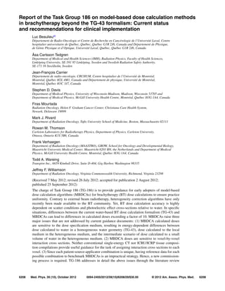
![6209 Beaulieu et al.: TG-186: Model-based dose calculation techniques in brachytherapy 6209
and provides explicit recommendations based on the current state of knowledge. TG-43-based dose
prescription and dose calculation remain in effect, with MBDCA dose reporting performed in par-
allel when available. In using MBDCAs, it is recommended that the radiation transport should be
performed in the heterogeneous medium and, at minimum, the dose to the local medium be reported
along with the TG-43 calculated doses. Assignments of voxel-by-voxel cross sections represent a
particular challenge. Electron density information is readily extracted from CT imaging, but cannot
be used to distinguish between different materials having the same density. Therefore, a recommen-
dation is made to use a number of standardized materials to maintain uniformity across institutions.
Sensitivity analysis shows that this recommendation offers increased accuracy over TG-43. MBDCA
commissioning will share commonalities with current TG-43-based systems, but in addition there
will be algorithm-specific tasks. Two levels of commissioning are recommended: reproducing TG-43
dose parameters and testing the advanced capabilities of MBDCAs. For validation of heterogeneity
and scatter conditions, MBDCAs should mimic the 3D dose distributions from reference virtual ge-
ometries. Potential changes in BT dose prescriptions and MBDCA limitations are discussed. When
data required for full MBDCA implementation are insufficient, interim recommendations are made
and potential areas of research are identified. Application of TG-186 guidance should retain practice
uniformity in transitioning from the TG-43 to the MBDCA approach. © 2012 American Association
of Physicists in Medicine. [http://dx.doi.org/10.1118/1.4747264]
Key words: brachytherapy, treatment planning, dose calculation, radiation transport, commissioning,
dose scoring, heterogeneities, material assignments
TABLE OF CONTENTS
I INTRODUCTION . . . . . . . . . . . . . . . . . . . . . . . . . . . . . 6210
I.A Problem description . . . . . . . . . . . . . . . . . . . . . 6210
I.B Report overview and rationale . . . . . . . . . . . . 6210
I.C Review of tissue and applicator material
heterogeneity effects . . . . . . . . . . . . . . . . . . . . 6211
I.C.1 Low energy regime, seeds, and
miniature
x-ray sources . . . . . . . . . . . . . . . . . . . 6211
I.C.2 Intermediate energies: 169
Yb . . . . . 6212
I.C.3 Higher energies: 192
Ir . . . . . . . . . . . . 6213
I.C.4 Cone beam computed tomography 6213
II REVIEW OF MODEL-BASED
BRACHYTHERAPY DOSE-CALCULATION
ALGORITHMS . . . . . . . . . . . . . . . . . . . . . . . . . . . . . . . 6213
II.A Semiempirical approaches . . . . . . . . . . . . . . . 6213
II.B Model-based algorithms . . . . . . . . . . . . . . . . . 6214
II.B.1 Collapsed-cone
superposition/convolution method 6214
II.B.2 Deterministic solutions to the
linear Boltzmann transport
equation . . . . . . . . . . . . . . . . . . . . . . . . 6214
II.B.3 Monte Carlo simulations . . . . . . . . . 6214
III DOSE SPECIFICATION MEDIUM SELECTION 6214
III.A Relationship between Dm,m and Dw,m in the
large cavity regime . . . . . . . . . . . . . . . . . . . . . . 6216
III.B Dm,m and Dw,m relationships in the small
and intermediate cavity regimes . . . . . . . . . . 6216
III.C Recommendations . . . . . . . . . . . . . . . . . . . . . . 6217
III.D Areas of research . . . . . . . . . . . . . . . . . . . . . . . 6218
IV CT IMAGING AND PATIENT MODELING . . . . 6218
IV.A Literature review . . . . . . . . . . . . . . . . . . . . . . . 6218
IV.A.1 Material characterization . . . . . . . . . 6219
IV.A.2 CT segmentation . . . . . . . . . . . . . . . . 6220
IV.A.3 CBCT segmentation . . . . . . . . . . . . . 6220
IV.A.4 Dual energy CT and spectral CT . . 6220
IV.A.5 CT artifacts . . . . . . . . . . . . . . . . . . . . . 6221
IV.A.6 Other imaging modalities . . . . . . . . 6221
IV.B Recommendations . . . . . . . . . . . . . . . . . . . . . . 6221
IV.B.1 Consensus material definition . . . . 6221
IV.B.1.a Prostate . . . . . . . . . . . . . . . . 6221
IV.B.1.b Breast . . . . . . . . . . . . . . . . . 6222
IV.B.1.c Calcifications . . . . . . . . . . . 6222
IV.B.1.d Other materials . . . . . . . . . 6222
IV.B.1.e Applicators, sources, and
other devices . . . . . . . . . . . 6222
IV.B.2 Material assignment method . . . . . . 6223
IV.B.2.a Use of other imaging
modalities . . . . . . . . . . . . . . 6224
IV.B.3 CT/CBCT artifact removal . . . . . . . 6224
IV.C Areas of research . . . . . . . . . . . . . . . . . . . . . . . 6224
IV.C.1 Determination of tissue
composition . . . . . . . . . . . . . . . . . . . . 6224
IV.C.2 Segmentation methods . . . . . . . . . . . 6224
IV.C.3 CT artifact removal . . . . . . . . . . . . . . 6224
V MBDCA COMMISSIONING . . . . . . . . . . . . . . . . . . 6225
V.A Literature review . . . . . . . . . . . . . . . . . . . . . . . 6225
V.B From TG-43 to MBDCA-based
commissioning . . . . . . . . . . . . . . . . . . . . . . . . . 6225
V.B.1 MBDCA commissioning level 1 . . 6225
V.B.2 MBDCA commissioning level 2 . . 6225
V.B.3 MBDCA commissioning workflow 6226
V.B.3.a Test case plans and data
availability . . . . . . . . . . . . . 6226
V.B.3.b DICOM test case
importing . . . . . . . . . . . . . . 6227
V.B.3.c TG-43-based dose
calculation tests . . . . . . . . . 6227
Medical Physics, Vol. 39, No. 10, October 2012](data:image/gif;base64,R0lGODlhAQABAIAAAAAAAP///yH5BAEAAAAALAAAAAABAAEAAAIBRAA7)