2. model 200 source of 103
Pd and the Best Medical 192
Ir source
͑certain commercial equipment, instruments, and materials
are identified in this work in order to specify adequately the
experimental procedure. Such identification does not imply
recommendation nor endorsement by either the AAPM or
National Institute of Standards and Technology ͑NIST͒, nor
does it imply that the material or equipment identified is
necessarily the best available for these purposes͒. In the 2004
update, the AAPM updated the data on the 125
I and 103
Pd
sources included in the original report and included data on
six other interstitial brachytherapy sources. All of the follow-
ing eight sources met the AAPM dosimetric prerequisites7
and the AAPM Calibration Laboratory Accreditation ͑CLA͒
subcommittee requirements8
as of July 15, 2001 and were
presented in the AAPM TG-43U1 report:
1 Amersham-Health model 6702 125
I source,
2. Amersham-Health model 6711 125
I source,
3. Best Medical model 2301 125
I source,
4. North American Scientific Inc. ͑NASI͒ model
MED3631-A/M 125
I source,
5. Bebig/Theragenics model I25.SO6 125
I source,
6. Imagyn isostar model IS-12501 125
I source ͑note that the
Imagyn isostar model IS-12501 125
I source which was
included in the 2004 AAPM TG-43U1 report has been
removed from the online Joint AAPM/RPC Source Reg-
istry due to discontinuation by the manufacturer͒,
7. Theragenics Corporation model 200 103
Pd source, and
8. NASI model MED3633 103
Pd source.
Since July 15, 2001 several additional sources have been
introduced in the market and have met the AAPM dosimetric
prerequisites and the CLA subcommittee requirements. As
planned during the writing of TG-43U1, a supplement was
needed to present consensus datasets for these newer
sources. This supplement is termed TG-43U1S1, and in-
cludes the following sources which met the criteria men-
tioned above as of January 10, 2005:
1. Amersham model 6733 125
I source,
2. DraxImage model LS-1 125
I source,
3. Implant Sciences model 3500 125
I source,
4. IBt model 1251L 125
I source,
5. IsoAid model IAI-125A 125
I source,
6. Mills Biopharmaceuticals model SL-125/SH-125 125
I
source,
7. SourceTech Medical model STM1251 125
I source, and
8. Best Medical model 2335 103
Pd source.
Manufacturers, dosimetry investigators, and end users
have generally adhered to AAPM recommendations given in
the TG-43U1 and CLA subcommittee reports. The source
models reviewed in this supplement ͑Fig. 1͒ satisfied AAPM
recommendations ͑dosimetric parameters accepted for publi-
cation in a scientific, peer-reviewed journal and metrologi-
cally acceptable source calibration procedures͒ on or before
January 10, 2005. After review and approval, these data were
posted on the online Joint AAPM/RPC Source Registry.9
As
stated in the AAPM TG-43U1 report, publications may re-
port dosimetry parameters using Monte Carlo, experimental
methods, or both techniques in the same publication. It is
also worth stressing that special care is needed to address
concerns for independence of various investigations included
in the development of consensus datasets. The independence
policy is described in detail in Sec. V F of the AAPM TG-
43U1
report.
II. CONSENSUS DATASETS FOR CLINICAL
IMPLEMENTATION
As presented in the TG-43U1 report, criteria used to
evaluate dosimetry parameters for each source model in-
cluded in this TG-43U1S1 report were:
1. Internal source geometry and a description of the source,
2. review of the pertinent literature for the source,
3. correction to ⌳ values due to the 1999 anomaly in NIST
air-kerma strength measurements ͑if applicable͒,
4. solid water-to-liquid water corrections,
5. experimental method used: TLD or diode,
6. active length assumed for the geometry function line-
source approximation,
7. name and version of the Monte Carlo transport code,
8. cross-section library used by the Monte Carlo simula-
tion,
9. Monte Carlo estimator used to score kerma or dose, and
10. agreement between Monte Carlo calculations and ex-
perimental measurement.
AAPM-approved consensus datasets are provided in
Tables I–X below with calculated dose rates using the one-
dimensional ͑1D͒ formalism in Table XI as similar to the
2004 AAPM TG-43U1 report. Descriptions of each source
and details used for obtaining the consensus datasets are
available in Appendix A. If essential items critical to the
evaluation of a given source were omitted from the salient
publications, then dosimetry investigators were contacted for
additional information and/or clarification. Fortunately, in re-
cent publications, analysis for some of these source models
benefited from adherence by dosimetry investigators to rec-
ommendations provided in Secs. V D and V E of the AAPM
TG-43U1 report. Data were italicized if they were not di-
rectly confirmed by other measurements or calculations;
boldface values indicate that data were interpolated towards
presenting data sets of all sources on a common mesh; ex-
trapolated data are underlined. As in the 2004 report, data
sets were thinned so as to minimize the amount of data while
maintaining interpolation errors ഛ2% for the purposes of
calculating dose rate distributions. Due to differences in
source construction, appropriate angular resolution for
F͑r,͒ was used to keep bilinear interpolation errors ഛ2%.
Additionally, the AAPM TG-43U1 report recommended a
mass density of 0.001 20 g cm−3
for both moist and dry air.
Upon analyzing the impact of relative humidity from 0% to
100%, a value of 0.001 19 g cm−3
is more appropriate and
should be used in conjunction with the recommended rela-
tive humidity of 40%.
2188 Rivard et al.: Supplement to AAPM TG-43 update 2188
Medical Physics, Vol. 34, No. 6, June 2007
3. III. CLARIFICATIONS ON RECOMMENDED
INTERPOLATION AND EXTRAPOLATION METHODS
While a sampling space with uniform increments for g͑r͒
and either F͑r,͒ or an͑r͒ is desired, the published data
indicate that authors have used a variety of spatial and angu-
lar increments and ranges. Therefore, interpolation or ex-
trapolation may be required to determine dose rate distribu-
tions at spatial locations not explicitly included in published
dosimetry-parameter tables. Methods for determining dose
rates at positions not characterized by the available datasets
or related publications were specified in the 2004 AAPM
TG-43U1 report. Interpolation methods for 2D and 1D do-
simetry parameters were provided in Sec. IV. ͑g͒ of the 2004
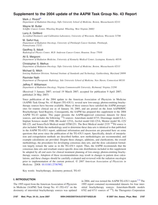
FIG. 1. Brachytherapy seeds examined
in this report: ͑a͒ Amersham model
6733 source, ͑b͒ DraxImage model
LS-1 source, ͑c͒ Implant Sciences
model 3500 source, ͑d͒ International
Brachytherapy model 1251L source,
͑e͒ IsoAid model IAI-125A source, ͑f͒
Mills Biopharmaceuticals Corporation
model SL-125/SH-125 source, ͑g͒
Source Tech Medical model STM
1251 source, and ͑h͒ Best Medical
model 2335 source. Titanium capsule
wall thicknesses are 0.08, 0.07, and
0.04 mm for the SourceTech Medical,
Best, and IBt seeds, respectively. Cap-
sule wall thickness for the remaining
seeds is 0.05 mm.
TABLE I. NIST standard WAFAC calibration dates for air-kerma strength for each manufacturer, and dose rate
constant values.
Manufacturer and source type
Date used by NIST and ADCLs
for calibration
CON⌳
͓cGy·h−1
·U−1
͔
Amersham 6733 125
I February 15, 2001 0.980
Draximage LS-1 125
I January 13, 2001 0.972
Implant Sciences 3500 125
I April 22, 2000 1.014
IBt 1251L 125
I May 17, 2000 1.038
IsoAid IAI-125A 125
I April 15, 2001 0.981
MBI SL-125/SH-125 125
I July 5, 2001 0.953
SourceTech Medical STM1251 125
I June 2, 2000 1.018
Best Medical 2335 103
Pd September 2, 2000 0.685
2189 Rivard et al.: Supplement to AAPM TG-43 update 2189
Medical Physics, Vol. 34, No. 6, June 2007
4. AAPM TG-43U1 report, and extrapolation methods for these
same parameters were provided in its Appendix C. Linear-
linear interpolation was recommended for F͑r,͒, and log-
linear interpolation was recommended for g͑r͒. However,
specific guidance on implementation of these recommenda-
tions by medical physicists or treatment planning software
manufacturers was limited. The brachytherapy dosimetry for-
malism should minimize the contribution of interpolation
and extrapolation errors to overall dose-calculation uncer-
tainty. Therefore, we consider the physical effects that gov-
ern the two-dimensional ͑2D͒ and one-dimensional ͑1D͒ an-
isotropy functions and the radial dose function, and aim to
clarify the recommended approaches towards ensuring im-
proved interpolation or extrapolation accuracy. Below are
presented the rationale and recommended methods for inter-
polation, rϽrmin extrapolation, and rϾrmax extrapolation of
F͑r,͒, an͑r͒, and gL͑r͒. Note that rmin and rmax are the
smallest and largest radii for a set of reported dosimetry pa-
TABLE II. AAPM Consensus L, gL͑r͒, and gP͑r͒ values for seven 125
I sources and one 103
Pd source ͑i.e., Best Medical model 2335͒. As used later in Table XI,
an͑r͒ data are given in the lowest five rows. Interpolated data are boldface, extrapolated data are underlined, and italicized data are obtained from candidate
datasets.
Line source approximation
L ͑mm͒ 3.0 4.1 3.76 4.35 3.0 3.0 3.81 4.55
r͑cm͒
Amersham
EchoSeed
6733
Draximage
BrachySeed
LS-1
Implant
Sciences
3500
IBt
1251L
IsoAid
advantage
IAI-125A
MBI
SL-125
SH-125
Source
Tech
STM1251
Best
Medical
2335
0.10 1.050 0.182 0.997 0.757 1.040 1.101 0.941 0.826
0.15 1.076 0.323 1.011 0.841 1.053 1.101 0.972 1.066
0.25 1.085 0.741 1.021 0.963 1.066 1.101 1.013 1.236
0.50 1.069 0.964 1.030 1.021 1.080 1.084 1.033 1.307
0.75 1.045 1.004 1.026 1.024 1.035 1.041 1.022 1.128
1.00 1.000 1.000 1.000 1.000 1.000 1.000 1.000 1.000
1.50 0.912 0.937 0.932 0.937 0.902 0.898 0.937 0.742
2.00 0.821 0.853 0.854 0.859 0.800 0.795 0.856 0.533
3.00 0.656 0.680 0.681 0.700 0.611 0.610 0.691 0.296
4.00 0.495 0.527 0.532 0.554 0.468 0.456 0.540 0.158
5.00 0.379 0.400 0.407 0.425 0.368 0.338 0.415 0.0920
6.00 0.285 0.300 0.308 0.323 0.294 0.250 0.314 0.0529
7.00 0.214 0.223 0.230 0.240 0.227 0.183 0.236 0.0309
8.00 0.155 0.166 0.171 0.180 0.165 0.134 0.176 0.0180
9.00 0.119 0.122 0.127 0.138 0.141 0.098 0.131 0.0105
10.00 0.0840 0.0900 0.0936 0.101 0.090 0.072 0.0969 0.0062
Point source approximation
0.10 0.693 0.100 0.576 0.403 0.686 0.727 0.544 0.427
0.15 0.851 0.225 0.732 0.569 0.833 0.871 0.700 0.706
0.25 0.985 0.629 0.886 0.805 0.967 0.999 0.876 1.020
0.50 1.046 0.928 0.997 0.978 1.056 1.061 0.999 1.247
0.75 1.039 0.994 1.017 1.012 1.029 1.035 1.013 1.114
1.00 1.000 1.000 1.000 1.000 1.000 1.000 1.000 1.000
1.50 0.916 0.944 0.938 0.945 0.906 0.901 0.943 0.749
2.00 0.826 0.862 0.862 0.869 0.804 0.799 0.864 0.539
3.00 0.660 0.688 0.688 0.710 0.615 0.614 0.698 0.300
4.00 0.498 0.534 0.538 0.562 0.471 0.459 0.546 0.161
5.00 0.382 0.405 0.412 0.432 0.371 0.340 0.420 0.0935
6.00 0.287 0.304 0.312 0.328 0.296 0.252 0.318 0.0538
7.00 0.216 0.226 0.233 0.244 0.229 0.184 0.239 0.0314
8.00 0.156 0.168 0.173 0.183 0.166 0.135 0.178 0.0184
9.00 0.120 0.124 0.129 0.141 0.142 0.099 0.133 0.0107
10.00 0.0846 0.0912 0.0947 0.102 0.091 0.072 0.0980 0.0063
an͑0.10͒ 1.173 2.004 1.129 1.162 1.127 1.091 1.172 1.052
an͑0.15͒ 1.246 2.275 1.268 1.327 1.197 1.159 1.317 1.205
an͑0.25͒ 1.112 2.152 1.164 1.296 1.069 1.035 1.210 1.213
an͑0.50͒ 0.996 1.150 0.973 1.028 0.957 0.927 0.982 0.938
an͑0.75͒ 0.974 1.030 0.942 0.992 0.962 0.907 0.962 0.894
2190 Rivard et al.: Supplement to AAPM TG-43 update 2190
Medical Physics, Vol. 34, No. 6, June 2007
5. rameters, respectively. For example, if g͑r͒ is reported for
r=͕0.5,1,2,3,4, and 5 cm͖, then rmin=0.5 cm and rmax
=5 cm.
A. F„r,… 2D anisotropy function
The 2D anisotropy function is a function of polar angle
for a specified radius and is normalized to unity at 0ϵ90°.
For all angles except 0, F͑r,͒ values generally trend to
asymptotically approach unity with increasing radial dis-
tance. The geometry function, G͑r,͒, accounts for dose dis-
tribution variations attributed to distance-dependent changes
in the solid angle and distribution of radioactivity, assuming
a uniform radioactive distribution. Therefore, nonunity val-
ues of the 2D anisotropy function are due to nonuniform
radionuclide distribution and to attenuation and scatter by the
source encapsulation and internal components. As a function
of polar angle, both of these effects generally change linearly
over small changes in radius or angle. Dose distributions at
10° ϽϽ170° for 0.5-cm-long capsules are primarily af-
fected by attenuation as a function of polar angle through the
cylindrical capsule wall. Dose distributions at other angles
are primarily affected by attenuation through encapsulation
end welds and radiation source carriers. Away from the
source long axis, F͑r,͒ behavior may be considered as a
combination of primary dose and dose due to photons scat-
tered in the surrounding medium where the proportion of
scattered radiation generally increases with increasing r. For
the sources included in this current report and the 2004
AAPM TG-43U1 report,2
variations in F͑r,Ͻ10°͒ or
F͑r,Ͼ170°͒ are largely due to photon attenuation by end
welds and capsule internal components. While these varia-
tions may exceed 50%, points within these volumes, i.e.,
P͑r,Ͻ10°͒ and P͑r,Ͼ170°͒, subtend ϳ1% of the solid-
angle weighted dose rate distribution around a source. F͑r,͒
may be accurately determined in general using linear inter-
polation. However, some sources have F͑r,͒ that signifi-
cantly exceed unity, e.g. the Draximage model LS-1 125
I
source, due to the geometry function not readily approximat-
ing the particle streaming function ͑i.e., in vacuo photon en-
ergy fluence͒.10
Thus, a linear-linear interpolation method for
TABLE III. F͑r,͒ for Amersham model 6733 taken directly from Sowards
and Meigooni ͑Ref. 15͒.
Polar angle
͑degrees͒
r ͓cm͔
1 2 3 4 5 6 7
0 0.305 0.397 0.451 0.502 0.533 0.551 0.565
5 0.386 0.468 0.510 0.557 0.586 0.595 0.611
10 0.507 0.570 0.609 0.634 0.660 0.669 0.685
15 0.621 0.663 0.680 0.712 0.717 0.726 0.719
20 0.714 0.738 0.743 0.774 0.769 0.779 0.785
30 0.848 0.851 0.849 0.873 0.859 0.860 0.880
40 0.944 0.933 0.918 0.932 0.921 0.912 0.924
50 0.999 0.985 0.969 0.983 0.953 0.965 0.949
60 1.029 1.015 0.995 1.012 0.985 1.003 0.982
70 1.038 1.033 1.015 1.022 1.001 0.994 1.019
80 1.026 1.034 1.014 1.026 1.009 0.999 1.000
90 1.000 1.000 1.000 1.000 1.000 1.000 1.000
an͑r͒ 0.967 0.964 0.953 0.966 0.953 0.948 0.955
TABLE IV. F͑r,͒ for Draximage model LS-1 taken directly from Chan, Nath, and Williamson ͑Ref. 24͒.
Polar angle
͑degrees͒
r ͓cm͔
0.25 0.5 0.75 1 1.5 2 3 5 10
0 3.459 1.261 0.979 0.872 0.799 0.775 0.765 0.766 0.781
10 3.312 1.246 0.977 0.877 0.808 0.787 0.775 0.778 0.786
20 2.755 1.219 0.988 0.901 0.841 0.821 0.811 0.816 0.822
30 2.130 1.178 0.994 0.925 0.877 0.861 0.854 0.864 0.873
40 1.675 1.125 0.999 0.950 0.912 0.902 0.898 0.909 0.899
50 1.380 1.073 0.998 0.967 0.945 0.938 0.934 0.940 0.935
60 1.194 1.032 0.996 0.981 0.970 0.968 0.967 0.968 0.964
70 1.085 1.007 0.998 0.994 0.990 0.989 0.988 0.991 0.993
80 1.024 0.999 1.001 1.001 0.999 0.999 0.998 1.004 0.982
90 1.000 1.000 1.000 1.000 1.000 1.000 1.000 1.000 1.000
an͑r͒ 2.152 1.150 1.030 0.987 0.958 0.949 0.943 0.947 0.942
TABLE V. F͑r,͒ for Implant Sciences model 3500 taken directly from Ri-
vard where higher resolution an͑r͒ data were published ͑Ref. 28͒.
Polar angle
͑degrees͒
r ͓cm͔
0.25 0.5 1 2 5 10
0 0.494 0.610 0.580 0.652 0.690 0.709
10 0.574 0.513 0.561 0.626 0.700 0.742
20 0.785 0.679 0.705 0.743 0.789 0.815
30 0.899 0.808 0.813 0.830 0.854 0.872
40 0.943 0.892 0.885 0.893 0.905 0.912
50 0.967 0.944 0.933 0.934 0.941 0.947
60 0.986 0.974 0.967 0.967 0.968 0.972
70 0.995 0.990 0.987 0.987 0.986 0.990
80 1.000 0.997 0.997 0.997 0.996 0.997
90 1.000 1.000 1.000 1.000 1.000 1.000
an͑r͒ 1.164 0.973 0.933 0.931 0.938 0.948
2191 Rivard et al.: Supplement to AAPM TG-43 update 2191
Medical Physics, Vol. 34, No. 6, June 2007
6. F͑r,͒ as a function of r and is appropriate, and should be
based on the two data points for each variable located imme-
diately adjacent to the interpolated point of interest. This
approach is identical to that recommended by the 2004
AAPM TG-43U1 report.2
When there is a need to extrapolate F͑r,͒ data outside of
the range of tabulated data, the 2004 AAPM TG-43U1
method ͑Appendix C 1͒ of using a nearest-neighbor or
zeroth-order approach is still recommended since differing
trends between different radionuclides do not warrant a dif-
ferent extrapolation methodology. Specifically, the nearest-
neighbor or zeroth-order approach presented in Appendix C
of the 2004 AAPM TG-43U1 report is still recommended for
F͑r,͒ extrapolation for rϽrmin and also for rϾrmax. Re-
garding need for F͑r,͒ extrapolation on polar angle, it ap-
pears that all sources have been characterized over the full
angular range of 0° ഛഛ90°. However, for example, if
F͑7,45°͒ were sought and data were available at F͑6,40°͒
and F͑6,50°͒ data where rmax=6 cm, one should first per-
form linear interpolation to obtain F͑6,45°͒ then extrapolate
͑zeroth order͒ to obtain F͑7,45°͒.
We advise Monte Carlo dosimetry investigators to exploit
continuously increasing computational and geometric model-
ing capabilities to estimate the dose rate distributions, includ-
ing F͑r,͒, as close to the source as possible and with fine
angular resolution. For typical low-energy photon-emitting
brachytherapy seeds which are 5 mm long and 0.8 mm in
diameter capsule, it is reasonable to calculate F͑r,͒ for r
ഛ2.5 mm for the limited range of theta values that place
calculation voxels outside of the source capsule and in the
range of dose calculation points relevant to specialized clini-
cal applications such as eye plaques.
TABLE VI. F͑r,͒ for IBt model 1251L taken from Reniers, and reprocessed
using Leff=4.35 mm.
Polar angle
͑degrees͒
r ͓cm͔
0.5 1 2 3 5
0 0.476 0.544 0.653 0.680 0.703
5 0.645 0.626 0.656 0.713 0.718
10 0.725 0.699 0.709 0.736 0.751
20 0.810 0.783 0.789 0.810 0.817
30 0.867 0.849 0.849 0.859 0.854
40 0.923 0.900 0.910 0.911 0.911
50 0.966 0.946 0.946 0.949 0.954
60 0.991 0.979 0.971 0.976 0.968
70 0.998 0.988 0.991 0.996 0.988
80 1.002 0.996 0.997 0.995 0.988
90 1.000 1.000 1.000 1.000 1.000
an͑r͒ 1.028 0.958 0.945 0.948 0.945
TABLE VII. F͑r,͒ for IsoAid IAI-125A taken directly from Solberg et al.
͑Ref. 36͒.
Polar angle
͑degrees͒
r ͓cm͔
0.5 1 2 3 5 7
0 0.352 0.406 0.493 0.520 0.578 0.612
5 0.411 0.465 0.545 0.584 0.658 0.701
10 0.481 0.527 0.601 0.642 0.704 0.726
20 0.699 0.719 0.757 0.775 0.794 0.799
30 0.848 0.846 0.862 0.862 0.869 0.879
40 0.948 0.936 0.932 0.916 0.937 0.969
50 1.002 0.986 0.974 0.961 0.963 0.971
60 1.029 1.024 1.008 0.993 0.990 1.001
70 1.029 1.039 1.027 1.006 1.016 1.010
80 0.999 1.025 1.024 1.023 1.009 1.025
90 1.000 1.000 1.000 1.000 1.000 1.000
an͑r͒ 0.957 0.968 0.964 0.955 0.959 0.955
TABLE VIII. F͑r,͒ for Mills Biopharmaceuticals model SL-125/SH-125
taken from Li ͑Ref. 45͒ and reprocessed using Leff=3.0 mm.
Polar angle
͑degrees͒
r ͓cm͔
1 2 3 4 5
0 0.359 0.424 0.471 0.501 0.520
10 0.429 0.493 0.535 0.563 0.574
20 0.568 0.610 0.643 0.672 0.670
30 0.710 0.744 0.759 0.771 0.762
40 0.823 0.842 0.852 0.863 0.857
50 0.918 0.926 0.936 0.937 0.921
60 0.973 0.972 0.980 0.986 0.974
70 0.985 0.987 0.989 0.993 0.993
80 0.991 1.000 1.013 1.002 0.993
90 1.000 1.000 1.000 1.000 1.000
an͑r͒ 0.900 0.907 0.916 0.921 0.914
TABLE IX. F͑r,͒ for Source Tech Medical model STM1251 taken directly
from Kirov and Williamson erratum ͑Ref. 48͒.
Polar angle
͑degrees͒
r ͓cm͔
0.25 0.5 1 2 3 5 7
0 0.863 0.524 0.423 0.453 0.500 0.564 0.607
2 0.865 0.489 0.616 0.701 0.702 0.706 0.720
5 0.784 0.668 0.599 0.611 0.637 0.657 0.682
7 0.861 0.588 0.575 0.603 0.632 0.655 0.682
10 0.778 0.562 0.579 0.617 0.649 0.672 0.700
20 0.889 0.688 0.698 0.722 0.750 0.761 0.781
30 0.949 0.816 0.808 0.819 0.841 0.838 0.845
40 0.979 0.898 0.888 0.891 0.903 0.901 0.912
50 0.959 0.956 0.943 0.941 0.950 0.941 0.945
60 0.980 0.988 0.982 0.980 0.985 0.973 0.982
70 0.989 0.973 1.005 1.002 1.011 0.995 0.998
80 0.994 0.994 0.989 1.015 1.018 1.003 1.011
90 1.000 1.000 1.000 1.000 1.000 1.000 1.000
an͑r͒ 1.210 0.982 0.942 0.937 0.947 0.938 0.944
2192 Rivard et al.: Supplement to AAPM TG-43 update 2192
Medical Physics, Vol. 34, No. 6, June 2007
7. B. an„r… 1D anisotropy function
The recommended an͑r͒ data sets were derived from
solid-angle weighted dose rates based on F͑r,͒ datasets,
removing effects of the geometry function. These an͑r͒ data
sets demonstrated nearly constant or linear behavior for r
ജ1 cm, especially for quasi mono-energetic photon sources
such as 125
I. For rϽ1 cm, an͑r͒ values significantly in-
creased with decreasing r as illustrated by Rivard, Melhus,
and Kirk for a general 103
Pd source.11
This behavior is
caused by volume averaging of larger dose rates near the
source long-axis due to the increasing ellipsoidal shape of
isodose distributions in comparison to the dose rate at the
same r value along the transverse plane. Based on increased
availability of high-resolution an͑r͒ data determined over a
wide range of distances, we recommend a log-linear ap-
proach to interpolating an͑r͒ data. The interpolation should
be based on the two data points located immediately adjacent
to the interpolated point of interest. This log-linear approach
differs from the 2004 AAPM TG-43U1 report which previ-
ously recommended that “linear interpolation may be used to
match the grid spacing of gX͑r͒ with the grid spacing of
an͑r͒.” In light of the general behavior of an͑r͒ observed in
multiple high-resolution datasets, it is recommended that
dosimetry investigators provide sufficient spatial sampling of
an͑rϽ1 cm͒ and for suitably large r to minimize the need
to extrapolate. This is especially convenient using Monte
Carlo techniques. Additionally, having a common high-
resolution sampling space for both an͑r͒ and g͑r͒ is crucial
for implementation of the simple 1D formalism of Eq. ͑9͒ of
TG-43U1.2
Appendix C of the 2004 AAPM TG-43U1 report recom-
mended using Eq. ͑1͒ below, Eq. ͑C3͒ of the 2004 TG-43U1
report,2
for extrapolating an͑r͒ for distances rϽrmin, where
rmin is the shortest distance for which an͑r͒ data are pro-
vided
an͑r͒ Ϸ
an͑rmin͒
r2
GL͑r,0͒
for r Ͻ rmin. ͑1͒
In this report, we recommend replacing the aforementioned
extrapolation procedure with a more accurate approach that
approximates the short distance behavior of an͑r͒ at r
Ͻrmin by the solid-angle ͑⍀͒ weighted integral of the line-
source geometry function correction
TABLE X. F͑r,͒ for Best Medical model 2335 taken from Meigooni et al.
͑Ref. 54͒ and reprocessed using Leff=4.55 mm.
Polar angle
͑degrees͒
r ͓cm͔
1 2 3 4 5 6 7
0 0.797 0.690 0.674 0.672 0.663 0.675 0.630
5 0.801 0.696 0.683 0.669 0.666 0.679 0.645
10 0.790 0.690 0.673 0.675 0.665 0.690 0.644
15 0.675 0.613 0.608 0.604 0.626 0.620 0.581
20 0.608 0.591 0.596 0.601 0.616 0.647 0.595
25 0.675 0.639 0.637 0.659 0.653 0.706 0.651
30 0.681 0.660 0.679 0.694 0.694 0.717 0.672
35 0.725 0.693 0.705 0.721 0.703 0.730 0.687
40 0.762 0.736 0.750 0.747 0.741 0.775 0.720
45 0.792 0.807 0.846 0.847 0.866 0.876 0.804
50 0.885 0.880 0.885 0.887 0.909 0.907 0.835
60 0.915 0.929 0.944 0.936 0.965 1.001 0.912
70 0.932 0.960 0.972 0.965 0.975 1.014 0.916
80 0.941 0.975 0.986 0.985 0.999 1.017 0.915
90 1.000 1.000 1.000 1.000 1.000 1.000 1.000
an͑r͒ 0.879 0.872 0.881 0.881 0.890 0.909 0.845
TABLE XI. Transverse plane dose rates ͑cGy·h−1
·U−1
͒ as a function of distance for the 8 brachytherapy sources included in this report using gL͑r͒ and an͑r͒,
and the 1-D formalism of Eq. ͑11͒ from the 2004 AAPM TG-43U1. Results using interpolated gL͑r͒ or an͑r͒ data are highlighted in boldface while
extrapolated results based on gL͑r͒ and/or an͑r͒ data are underlined.
r ͓cm͔
Amersham
EchoSeed
6733
Draximage
BrachySeed
LS-1
Implant
Sciences
3500
IBt
1251L
IsoAid
Advantage
IAI-125A
MBI
SL-125
SH-125
Source
Tech
STM1251
Best
Medical
2335
0.10 7.97E+1 1.96E+1 6.56E+1 4.86E+1 7.59E+1 7.56E+1 6.48E+1 3.08E+1
0.15 4.62E+1 2.21E+1 4.17E+1 3.49E+1 4.35E+1 4.28E+1 4.16E+1 2.59E+1
0.25 1.72E+1 2.11E+1 1.67E+1 1.73E+1 1.62E+1 1.58E+1 1.73E+1 1.26E+1
0.50 4.09E+0 4.15E+0 3.93E+0 4.18E+0 3.97E+0 3.75E+0 3.99E+0 3.21E+0
0.75 1.76E+0 1.77E+0 1.73E+0 1.85E+0 1.73E+0 1.59E+0 1.76E+0 1.21E+0
1.0 9.48E−1 9.59E−1 9.46E−1 9.94E−1 9.50E−1 8.58E−1 9.58E−1 6.02E−1
1.5 3.85E−1 3.90E−1 3.94E−1 4.15E−1 3.81E−1 3.45E−1 4.01E−1 2.00E−1
2.0 1.95E−1 1.99E−1 2.03E−1 2.13E−1 1.90E−1 1.73E−1 2.06E−1 8.06E−2
3.0 6.85E−2 7.01E−2 7.24E−2 7.76E−2 6.40E−2 5.96E−2 7.47E−2 2.01E−2
4.0 2.95E−2 3.06E−2 3.19E−2 3.45E−2 2.77E−2 2.52E−2 3.27E−2 6.05E−3
5.0 1.43E−2 1.49E−2 1.57E−2 1.69E−2 1.39E−2 1.19E−2 1.60E−2 2.28E−3
6.0 7.41E−3 7.81E−3 8.24E−3 8.93E−3 7.72E−3 6.09E−3 8.44E−3 9.30E−4
7.0 4.12E−3 4.29E−3 4.54E−3 4.88E−3 4.37E−3 3.28E−3 4.68E−3 3.71E−4
8.0 2.28E−3 2.43E−3 2.59E−3 2.81E−3 2.43E−3 1.84E−3 2.67E−3 1.66E−4
9.0 1.39E−3 1.41E−3 1.52E−3 1.70E−3 1.64E−3 1.06E−3 1.57E−3 7.66E−5
10.0 7.92E−4 8.34E−4 9.10E−4 1.00E−3 8.49E−4 6.30E−4 9.41E−4 3.62E−5
2193 Rivard et al.: Supplement to AAPM TG-43 update 2193
Medical Physics, Vol. 34, No. 6, June 2007
8. an͑r͒ Ϸ an͑rmin͒
͐4GL͑r,0͒d⍀
͐4GL͑rmin,0͒d⍀
for r Ͻ rmin. ͑2͒
When using Eq. ͑2͒ instead of Eq. ͑1͒ for sources in this
report and the TG-43U1 report, extrapolating from an͑rmin
=1 cm͒ to an͑0.25͒ and an͑0.50͒ improved the average
extrapolation accuracy by 8.8% and 0.2%, respectively, as
shown comparing data at the bottom of the last two columns
in Table XII. Extrapolated an͑r͒ values are given in Table II
as used in Table XI. Care should be taken when extrapolating
an͑r͒ to distances smaller than half the capsule length since
dose rates at these distances for some polar angles are lo-
cated within the source and are clinically irrelevant. For in-
stance, for the 0.4 mm radius source capsules presented in
this report, an͑0.10͒ was integrated over 23.6° ഛ
ഛ156.4° and an͑0.15͒ over 15.5° ഛഛ164.5°, both with
0.1° increments.
There were no specific recommendations given in the
2004 AAPM TG-43U1 report on how to extrapolate an͑r͒ at
distances where rϾrmax.2
While poly-energetic sources such
as 103
Pd exhibit significantly diminished anisotropy at dis-
tances greater than 10 cm in liquid water due to contribu-
tions from the weakly abundant high-energy photon emis-
sions ͑i.e., E␥ജ0.3 MeV͒, at this time a radionuclide-
specific approach is not recommended. Conservatively, a
nearest neighbor or zeroth-order extrapolation approach is
recommended until more results at larger distances become
available. Consequently, brachytherapy dosimetry investiga-
tors are advised to determine dose rate distributions and sub-
sequently publish F͑r,͒ and an͑r͒ values at distances as
large as reasonably achievable. For 125
I and 103
Pd, character-
ization out to distances exceeding 10 cm is possible with
acceptable statistical precision using modern codes. How-
ever, the investigators should limit their published results to
those data where contributions from scattered radiation ap-
proximate those of an infinitely large phantom.12,13
C. Radial dose function
The physical effects that govern the behavior of g͑r͒ are
based on attenuation and scatter in a recommended 15 cm
radius liquid water medium, where broad beam attenuation is
based on Ј/ and absorbed dose is based on en/. For
points further than a few cm from the sphere surface yet
beyond 1 cm for an 125
I or 103
Pd source, g͑r͒ should de-
crease approximately exponentially as a function of increas-
ing r. Consequently, a log-linear function for g͑r͒ interpola-
tion was recommended in the 2004 AAPM TG-43U1 report.2
Upon additional examination, any logarithmic function such
as log10 or ln ͑i.e., loge͒ will suffice to interpolate gL͑r͒ data
in a log-linear manner since differences are expressed as
changes in slope and offset. Additionally, it is recommended
that gL͑r͒ data be interpolated instead of gP͑r͒ since this
latter function changes more rapidly for rϽ1 cm due to im-
proved approximation of the particle streaming function by
GL͑r,͒.10
The log-linear interpolation should be performed
using data points immediately adjacent to the radius of inter-
est. Equation ͑3͒ may be used to solve for gL͑r2͒ where r1
Ͻr2Ͻr3 given gL͑r1͒ and gL͑r3͒.
TABLE XII. Extrapolation of an͑r͒ from r=1 cm to r=0.25 cm and r=0.50 cm. Equation ͑1͒ uses the ratio of point- and line-source geometry functions
applied to an͑rmin͒ to extrapolate to smaller distances. Equation ͑2͒ uses a solid-angle weighted line-source geometry function to extrapolate an͑rmin͒ to
smaller distances. These extrapolation approaches are tested on consensus an͑0.25͒, an͑0.50͒, and an͑1.00͒ data for six brachytherapy sources. The
percentage error relative to the consensus an͑r͒ data when using Eq. ͑1͒ and Eq. ͑2͒ is indicated by an͑r͒error1 and an͑r͒error2, respectively. From the
summary in the lower right of this table, it is apparent that an͑r͒ extrapolation for rϽrmin is significantly better using Eq. ͑2͒.
Source model r ͓cm͔ Consensus an͑r͒ Eq. ͑1͒ Eq. ͑2͒ an͑r͒error1 ͓%͔ an͑r͒error2 ͓%͔
Implant Sciences 0.25 1.164 1.078 1.160 −8.0 −0.3
3500 125
I 0.50 0.973 0.965 0.977 −0.9 0.4
AAPM TG-43U1S1 1.00 0.933 0.933 0.933 NA NA
Source Tech Medical 0.25 1.210 1.089 1.172 −11.1 −3.3
STM1251 125
I 0.50 0.982 0.974 0.986 −0.8 0.4
AAPM TG-43U1S1 1.00 0.942 0.942 0.942 NA NA
North American Scientific 0.25 1.288 1.128 1.241 −14.2 −3.8
MED3631-A/M 125
I 0.50 1.008 0.991 1.007 −1.7 −0.1
AAPM TG-43U1 1.00 0.952 0.952 0.952 NA NA
Bebig/Theragenics 0.25 1.122 1.065 1.131 −5.3 0.8
I25.S06 125
I 0.50 0.968 0.966 0.977 −0.2 0.9
AAPM TG-43U1 1.00 0.939 0.939 0.939 NA NA
Theragenics 0.25 1.130 1.015 1.119 −11.3 −1.0
200 125
I 0.50 0.880 0.891 0.905 1.2 2.7
AAPM TG-43U1 1.00 0.855 0.855 0.855 NA NA
North American Scientific 0.25 1.257 1.070 1.177 −17.5 −6.8
MED3633 103
Pd 0.50 0.962 0.940 0.955 −2.3 −0.8
AAPM TG-43U1 1.00 0.903 0.903 0.903 NA NA
Average at r=0.25 cm an͑0.25͒error1 =−11.2% an͑0.25͒error2 =−2.4%
Average at r=0.50 cm an͑0.50͒error1 =−0.8% an͑0.50͒error2 =0.6%
2194 Rivard et al.: Supplement to AAPM TG-43 update 2194
Medical Physics, Vol. 34, No. 6, June 2007
9. gL͑r2͒ = gL͑r1͒e͑r2−r1͒/͑r3−r1͒͑ln͓gL͑r3͔͒−ln͓gL͑r1͔͒͒
for r1 Ͻ r2 Ͻ r3. ͑3͒
For example, if gL͑r1͒=1.000 and gL͑r3͒=0.800 where r1
=1 cm, r2=1.5 cm, and r3=2.0 cm, one may obtain gL͑r2͒
=0.894 using Eq. ͑3͒.
Appendix C 3 of the 2004 AAPM TG-43U1 report clearly
specified an extrapolation method ͑nearest neighbor or zeroth
order͒ for 1D dose rate distributions when rϽrmin for
g͑rmin͒.2
Due to the great variability in g͑r͒ based on choice
of L and features of source construction, use of nearest-
neighbor or zeroth-order data is still recommended for ex-
trapolation of gL͑r͒ for rϽrmin. However, the gP͑r͒ data
should then be determined by applying the ratio of the point-
and line-source geometry functions to gL͑r͒ as previously
explained.
The 2004 AAPM TG-43U1 report was not explicit for
extrapolating beyond g͑rmax͒ where rϾrmax.2
Consequently,
we have revisited the g͑r͒ extrapolation methodology, and
considered a variety of fitting functions such as the bi-
exponential fit as suggested by Furhang and Anderson.14
The
AAPM now recommends adoption of a single exponential
function based on fitting gL͑r͒ data points for the largest two
consensus r values in a similar vein as Eq. ͑3͒. Specifically, a
log-linear extrapolation as illustrated in Eq. ͑4͒ may be used
to solve for gL͑r3͒ where r2=rmax, r1Ͻr2Ͻr3, and given
gL͑r1͒ and gL͑r2͒.
gL͑r3͒ = gL͑r1͒e͑r3−r1͒/͑r2−r1͒͑ln͓gL͑r2͔͒−ln͓gL͑r1͔͒͒
for r1 Ͻ r2 Ͻ r3. ͑4͒
For example, if gL͑r1͒=0.510 and gL͑r2͒=0.391 where r1
=4 cm, r2=5 cm, and r3=6 cm, one may obtain gL͑r3͒
=0.300 using Eq. ͑4͒. Using gL͑rmax͒ as a test for extrapolat-
ing gL͑r͒ data for the sources included in this report, the
single exponential function extrapolation technique reduces
gL͑r͒ extrapolation errors by over 40% as compared to
zeroth-order extrapolation, with negligible differences in
comparison to more complex fits such as a three-point linear
regression. Therefore, the AAPM recommends that treatment
planning software manufacturers no longer employ a zeroth-
order approach for determining gL͑r͒ extrapolated values be-
yond gL͑rmax͒, and that they immediately use a single expo-
nential fit to extrapolate gL͑r͒ values based on the furthest
two consensus data points. Following this guidance, Table II
includes gL͑r͒ and gP͑r͒ extrapolated beyond rmax for the
sources included in this report.
To provide practical data for treatment planning quality
assurance that typically uses gP͑r͒ instead of gL͑r͒, values in
Table XI include extrapolated an͑r͒ or gP͑r͒ data. These
latter data were converted from extrapolated gL͑r͒ data since
gP͑r͒ changes more rapidly and may be derived from gL͑r͒
using the ratio of the point- and line-source geometry func-
tions. It is also noteworthy to point out that these interpola-
tion and extrapolation techniques may be extended to the
dosimetry parameters in the 2004 AAPM TG-43U1 report or
other brachytherapy sources in general.
IV. SUMMARY
As stated in the AAPM TG-43U1 report, the AAPM rec-
ommends that the revised dose-calculation protocol and re-
vised source-specific dose-rate distributions be adopted by
all end users for clinical treatment planning of low-energy
brachytherapy using interstitial sources. Depending upon the
dose-calculation protocol and parameters currently used by
individual physicists, adoption of this protocol may result in
changes to patient dose calculations. These changes should
be carefully evaluated and reviewed with the radiation on-
cologist preceding implementation of the current protocol.
ACKNOWLEDGMENTS
The authors wish to thank Frank A. Ibbott for creation of
the artwork in Fig. 1, which was supported by the AAPM
Therapy Physics Committee ͑TPC͒. Also, we extend our ap-
preciation to Sujat Suthankar of Rosses Medical Systems
͑now at Implant Sciences͒ for discussions on Sec. III. Fur-
thermore, we thank Janelle A. Molloy for AAPM TPC re-
view and Zuofeng Li, Ning J. Yue, and Bruce Thomadsen of
the AAPM BTSC for their constructive comments and care-
ful review of this report. We also wish to thank David W. O.
Rogers for bringing the air mass density correction to our
attention. Some of the authors ͑M.J.R.; A.S.M.; R.N.; J.F.W.͒
have received research support to perform dosimetry studies
for the sources included herein ͑Implant Sciences Corp.,
Mills Biopharmaceuticals Corp.; International Brachy-
therapy, IsoAid Corp., and Best Medical Inc.; DraxImage
Inc.; DraxImage Inc. and SourceTech Medical, respectively͒.
APPENDIX: MODEL-SPECIFIC SOURCE
DOSIMETRY DATA
The following sections summarize the dosimetry param-
eters for each source, listed alphabetically. A description of
the source and its references are first provided. Afterwards
each dosimetry parameter is discussed briefly.
A. Amersham model 6733 125
I source
The EchoSeed™ model 6733 source was introduced in
2001, and is similar to the model 6711 source. The model
6733 consists of a 4.5 mm welded titanium capsule with its
external surface having several circular grooves, 0.8 mm in
diameter, and a titanium wall 0.05 mm thick, with welded
end caps. The grooves are to enhance the ultrasound visual-
ization of the sources. The capsule contains a 3.0-mm-long,
0.5-mm-diam silver rod onto which 125
I is adsorbed. ͑Fig. 1͒.
The active length for the geometry function line-source ap-
proximation is L=3.0 mm.
There are two published papers for this model; one deal-
ing with Monte Carlo determination by Sowards and
Meigooni,15
and the other by Meigooni et al. dealing with
experimental dose determinations using TLDs.16
Both of
these papers report values for all the TG-43 parameters. The
Monte Carlo calculations were performed both in SolidWa-
ter™ ͑model 457 by Radiation Measurements Inc., of
Middletown, WI͒ and in liquid water, and used the PTRAN
2195 Rivard et al.: Supplement to AAPM TG-43 update 2195
Medical Physics, Vol. 34, No. 6, June 2007
10. version 7.43 Monte Carlo code. Photon cross sections used
were from DLC-99 with mass energy absorption coefficients
from Hubbell and Seltzer.17
Experimental results were ob-
tained using TLD-100 chip dosimeters ͑Harshaw/Bicron of
Solon, OH͒ in a SolidWater™ phantom. The calibration of
the TLD chips was performed using a 6 MV beam and an
energy correction factor of 1.4 was used. Correction from
SolidWater™ to water was done with a factor of 1.05 bor-
rowed from Williamson.18
The standard deviation from 16
chips was 5%. These measurements in SolidWater™ were
compared with measurements by Meigooni et al. in
SolidWater™.16
1. 6733 Λ
Values for the Monte Carlo dose rate constant were ob-
tained at a point on the transverse plane in both liquid water
and SolidWater™. Using the liquid water results, MC⌳
=0.99 cGy h−1
U−1
. The air-kerma strength was measured at
NIST in Spring 2001 with EXP⌳ measured in SolidWater™
using TLDs. After correction to liquid water, the value of
EXP⌳ was 0.97 cGy h−1
U−1
. Averaging these values gives a
CON⌳=0.98 cGy h−1
U−1
as in Table I.
2. 6733 g„r…
The Monte Carlo and measured values for rജ1 cm for
the radial dose function agree within 5%, which is within the
experimental uncertainties. Therefore, Table II shows
CONg͑r͒ as taken from the Monte Carlo data set in liquid
water.
3. 6733 F„r,…
Experimental and Monte Carlo results agree within 5%
for angles greater than 20°. The experimental and Monte
Carlo results agree within 5% for distances 5 cm or greater,
but have greater differences at 0° for a distance of 2 cm
because of uncertainties in the TLD measurement. Table III
presents the consensus model 6733 F͑r,͒ data taken di-
rectly from Sowards and Meigooni.15
B. Draximage model LS-1 125
I source
The BrachySeed™ model LS-1 source was approved by
the U.S. Food and Drug Administration in October 2000 and
introduced to the North American market in 2001 by Cyto-
gen Corporation ͑Princeton, NJ͒, under license from
DRAXIS Health Inc. ͑Mississauga, Ontario, Canada͒. The
BrachySeed™ was distributed by Draximage Inc. ͑Kirkland,
Quebec, Canada͒, a subsidiary of DRAXIS Health Inc. Pro-
duction stopped in February 2006, but these data are of in-
terest to dosimetry investigators and interpretation of clinical
trial results.
The model LS-1 features a two-bead geometry and unique
laser weld about the center of the 4.4-mm-long and
0.8-mm-diam seed. 125
I is uniformly impregnated in 0.5 mm
diameter ceramic ͑alumina-silicate͒ beads, separated by a
2.97-mm-long Pt/Ir radio-opaque marker ͑Fig. 1͒. A medial
Ti spacer is included to center the x-ray marker and provide
a surface for the central weld of the two end capsules, which
have a wall thickness of 0.05 mm. There are four peer-
reviewed papers that assess the 2D dosimetry parameters of
the BrachySeed™ model LS-1, and a fifth publication de-
scribes a consensus dataset methodology using the Brachy-
Seed™ publications as examples.
Nath and Yue measured 2D brachytherapy dosimetry pa-
rameters in a water-equivalent phantom using 1 mm3
TLD
rods and TLD-specific calibration factors.19
A SolidWater™
to liquid water correction factor of 1.043 obtained by
Williamson18
and a TLD energy dependence correction of
1.41 published by Meigooni et al.20
were used to calculate ⌳.
On the transverse plane, radial distances are listed for a range
of 0.5–7 cm, and 2D measurements are reported between 1
and 6 cm. A correction was made to account for the 1999
NIST WAFAC anomaly, which impacted measurements of ⌳
by +6.8%. Towards the calculation of 2D dose distributions,
results were presented for both a line source model ͑Leff
=4.1 mm͒ and a two-point source model ͑separation
=3.6 mm͒.
Chan and Prestwich measured and calculated dosimetry
parameters for the model LS-1 source.21
Measurements were
performed using GafChromic MD-55-2 film, which is cur-
rently not a well-established method for determining single-
seed brachytherapy dose distributions, and the data were not
included in the consensus. Chan and Prestwich used the In-
tegrated Tiger Series CYLTRAN ͑version 3.0͒ with photon
cross sections published by NIST17
to perform Monte Carlo
photon transport simulations. The CYLTRAN code has been
benchmarked using the MED3631-A/M 125
I source. The au-
thors state that the source geometry was modeled exactly
with the exception of the capsule ends, which were given a
flat thickness of 60 m instead of modeling a spherical shell
with thickness 65 m. sK was estimated using a cylindrical
volume of air and a 5 keV photon energy cutoff to simulate
the NIST WAFAC. Material densities and compositions were
not explicitly stated, and the calculation geometry was de-
scribed as a series of concentric cylinders. A two-point
source model with 3.6 mm separation was employed for re-
constructing 2D brachytherapy dose distributions. The num-
ber of particle histories was chosen to ensure that 1 stan-
dard uncertainty about the mean was less than 1%.
Williamson22
published calculated single-seed brachy-
therapy dosimetry parameters for the model LS-1 125
I seed
using the PTRAN code ͑PTRANគCCG, version 7.43͒, the
DLC-146 photon cross-section library, and the mass-energy
absorption coefficients of Hubbell and Seltzer.17
The colli-
sion kerma rate at a given geometric location was calculated
using the bounded next flight estimator, and for distances
less than 3 mm, a once-more collided flux point-estimator
was employed. Results for g͑r͒ were evaluated over
0.1–14 cm in radial distance. F͑r,͒ was evaluated from
0.25 to 10 cm in distance range and over 0° ഛഛ180° at 34
angular increments with a maximum 5° spacing, although,
data were presented graphically. Towards calculation of ⌳,
SK was estimated by simulating the measurement geometry
of the NIST WAFAC. Ti characteristic x-ray production was
2196 Rivard et al.: Supplement to AAPM TG-43 update 2196
Medical Physics, Vol. 34, No. 6, June 2007
11. suppressed in the PTRAN code to simulate the function of
the aluminum filter in the NIST WAFAC. Three geometry
functions were included in the 2D dosimetry calculations: a
point source model, a two-point source model ͑separation
=3.6 mm͒, and a line source model of length 7.2 mm. Addi-
tional simulations were performed to assess the variation of
g͑r͒ with respect to internal motion of the active beads. The
number of starting particles was chosen to provide statistical
standard errors of the mean between 0.2% and 2%.
For the BrachySeed™ model LS-1 125
I source, Wang and
Sloboda23
calculated the 2D dosimetry parameters using
EGS4 and an associated user code, DOSCGC. A track-length
estimator was used to score photon energy fluence and then
combined with appropriate mass-energy absorption coeffi-
cients from Hubbell and Seltzer to estimate absorbed dose.
Furthermore, the EGS4/DOSCGC code was verified by the
authors using the model 6702 and 6711 125
I brachytherapy
seeds, among other radiation sources. As EGS4 does not
simulate the production of characteristic x rays, three meth-
ods were used to model characteristic x-ray production from
the Ag doped into the ceramic beads. Published results of
Wang and Sloboda include the method that simulates char-
acteristic x-ray production by determining the probability of
interaction between the principal 125
I photons and Ag.23
Re-
sults were evaluated between 0.1 and 14 cm of radial dis-
tance for g͑r͒ and over 0.25–10 cm for F͑r,͒ using a
spherical coordinate geometry. Ti characteristic x-ray contri-
butions were removed to simulate NIST WAFAC measure-
ment. Assuming a source separation of 3.6 mm, a double-
point model was used in the reconstruction of the 2D dose
distributions.
Chan, Nath, and Williamson24
published a methodology
for constructing consensus reference dosimetry parameters
for a single brachytherapy source, and used the four afore-
mentioned BrachySeed™ publications as an example. Addi-
tionally, Chan, Nath, and Williamson included minor correc-
tions or clarifications of results published by Chan and
Prestwich and by Williamson,22
and a detailed table of Wil-
liamson’s F͑r,͒ data is included, which was not present in
the original publication. The recommended consensus values
in Chan and co-workers24
are similar to those published here,
with specific differences listed below. However, the 1D dose
rate per unit air-kerma strength values published in Table IV
of Chan et al. are not in agreement with the recommended
dosimetry data of Chan et al.24
For example, a value of
0.9673 cGy h−1
·U−1
is published for 1 cm, while a value of
0.9594 cGy h−1
·U−1
is expected ͑CON⌳=0.972 cGy h−1
·U−1
;
an͑r͒=0.987; and, gP͑r͒=1͒. Because of this discrepancy
and because Chan et al. do not describe how the values were
generated, use of the 1D dose rate per unit air-kerma strength
values in Table IV of Chan et al.24
to validate the entry of
consensus dosimetry data into a given treatment planning
system is not recommended.
1. LS-1 Λ
Nath and Yue19
published a measured ⌳ value of
1.02±0.07 cGy h−1
·U−1
that includes correction for the 1999
NIST WAFAC anomaly using TLDs in a SolidWater™ phan-
tom. The GafChromic film measurement of Chan and
Prestwich21
yielded 0.98±0.06 cGy h−1
·U−1
, but is not in-
cluded in the CON⌳ derivation since radiochromic film is still
considered an experimental method for determining low-
energy photon dosimetry characteristics. Thus, EXP⌳ is
1.02 cGy h−1
·U−1
. Chan and Prestwich published21
⌳
=0.90±0.03 cGy h−1
·U−1
using the CYLTRAN code, but up-
dated the value in Chan et al.24
after improved source mod-
eling to be ⌳=0.918 cGy h−1
·U−1
.24
Williamson22
published
⌳=0.935 cGy h−1
·U−1
using the PTRAN code and the WAFAC
geometry for solid-angle averaging, and Wang and Sloboda23
published ⌳=0.932±0.003 cGy h−1
·U−1
using EGS4 at a
point on the transverse plane. Consequently, MC⌳
=0.928 cGy h−1
·U−1
was obtained, and CON⌳
=0.972 cGy h−1
·U−1
͑Table I͒.
2. LS-1 g„r…
The Monte Carlo results of Williamson22
and of Wang and
Sloboda23
covered the largest radial distance range and came
closest to the source. After both datasets were converted to a
common effective length of 4.1 mm, agreement in gL͑r͒ be-
tween the two reports was Ͻ2% within 5 cm, increases to
6% at 10 cm, and is 10% at 14 cm. Because the
Williamson22
result included greater sampling at large radial
distances, the g͑r͒ results generated using PTRAN are recom-
mended for CONg͑r͒ data ͑Table II͒. Note that the experimen-
tal data of Nath and Yue19
and the Monte Carlo result of
Chan and Prestwich,21
corrected to Leff=4.1 mm, were also
in good agreement, often within 5%. The publication by
Williamson22
contains a rounding error in its Table III, where
the g͑r͒ values listed at 0.8 cm were actually calculated for a
radial distance of 0.75 cm.25
This error was corrected in
Chan, Nath, and Williamson24
by publishing data at a radial
distance of 0.8 cm, although, Chan et al. do not acknowledge
the error or publication of new data. The corrected value for
0.75 cm is included in CONg͑r͒.
3. LS-1 F„r,…
Monte Carlo results of Williamson,22
published in Chan,
Nath, and Williamson24
were chosen to be the consensus
F͑r,͒ dataset because they featured finer radial distance
range resolution below 2 cm and higher angular resolution
near =0° and =90° compared to that of Wang and
Sloboda.23
The data were compared with Monte Carlo results
by Chan and Prestwich,21
and with TLD results by Nath and
Yue19
at common radial distances of 1, 2, and 5 cm. Over
these radii, the Chan and Prestwich21
results agreed with
Williamson’s22
data within ±2% ͓maximum was −8.5% at
F͑5,0°͔͒. Nath and Yue results were generally +6% in com-
parison to those by Williamson,22
with a maximum differ-
ence of +11.5% at ͑5,50°͒.
Towards derivation of CONF͑r,͒, Williamson’s22
high-
angular resolution data were condensed using the recommen-
dations of TG-43U1 ͑Sec. V Part B.4͒ to simplify entry into
treatment planning systems, and results taken directly from
Chan, Nath, and Williamson24
in Table IV are presented us-
2197 Rivard et al.: Supplement to AAPM TG-43 update 2197
Medical Physics, Vol. 34, No. 6, June 2007
12. ing a 10° sampling space. Calculation of an͑r͒ using the
condensed CONF͑r,͒ data yields results within 0.1% of
Williamson’s,22
except at 0.25 cm where the discrepancy is
−1.3%; thus the an͑r͒ data from Williamson in Chan and
co-workers24
are used herein.
C. Implant Sciences model 3500 125
I source
The model 3500 I-Plant™ source was first marketed in
August 2000 and is notable for a novel manufacturing pro-
cess involving ion implantation with 124
Xe. The outer surface
of a 0.64-mm-diam quartz tube is coated with a 16 m layer
of silicon into which approximately 1017 124
Xe ions are im-
planted. A 5-m-thick layer of SiO2 is then applied as an
overcoat to contain the xenon and later the radioactive 125
I.
These nonradioactive-doped quartz tubes are then stored un-
til 125
I seeds are needed. At that time, the 125
Xe is neutron
activated to 125
I, and the assembly, consisting of the quartz
tube and a conical ended silver radiographic marker inside
the tube, is sealed in a laser-welded titanium capsule. Fig. 1
illustrates the assembled source. The wall thickness of the
0.8-mm-diam titanium capsule is 0.05 mm, and the end
welds are 0.25 mm thick. The overall seed length is 4.5 mm,
and the effective active source length, Leff, is taken as the
length of the glass tube, 3.76 mm.
Four published papers were reviewed to determine the full
consensus dataset for the model 3500 I-Plant™ source. Dug-
gan and Johnson26
measured dosimetry parameters using LiF
TLD rods in SolidWater™ for dose rate constant measure-
ments and in PlasticWater™ ͑CIRS PW2030͒ for radial dose
function and anisotropy measurements. The TLDs were cali-
brated against 60
Co, and the distance-dependent phantom to
water correction was calculated from the MCNP4B Monte
Carlo code based on the NIST measured photon spectrum of
the model 3500 source. The PlasticWater™ to liquid water
correction varied from 0.99 at 0.5 cm to 1.07 at 7 cm. Three
separate measurements of the dose rate constant, each mea-
surement based on six TLD rods, were made, but the actual
determination was by cross calibration relative to Accredited
Dosimetry Calibration Laboratory ͑ADCL͒ calibrated Amer-
sham model 6711 seeds. Radial dose function measurements
were made at 0.5 cm increments from 0.5 to 5.0 cm and at
6.0 and 7.0 cm. Anisotropy was measured at 9 or 10 angles
in a given quadrant at distances from 1–4 cm in 0.5 cm
increments and at 5, 6, and 7 cm.
Wallace27
also determined dosimetry parameters, and
used LiF TLD rods in plastic water phantoms ͑CIRS
PW2030͒ measuring 30ϫ30ϫ7 cm3
. The TLDs were cali-
brated against 60
Co, and corrections for the plastic phantom,
finite TLD volume, and energy response were applied to the
TLD readings. Twelve evaluations of the dose rate constant,
each based on ten TLD rods at 1 cm from one of two seeds
with NIST traceable calibrations, were made with an esti-
mated net uncertainty of 6% ͑k=2͒. Wallace measured the
radial dose function at 0.5 cm increments from 0.5 to 6.0 cm
and at 1.0 cm increments from 7.0 to 10.0 cm plus some in-
termediate distances.27
Two-dimensional anisotropy was
measured in 10° increments from 0° to 90° at distances from
1 to 6 cm in 1.0 cm increments and at 0.5, 0.75, and 1.5 cm.
With at most 3.5 cm of top/bottom scattering material, these
results may be lower than expected values by at least a few
percent due to differences from an infinite scattering environ-
ment. Therefore, these data are not recommended.
Two papers reported Monte Carlo calculated dosimetry
parameters for this source. Rivard28
also used the MCNP4B
software and photon and electron cross sections from the
supplied DLC-189 library with the source in a 30-cm-diam
phantom. Dose rates were calculated from the MCNP pulse
height tally, and the dose rates extrapolated to zero distance
͑to remove effects of air attenuation͒ after subtraction of ti-
tanium fluorescent x-ray contributions to calculate the dose
rate constant. Each calculation of ⌳, g͑r͒, and F͑r,͒ in-
volved 2ϫ109
photon histories. The radial dose function was
calculated at distances from 0.05 to 10 cm with a standard
deviation typically less than 0.3%. Two-dimensional aniso-
tropy function was reported in 5° increments from 0° to 90°
at distances from 0.05 to 10 cm. The statistical uncertainty
in these calculations was angle dependent, ranging from
Ͻ0.3% at 90° to 3% at 0°.
While the Monte Carlo calculations by Duggan29
also
used MCNP to calculate the radial dose function of the
model 3500 source, the impact of using versions 4C2 and 5
was examined. The latter version includes completely re-
vised low-energy photon cross-section data. Each simulation
consisted of four-batches of 3ϫ108
histories. Using an ef-
fective source length, L=4 mm, the radial dose function was
calculated at distances from 0.25 to 10 cm with a standard
deviation ഛ0.3% in the range 0.5–8 cm.
1. 3500 Λ
Because Duggan and Johnson26
used a relative methodol-
ogy comparing the air-kerma strength adjusted dose rates of
the model 3500 with that of a measured Amersham model
6711 source, their value of ⌳ was not included in the average
of EXP⌳. The Wallace27
value of the dose rate constant in
water, 1.01±0.005 cGy h−1
U−1
, was taken as EXP⌳. The
MCNP derived value of the dose rate constant from Rivard28
of 1.017±0.04 cGy h−1
U−1
, obtained by extrapolating to
zero distance on the transverse plane, was taken as MC⌳.
Averaging these two values gives a CON⌳ of
1.014 cGy h−1
U−1
as in Table I.
2. 3500 g„r…
The Monte Carlo values of g͑r͒ from Duggan29
in the
range 0.5–10 cm were converted to Leff=3.76 mm and are
listed as CON⌳g͑r͒ in Table II. These values were chosen
because the updated low-energy photon cross sections used
by Duggan29
are considered more accurate than those used
by Rivard,28
particularly at greater distances. Values at r
Ͻ0.5 cm from the source, where differences in the photo-
electric interaction cross sections are less important, are
taken from Rivard.28
2198 Rivard et al.: Supplement to AAPM TG-43 update 2198
Medical Physics, Vol. 34, No. 6, June 2007
13. 3. 3500 F„r,…
Comparing F͑r,͒ at the common radial distances of 1, 2,
and 5 cm, the average pair wise difference between the three
published values is less than 6%. The maximum difference is
24.8% between the Monte Carlo results and Wallace’s27
val-
ues at F͑1,10°͒. The maximum difference between the two
TLD studies is 14.5% at F͑5,20°͒. Because the MCNP-
derived values of F͑r,͒ from Rivard28
are of finer angular
resolution and finer distance resolution less than 2 cm from
the source than those of the TLD based measurements,26,27
these are chosen as CONF͑r,͒ in Table V.
D. IBt model 1251L 125
I source
A double walled encapsulated source of radioactive 125
I
was developed in 2000 for interstitial brachytherapy by In-
ternational Brachytherapy ͑IBt, SA Zone Industrielle C, Sen-
effe, Belgium 7180͒. The source is marketed as
InterSource125
model 1251L and is composed of two concen-
tric titanium tubes of 0.04 mm wall thickness, laser welded
at the edges ͑Fig. 1͒. The capsule diameter is 0.8 mm, and
capsule length is 4.5 mm. An x-ray marker composed of
0.045-mm-thick 90% platinum/10% iridium alloy is attached
to the inner tube. The radioactive iodine is deposited on the
inner tube in three printed bands. The distance between the
outermost edges of the bands of activity is 3.7 mm. The
sources are available with air-kerma strengths between
0.254 U and 1.27 U. The source strength is determined by
comparison to the NIST WAFAC standard, developed in
1999 and revised in 2000. The lack of silver in this design
results in dosimetric characteristics that are very different
from those of the Amersham model 6711 seed, and similar
designs that incorporate silver. Instead, the dosimetry data
are more consistent with those of the Amersham model 6702
seed, which likewise did not incorporate silver.2
Dosimetry characteristics have been reported for this
source model by Reniers, Vynckier, and Scalliet30,31
and by
Meigooni et al.32
Both reports were based on the revised
1999 NIST standard. Both authors performed measurements
in a solid water-equivalent phantom with 1 mm3
LiF ther-
moluminescent dosimeters. Reniers et al. used material iden-
tified as “WT1” without further description. Meigooni used
SolidWater™. The TLDs were calibrated in a 6 MV accel-
erator beam by both authors. Reniers used an energy correc-
tion factor of 1.41 while Meigooni et al. reported using a
value of 1.4. Neither author adjusted the energy conversion
factor with distance from the source. Reniers, Vynckier, and
Scalliet30
performed Monte Carlo calculations using the
MCNP4B code, with antiquated photoelectric cross-section
libraries. Those calculations were later updated by Reniers,
Vynckier, and Scalliet31
using the more recent cross-section
data from EPDL97 and from XCOM. Meigooni used
PTRAN v.6.3 Monte Carlo code with the DLC-99 cross-
section libraries.32
Both authors performed calculations to
estimate the dose in water-equivalent plastic for comparison
with measurements. Additional calculations were performed
with liquid water as the medium.
1. 1251L Λ
Reniers, Vynckier, and Scalliet30
measured a dose rate
constant in WT1 of 1.03±0.07 cGy h−1
U−1
, and calculated a
corresponding value of 0.98±0.01 cGy h−1
U−1
͑where the
uncertainty of the Monte Carlo calculations is a reflection of
the statistical uncertainty only͒. They then calculated the
dose rate constant in water to be 1.02±0.01 cGy h−1
U−1
,
obtained at a distance of 5 cm on the transverse plane. Re-
niers and co-workers used the ratio of calculated values to
determine a correction factor for WT1.30
They reported a
value of 1.031, although the ratio is in fact 1.041. The
plastic-to-water correction factor was then applied to the
TLD measurements to estimate a measured value of dose
rate constant in water of 1.072 cGy h−1
U−1
, although Re-
niers and co-workers reported this value to be
1.05±0.07 cGy h−1
U−1
.30
Meigooni et al. reported a measured dose rate constant in
SolidWater™ of 1.014±0.08 cGy h−1
U−1
.32
They also cal-
culated a value of 0.981±0.03 cGy h−1
U−1
, obtained at
5 cm by extrapolating to 1 cm on the transverse-plane. The
calculated dose rate constant in water medium was
1.013±0.03 cGy h−1
U−1
. Calculated dose rate constants
from Meigooni et al. can be used to determine a correction
factor for SolidWater™ of 1.033, leading to an estimated
measured value in liquid water of 1.047 cGy h−1
U−1
, al-
though Meigooni et al. did not report this value.32
Measured
and calculated values from both publications have been av-
eraged to yield CON⌳=1.038 cGy h−1
U−1
.
2. 1251L g„r…
All three publications30–32
considered the active length of
the source to be the distance between the outermost edges of
the bands of activity, or 3.7 mm. Recently, the AAPM rec-
ommended a value of 4.35 mm,6
which has been used here to
assure consistency among data sets. The data from Meigooni
et al.32
were selected to represent the consensus data due to
the smaller range of the Reniers et al. data and the use by
Reniers et al. of outdated cross-section libraries in their first
publication.30
In addition, the data from Meigooni et al. and
the recalculated data from Reniers et al. from the second
publication are in very close agreement.31
Monte Carlo cal-
culations by Meigooni et al. showed better agreement with
their measured data in comparison to the diminished internal
consistency of the first paper from Reniers
et al. calculations and measurements. This inconsistency in
the Reniers et al. data has been resolved in their next
publication.31,33
Thus, the data of Meigooni et al. were recal-
culated using Leff=4.35 mm and presented in Table II.
3. 1251L F„r,…
Reniers and co-workers30
and Meigooni et al.32
performed
measurements and calculations of anisotropy function. Mea-
surements by Reniers and co-workers30
were made in WT1
at 2, 3, and 5 cm, at increments of 10° around the source,
and corresponding calculations were performed for compari-
son. Calculations were performed in liquid water medium at
2199 Rivard et al.: Supplement to AAPM TG-43 update 2199
Medical Physics, Vol. 34, No. 6, June 2007
14. 0.5, 1, 2, 3, and 5 cm from the source, at increments of 5°.
TLD measurements of anisotropy by Meigooni et al.32
were
made in SolidWater™ at 2, 3, 5, and 7 cm, with 10° incre-
ments, and calculations were made at 2, 3, and 5 cm for
comparison. Meigooni et al. also performed calculations in
liquid water medium from 1 to 7 cm from the source in
1 cm increments and from 0° to 90° and with 10° incre-
ments. Monte Carlo calculations from Reniers and
co-workers,30
reprocessed using Leff=4.35 mm, were chosen
for CONF͑r,͒ ͑Table VI͒ because of their consistency with
measurements from Meigooni et al.32
and Reniers and
co-workers.30
Monte Carlo calculations by Meigooni et al.32
show nonphysical excursions at =0°, and values consider-
ably greater than unity at angles close to 90°.
E. IsoAid model IAI-125A 125
I source
The IsoAid ADVANTAGE™ 125
I model IAI-125A source
was introduced in the North American market in 2002. The
model consists of a cylindrical silver core, 3 mm long and
0.5 mm in diameter, onto which 125
I has been uniformly ad-
sorbed as a 1-m-thick coating of silver halide. The silver
core is sealed within cylindrical titanium housing with a
physical length of 4.5 mm and outer diameter of 0.8 mm
͑Fig. 1͒. The cylindrical portion of the titanium housing is
0.05 mm thick, with rounded titanium welds at each end.
There are two published papers for this model; both of these
papers report values for all the TG-43 parameters.34–36
In 2002, Meigooni et al. published the results of both
TLD measurements and Monte Carlo simulations of the do-
simetric characteristics of the model IAI-125A source.34,35
The measurements were performed in a SolidWater™ phan-
tom of dimension ͑25ϫ25ϫ20 cm3
͒, machined precisely to
accept LiF TLD-100 chips of dimensions ͑3.1ϫ3.1
ϫ0.8 mm3
͒ and ͑1.0ϫ1.0ϫ1.0mm3
͒. An energy response
correction factor between the 6 MV calibration energy and
125
I of 1.4 was used.37
Nonlinearity correction of the TLD
response for the given dose was included. Monte Carlo simu-
lations utilized the PTRAN code in both SolidWater™ and
water. Although unspecified, it was learned that the DLC-99
photon cross section library was employed. Simulation data
from 1.375ϫ106
histories ͑divided into 55 batches͒ were
combined using a distance and attenuation-average bounded
next flight point kerma estimator.38
This resulted in standard
errors about the mean ranging from 1.5% ͑near the source:
rϽ3 cm͒ to 5–6% ͑far from the source: rϾ5 cm͒. sK was
determined by calculating the air-kerma rate at a distance of
5 cm and subsequently correcting for inverse square law to
1 cm. The titanium characteristic x-ray production was sup-
pressed for the simulations of air-kerma rate in air.
Solberg et al.36
published the results of Monte Carlo cal-
culations and TLD measurements on the model IAI-125A
source in 2002. The measurements were performed in a Plas-
tic Water® phantom ͑model PW2030, Computerized Imag-
ing References Systems of Norfolk, Virginia͒ of dimensions
͑30ϫ30ϫ7 cm3
͒, machined precisely to accept LiF TLD-
100 rods of dimensions 6 mm long and 1 mm diameter. A
correction factor of 0.995, calculated for the phantom mate-
rial at 1 cm, was applied to TLD responses to arrive at the
dose rate constant in water. The IAI-125A source, used for
measurements, had a direct traceability to NIST ͑1999 stan-
dard͒. Total combined uncertainty of dose rate constant mea-
surement was estimated at 4.8%. The component uncertain-
ties that contribute to the combined uncertainty are an
assumed uncertainty of 0.5% for the air-kerma strength SK,
statistical uncertainty in the TLD responses of 4–5%, uncer-
tainty in the TLD energy correction factor of 1–2%, and a
phantom correction of 2%. These uncertainties were added in
quadrature to arrive at the combined estimated uncertainty of
4.8%. The radial dose function had a quoted uncertainty of
7–8% at the 95% confidence level and the net uncertainty of
the anisotropy data was quoted at 10% which results from
statistical uncertainty of the measurements of TLD re-
sponses. As above for the model 3500 125
I source for consis-
tency, these data were excluded due to lack of sufficient
backscattering material and these data are not recommended.
Monte Carlo simulations utilized the MCNP4C in liquid wa-
ter. The photoelectric cross section data were taken from
XCOM tabulations of Berger and Hubbell.39
The 125
I spec-
trum used for all calculations consisted of five energies
which were similar to those recommended in AAPM
TG-43U1.2
Dose rate was determined at 1 cm in a cylindri-
cal annulus 0.05 cm thickϫ0.05 cm deep. The MCNP *
F4
tally was used to score the energy fluence in the cylindrical
annulus; the energy fluence was converted to dose rate using
mass-energy absorption coefficients obtained from Seltzer.40
Air-kerma strength was scored in vacuum in a similar cylin-
drical geometry 0.2 cm thickϫ0.2 cm deep at a radial dis-
tance of 50 cm from the center of the source. For TLD mea-
surements, the geometry function was calculated using the
AAPM TG-43 approximation for a line source; for Monte
Carlo calculations, MCNP was used to determine the particle
streaming function.10,41–43
1. IsoAid IAI-125A Λ
Meigooni et al. published a measured ⌳ value of
1.02±0.08 cGy h−1
·U−1
;34,35
this was obtained by multiply-
ing the TLD measured dose rate constant ͑0.99͒ by the ratio
͑0.98/0.95͒ of the Monte Carlo simulated dose rate
constant in water to SolidWater™. Solberg et al. published a
measured value of ⌳=0.96±0.05 cGy h−1
·U−1
.36
Thus, EXP⌳=0.99 cGy h−1
·U−1
. Meigooni et al. published
⌳=0.98±0.03 cGy h−1
·U−1
in water using the PTRAN code
obtained at 5 cm by extrapolating to 1 cm on the trans-
verse plane.34,35
Solberg et al. published ⌳
=0.962±0.005 cGy h−1
·U−1
, using the MCNP code.36
Here,
air-kerma strength was determined at 50 cm on the trans-
verse plane with vacuum between the source and tally re-
gion. Consequently, MC⌳=0.971 cGy h−1
·U−1
was obtained
as an average of these two results, and CON⌳
=0.981 cGy h−1
·U−1
as shown in Table I.
2. IsoAid IAI-125A g„r…
Both Meigooni et al.34
and Solberg et al.36
obtained gL͑r͒
data using measurements and calculations. The ratio of Mei-
2200 Rivard et al.: Supplement to AAPM TG-43 update 2200
Medical Physics, Vol. 34, No. 6, June 2007
15. gooni et al. corrected measurements and calculations for liq-
uid water from r=0.5 to 6.0 cm was typically 0.95, and
ranged from 0.99 at 0.5 cm to 0.87 at 5.0 cm. Over the same
radial range, the ratios were typically 0.98 for Solberg
et al.,36
and ranged from 0.96 at 3.0 cm and 1.02 at 6.0 cm.
Comparisons of Monte Carlo results by Meigooni et al.34
and
Solberg et al.36
gave an average ratio of 1.06 from
0.5 to 6.0 cm, ranging from 0.97 at 0.5 cm and 1.12 at
4.0 cm. Comparisons of TLD results by Meigooni et al.34
and Solberg et al.36
gave an average ratio of 0.98 over the
same radial range, and ranged from 1.03 at 1.5 and 0.93 at
5.0 cm. Based on this analysis, there was good agreement
among the calculations of Solberg et al.,36
measurements by
Solberg et al.,36
and measurements by Meigooni et al.34
Thus, the Monte Carlo results of gL͑r͒ directly from the pub-
lication by Solberg et al.36
were chosen as the consensus data
set and listed in Table II, with italicized data indicating data
from Meigooni et al.34
to expand the radial range.
3. IsoAid IAI-125A F„r,…
Meigooni et al.34
calculated results using an end weld
thickness of 0.1 mm, while Solberg et al.36
calculated using
0.25 mm. Thus, it was expected that the anisotropy along the
long axis would be larger as calculated by Solberg et al.36
in
comparison to Meigooni et al.34
Solberg et al.36
also explic-
itly mentioned that the source geometry was per manufac-
turer provided specifications. Finally, results of Meigooni
et al.34
exhibited nonphysical behavior of anisotropy along
the long axis to generally decrease with increasing distance.
This should not be expected due to increased scatter for in-
creasing distances that would tend to reduce the effects of
anisotropy. Thus, the Monte Carlo results of Solberg et al.36
are recommended as the CONF͑r,͒ as in Table VII.
F. Mills Biopharmaceuticals Corporation model SL-
125/SH-125 125
I source
Mills Biopharmaceuticals originally introduced the model
SL-125 ͑ProstaSeed®͒ 125
I source in 1999 and was acquired
by Mentor Corporation in early 2003. The source is encap-
sulated in a 0.05-mm-thick Ti tube with a measured external
length of 4.5 mm, an average measured outer diameter of
0.8 mm, and an end-weld thickness of 0.3 mm ͑Fig. 1͒. In-
ternal source components include five 0.50-mm-diam silver
spheres upon which a mixture containing radioactive iodine
is adsorbed, similar to the process employed in production of
the Amersham model 6711 seed. The deposition of radioac-
tive iodine is nominally within several micrometers of the
surface of the Ag sphere. Two published papers were re-
viewed to determine the full consensus dataset for the Pros-
taSeed®. Wallace presents comprehensive experimental
measurements using lithium fluoride TLD 100 rods in
PW2030 plastic water.44
Li has published Monte Carlo cal-
culations using version 7.3 of the PTRAN Monte Carlo code
and the DLC-99 photon cross-section library for a 30ϫ30
ϫ30 cm3
liquid water phantom.45
1. SL-125/SH-125 Λ
Wallace determined EXP⌳ using two calibration
methods:44
a 60
Co standard with corrections for photon en-
ergy response46
and a cross calibration using NIST-traceable
Amersham model 6711 and 6702 125
I seeds. Due to the rela-
tive methodology employed, the cross-calibration results
were not included in EXP⌳. In addition, TLD measurements
of ⌳ utilized SK,N99 and were subject to the 1999 WAFAC
anomaly. Thus, a +3.1% correction was applied to give
EXP⌳=0.9805 cGy h−1
U−1
. Phantom correction factors were
taken from an unpublished manuscript by Wallace. However,
Wallace specified that correction factors varied between
1.002 at 0.5 cm and 0.99 at 10 cm and were 0.995 at 1 cm.
Li’s calculation of MC⌳ employed the once more collided
flux estimator for points adjacent to the seed ͑Ͻ5 mm͒ and
the bounded next flight dose estimator for points beyond
5 mm.45
In combination with the number of photon histories
simulated, these estimators resulted in statistical uncertain-
ties ͑1 ͒ within 1.3% for all calculation points and dis-
tances. For CON⌳, Wallace’s44
measured value ͑multiplied by
1.031 to reflect the 1999 WAFAC measurement anomaly͒
was averaged with Li’s45
Monte Carlo estimate, yielding the
0.953 cGy h−1
U−1
value given in Table I. These two values
agreed within 6%.
2. SL-125/SH-125 g„r…
Because Wallace44
used a five-point geometry function
and Li45
employed the maximum extent of the radioactivity
͑0.29 cm͒ assuming 0.1 cm spacing between the pellets,
gL͑r͒ results for both studies were recalculated using Leff
=3.0 mm according to Eq. ͑5͒ of the AAPM TG-43U1 re-
port. Except for rϽ0.5 cm, good agreement with measured
results by Wallace44
is achieved between 0.5 and 7 cm,
yielding maximum and minimum ratios of 1.13 and 0.90 at
4.0 and 7.0 cm, respectively. Due to the influence of volume-
averaging effects at short distances, the g͑r͒ Monte Carlo
data of Li45
are recommended as consensus data ͑Table II͒.
3. SL-125/SH-125 F„r,…
After conversion to a common Leff, the Li F͑r,͒ Monte
Carlo data45
were compared to the Wallace measured data.44
Good agreement of F͑r,͒ between Monte Carlo results and
measured results for radial distances of 1, 2, 3, 4, and 5 cm is
observed, often within 6%. The Li45
data covered the dis-
tance range from 1 to 5.0 cm and were smooth and continu-
ous in comparison to the measured result. Furthermore, the
measured F͑r,͒ data exhibited a different trend near the
transverse plane in comparison to the calculated result, e.g.,
average differences of 6%, 8%, and 7% at 60°, 70°, and 80°,
respectively. Thus, the Monte Carlo data of Li45
are recom-
mended as the consensus data set ͑Table VIII͒.
G. Source Tech Medical model STM1251 125
I source
The Model STM1251 125
I interstitial source was intro-
duced to the market in 2002 by Source Tech Medical, who
manufactured and marketed the source under the trade name
2201 Rivard et al.: Supplement to AAPM TG-43 update 2201
Medical Physics, Vol. 34, No. 6, June 2007
16. “125
Implant Seed.” In 2003, C.R. Bard acquired the assets of
Source Tech Medical and now markets the source. The
model STM1251 source core ͑see Fig. 1͒ consists of a right
circular cylinder of aluminum ͑0.51 mm diameter by
3.81 mm long͒ in which a 0.36-mm-diam gold radiographic
marker is embedded. The aluminum cylinder is coated with
2-m-thick inner and outer layers of nickel and copper, re-
spectively, upon which a very thin ͑17 nm͒ layer of radioac-
tive and cold iodine is deposited. The core is encapsulated in
a titanium shell that is somewhat thicker than usual
͑0.08 mm radial thickness͒ but with very thin 0.13-mm-thick
end caps. The external dimensions of the source are similar
to those of other seeds.
The first published paper on model STM1251 dosimetry
is a complete Monte Carlo study by Kirov and
Williamson47,48
in 2001 based upon the photon-transport
code, PTRANគCCG ͑version 7.44͒ using the DLC146 photon
cross-section library and the corresponding mass-energy ab-
sorption coefficients.17
The model was placed at the center of
a 30-cm-diam liquid-water sphere and the radial dose func-
tion calculated over the 0.1–14 cm distance range. The 2D
anisotropy functions were calculated over the 0.25–7 cm
distance at 1°–5° angular intervals. The dose-rate constant
was calculated using both extrapolation from transverse-
plane point kerma-rate estimates ͑⌳extr͒ and explicit simula-
tion of the WAFAC standard ͑⌳WFC͒ to estimate the air-
kerma strength/contained activity ratio. Two experimental
dosimetry studies, utilizing TLD-100 dosimeters in SolidWa-
ter™ phantom material, were subsequently published.49,50
The Li and Williamson study49
was based upon three seeds
calibrated against the SK,N99 standard ͑as revised in 2000͒ by
an ADCL. The study was limited to the transverse plane, and
the PTRAN calculational model used by Kirov and
Williamson47
was used to derive SolidWater™-to-liquid wa-
ter corrections based upon the vendor’s estimate of SolidWa-
ter™ composition. A standard distance-independent relative
energy response correction of 1.41 was used. The TLD in-
vestigation of Chiu-Tsao et al.50
included 2D anisotropy
function measurements at 1, 2, 3, and 5 cm distances as well
as partial measurements at 0.5 and 1.5 cm. Using the same
Monte Carlo simulation approach as Kirov and
Williamson,47
the relative energy response function, E͑r͒,
was calculated for the transverse axis measurement positions
based upon the measured chemical composition of their
SolidWater™ phantom ͑which had a calcium content about
10% lower than the vendor’s specified concentration͒. The
dependence of E͑r͒ on polar angle was not investigated.
Eight seeds, calibrated against the SK,N99 standard ͑as revised
in 2000͒, were used to measure the dose-rate constant. For
r=1 cm detector locations, 28 TLD readings from eight
seeds were obtained while 18 readings from three seeds were
obtained at other distances.
1. STM1251 Λ
Li and Williamson reported a measured ⌳ in water of
1.039±0.075 cGy h−1
U−1
excluding uncertainties associated
with SolidWater™ composition.49
Chiu-Tsao et al. reported a
somewhat higher value of 1.07±0.06 cGy h−1
U–1
although
their uncertainty analysis did not appear to include uncertain-
ties associated with E͑r͒.50
Based on the PTRAN calculations,
described above, Kirov and Williamson reported ⌳extr and
⌳WFC values of 1.041±0.026 and 0.982±0.025 cGy h−1
U−1
,
respectively.47,48
The discrepancy between the point-detector
extrapolation and WAFAC simulation methods was attributed
to the fact that the right cylindrically shaped core is coated
with a radio-opaque layer upon which the radioactive mate-
rial is deposited. Similar to the model 200 103
Pd and model
6711 125
I seeds,2,51
this induces polar anisotropy near the
transverse axis at typical calibration distances due to self-
absorption of radiation emitted by the radioactivity on the
circular end surfaces of the core. Because the high atomic
number Cu and Ni layers are so thin, they do not signifi-
cantly attenuate 125
I x rays at short distances of 1–3 cm. In
support of this explanation, the authors demonstrate that in-
air profiles at 30 cm reveal “anisotropy overshoot” of 5%
near the transverse axis. Polar dose profiles in medium also
revealed subtle discontinuities that could be explained by
screening of radioactivity on the core end surfaces. The error
analysis by Kirov and Williamson included the influence of
underlying cross-section uncertainties. To estimate a consen-
sus dose-rate constant, CON⌳, the two TLD measurements
were averaged to yield EXP⌳=1.055 cGy h−1
U−1
. This was
averaged with the Monte Carlo estimate of ⌳WFC, yielding
CON⌳=1.018 cGy h−1
U−1
.
2. STM1251 g„r…
All three studies47–50
used a simple line source model with
L=3.81 mm to evaluate the geometry function, GL͑r,͒.
Both Li and Williamson49
and Chiu-Tsao et al.50
corrected
their TLD readings for the line-source geometry function and
applied SolidWater™-to-liquid water corrections derived
from PTRAN Monte Carlo calculations using the same geo-
metric model of the seed. Li and Williamson49
estimated
gL͑r͒ uncertainties to range from 3% to 10% while Chiu-
Tsao et al.50
claimed that g͑r͒ and F͑r,͒ functions derived
from TLD measurements had uncertainties of 2% at all dis-
tances. The Chiu-Tsao et al.50
article also states that overall
measurement uncertainty was 8% or less at all detector loca-
tions. Both reports49,50
provided gL͑r͒ at distances of
0.5–5 cm. A comparison of the three datasets reveals mod-
erately good agreement between TLD measurements and the
Monte Carlo calculations. At distances greater than 2 cm, Li
and Williamson’s49
radial dose function is systematically
more penetrating than that derived from the Monte Carlo
calculations, by 5% at 2 cm to 13% at 5 cm. Li and
Williamson49
hypothesized that this discrepancy was due to
errors in the solid-to-liquid correction function, which was
based upon the vendor’s specified composition which others
have shown overestimates SolidWater™ calcium
content.52,53
Results by Chiu-Tsao et al.50
for gL͑r͒ are also
larger than the Monte Carlo counterpart by 5%–6% in the
3–5 cm distance range. However, Kirov and
Williamson’s47,48
gL͑r͒ function agrees with the consensus
radial dose function for the model 6702 seed,2
also based
2202 Rivard et al.: Supplement to AAPM TG-43 update 2202
Medical Physics, Vol. 34, No. 6, June 2007
17. upon PTRAN Monte Carlo calculations, within 2%. This was
an expected finding since there is no reason to believe that
the STM1251 photon spectrum should significantly differ
from that of the model 6702 seed. Thus Kirov and William-
son’s Monte Carlo data are recommended for CONg͑r͒.47
3. STM1251 F„r,…
A comparison of F͑r,͒ data from Chiu-Tsao et al.50
with
the corrected Monte Carlo data published by Kirov and
Williamson48
shows excellent agreement ͑2%–5%͒ at all
angles and distances except the longitudinal axis ͑=0͒. The
agreement is especially good at 1 cm, which is Chiu-Tsao
et al.’s highest precision dataset.50
The poor agreement
͑19%–46% differences͒ on the longitudinal axis may be due
to the very large dose gradients in this region ͑30%–40%
dose reduction in a 1° interval, corresponding to a 0.2–1 mm
spatial increment depending on distance͒, which are caused
by self-absorption of the primary photons emitted from the
cylindrical surface of the core. The Monte Carlo simulation
used a point-kerma estimator to score dose and is able to
accurately resolve rapidly changing dose distributions.38
However, TLD measurements from Chiu-Tsao et al.50
were
corrected for volume averaging only on the transverse axis,
where gradients are much smaller. The TLD and Monte
Carlo 1D anisotropy functions agree within experimental un-
certainties. Thus the Monte Carlo data of Kirov and
Williamson,48
were selected for CONF͑r,͒.
H. Best Medical model 2335 103
Pd source
The model 2335 consists of 6 103
Pd-coated spherical poly-
mer ͑composition by weight percent: C: 89.73%, H: 7.85%,
O: 1.68%, and N: 0.74%͒ beads 0.56 mm in diameter, three
on each side of a 1.2-mm-long tungsten x-ray marker, all
contained within a double-wall titanium capsule of total
thickness 0.08 mm. As shown in Fig. 1, the outside dimen-
sions of the cylindrical capsule are 5 mm in length and
0.8 mm in diameter, where the rim of the outer capsule is
laser welded to the wall of the inner capsule.
In 2001, Meigooni et al. published the results of both
TLD measurements and Monte Carlo simulations of the do-
simetric characteristics of the model 2335 source.54
For the
measurements, a total of 12 seeds from three-different
batches were used to irradiate TLD chips ͑1.0ϫ1.0
ϫ1.0 mm3
and 3.1ϫ3.1ϫ0.8 mm3
͒ placed in holes ma-
chined in SolidWater™ blocks 25ϫ25ϫ20 cm3
. An energy
response correction factor between the 6 MV calibration en-
ergy and 103
Pd of 1.4 was used. Monte Carlo simulations
utilized the PTRAN v.6.3 code in both SolidWater™ and wa-
ter. The DLC-99 photon cross section library, Hubbell and
Seltzer mass-energy absorption coefficients,17
and NCRP Re-
port 58 primary photon spectrum ͑1985͒ were employed.
Simulation data from 3ϫ106
histories ͑divided into 75
batches͒ were combined using a distance and attenuation-
average bounded next flight point kerma estimator. The air-
kerma rate, sK, was calculated at a distance of 5 cm and
subsequently corrected for inverse square law to 1 cm.
In 2002, Peterson and Thomadsen published the results of
TLD measurements on the model 2335 source.53
The 103
Pd
source was mounted in the center of a Virtual Water™
͑MED-CAL, Inc.͒ phantom on a rotating insert, allowing the
source to be positioned at any angle with respect to the
TLDs. Six phantoms were constructed from pairs of 15.2
ϫ15.2ϫ5.0 cm3
blocks ͑28 TLD holes per block͒ which
could accommodate 12 TLD cubes 1.0ϫ1.0ϫ1.0 mm3
͑r
ഛ1 cm͒ and 16 TLD rods 1.0ϫ1.0ϫ3.0 mm3
͑rϾ1 cm͒.
Twenty three sources were used in 34 independent experi-
ments, with a total of 28 TLDs for each run ͑two for each
data point͒. Conversion factors from dose rate in
SolidWater™-to-liquid water were obtained from
Williamson.55
One factor was used for each source-to-TLD
distance, assumed to apply to Virtual Water due to the essen-
tially identical chemical formulas of SolidWater™ and Vir-
tual Water™. An energy response correction factor between
the 60
Co ͑used for TLD calibration͒ and 103
Pd of 1.41 was
used.
1. 2335 Λ
Meigooni et al.54
reported a TLD-measured value of ⌳ in
SolidWater™ of 0.67±0.054 cGy h−1
U−1
, as well as an es-
timated TLD value of ⌳ in water of
0.69±0.055 cGy h−1
U−1
. The latter was obtained by multi-
plying the measured value of ⌳ in SolidWater™ by 1.031,
the ratio of the calculated value of ⌳ in water,
0.67±0.02 cGy h−1
U−1
, to the calculated value of ⌳ in
SolidWater™, 0.65±0.02 cGy h−1
U−1
. Uncertainties in the
TLD determination of ⌳ were quoted as having a Type A
component of 4.0%, a Type B component of 5.5%, and a
2.5% uncertainty in SK for a combined standard uncertainty
of 7.2%. Uncertainty in the calculated values of ⌳ given
above was estimated to be 1.5%, not including the compo-
nent of uncertainty due to use of the DLC-99 cross section
library.
Peterson and Thomadsen reported a TLD-measured value
of 0.71±0.07 cGy h−1
U−1
which was not impacted by the
NIST WAFAC anomaly.53
Uncertainties in ⌳ were quoted as
having a Type A component of 10.0% ͑n=10͒ and a Type B
component of 6.0% for a combined standard uncertainty of
11.7%. TLD measurements by Meigooni et al. were per-
formed in SolidWater™, and produced in-phantom and in-
water ⌳ values of 0.67 cGy h−1
U−1
and 0.69 cGy h−1
U−1
,
respectively. As the calcium content ͑1.7% by mass͒ used for
the in-phantom correction was only 0.6% less than the ex-
pected value, no significant change in the Meigooni et al.
measured ⌳ values is expected beyond the experimental un-
certainties ͑8%͒.56
Therefore, EXP⌳=0.700 cGy h−1
U−1
is
based on the equally weighted average of Peterson and
Thomadsen53
and Meigooni et al.56
measured values. Since
the only calculated results ͑0.67 cGy h−1
U−1
͒ were from
Meigooni et al. obtained at 5 cm by extrapolating to 1 cm on
the transverse plane; these were used for MC⌳. Consequently,
CON⌳=0.685 cGy h−1
U−1
͑Table I͒.
2203 Rivard et al.: Supplement to AAPM TG-43 update 2203
Medical Physics, Vol. 34, No. 6, June 2007
18. 2. 2335 g„r…
For calculating G͑r,͒, Meigooni et al.54
used the line
source approximation with an effective length of Leff
=4.25 mm. To obtain g͑r͒, two sizes of TLDs were used at
different distance ranges: 0.5–2 cm ͑small chips, 0.5 cm in-
crements͒, 3–7 cm ͑large chips, 1 cm increments͒. The data
reported for each distance was the average of that from at
least eight TLD chips ͑Х5%͒. Monte Carlo calculations
were performed over a range of distances from 0.1 to 7 cm.
Peterson and Thomadsen determined G͑r,͒ based on
three different approximations of the radioactive material
distribution within the source:53
1. Line source approximation, where the activity was as-
sumed to be uniformly distributed over the length occu-
pied by all active spheres ͑Leff=4.55 mm͒,
2. Multi-point source approximation, where all active
spheres were modeled as point sources, and
3. Point source approximation, where the source was mod-
eled as a single point source. TLD measurements of g͑r͒
were made over a range of distances from 0.5 to 5 cm.
It was noted that the data appeared to be shifted from that of
Meigooni et al.,54
which lead Peterson and Thomadsen53
to
investigate the cause of the disagreement. The results of
phantom material chemical analysis performed by Peterson
and Thomadsen indicated a difference in calcium content
͑Virtual Water=2.4% vs. Solid Water™=1.7%, compared to
the expected value of 2.3%͒ between the two phantom ma-
terials. Phantom construction and G͑r,͒ were also noted as
additional factors which possibly contributed to disagree-
ment between the datasets.
To determine CONg͑r͒, the data measured using TLDs
from Meigooni et al.54
were first corrected from
SolidWater™-to-liquid water using factors provided by
Williamson.55
There were no details given by Meigooni
et al.54
as to how their value of Leff was determined ͑which
differed from that of Peterson and Thomadsen͒, whereas the
value given by Peterson and Thomadsen53
agreed with that
obtained using the calculation method published in
TG-43U1. Therefore, measured and calculated datasets from
Meigooni et al. were then reprocessed using Leff=4.55 mm.
Ratios of gL͑r͒ values from TLD measurements by Peterson
and Thomadsen53
and Monte Carlo calculations by Meigooni
et al.54
were within ±9% for all values of r. CONg͑r͒ was
formed by combining Monte Carlo-calculated values from
Meigooni et al. from r=0.1 to 0.4 cm and r=5.5–7 cm with
the Peterson and Thomadsen line source approximation
dataset from r=0.5 to 5 cm.53,57
3. 2335 F„r,…
The anisotropy function was measured by Meigooni
et al.54
using TLDs placed at distances of r=2, 3, and 5 cm
from the source, with in 10° intervals relative to the source
axis. Each point of F͑r,͒ was based on the average of data
from at least eight TLD chips. Their Monte Carlo calcula-
tions were conducted over a range of distances from r
=1 to 7 cm from the source, with in 5° increments relative
to the source axis. The uncertainty in the calculations ͑com-
ponent due to use of the DLC-99 cross section library not
included͒ was estimated to be 1.5% for rϽ3 cm, and
5%–6% for rϾ5 cm.
TLD measurements of F͑r,͒ by Peterson and Thomad-
sen used a range of angles from 0° to 165°.53
The line source
approximation for G͑r,͒ was used. The ratios of F͑r,͒
values from the TLD measurements of Peterson and Tho-
madsen and the Monte Carlo calculations of Meigooni
et al.54
were within ±13% for all values of r and . Due to
the finer angular resolution of the Monte Carlo calculated
values of F͑r,͒ by Meigooni et al. compared to both sets of
TLD measurements, CONF͑r,͒ was taken from Meigooni
et al. and reprocessed using Leff=4.55 mm.
a͒
Electronic mail: mrivard@tufts-nemc.org
1
R. Nath, L. L. Anderson, G. Luxton, K. A. Weaver, J. F. Williamson, and
A. S. Meigooni, “Dosimetry of interstitial brachytherapy sources: Recom-
mendations of the AAPM Radiation Therapy Committee Task Group No.
43,” Med. Phys. 22, 209–234 ͑1995͒.
2
M. J. Rivard, B. M. Coursey, L. A. DeWerd, W. F. Hanson, M. S. Huq, G.
S. Ibbott, M. G. Mitch, R. Nath, and J. F. Williamson, “Update of AAPM
Task Group No. 43 Report: A revised AAPM protocol for brachytherapy
dose calculations,” Med. Phys. 31, 633–674 ͑2004͒.
3
M. J. Rivard, W. M. Butler, L. A. DeWerd, M. S. Huq, G. S. Ibbott, Z. Li,
M. G. Mitch, R. Nath, and J. F. Williamson, “Erratum: Update of AAPM
Task Group No. 43 Report: A revised AAPM protocol for brachytherapy
dose calculations ͓Med. Phys. 31, 633–674 ͑2004͔͒,” Med. Phys. 31,
3532–3533 ͑2004͒.
4
J. F. Williamson, W. Butler, L. A. DeWerd, M. S. Huq, G. S. Ibbott, Z. Li,
M. G. Mitch, R. Nath, M. J. Rivard, and D. Todor, “Recommendations of
the American Association of Physicists in Medicine regarding the impact
of implementing the 2004 Task Group 43 report on dose specification for
103
Pd and 125
I interstitial brachytherapy,” Med. Phys. 32, 1424–1439
͑2005͒.
5
A. S. Meigooni, V. Rachabatthula, S. B. Awan, and R. A. Koona, “Com-
ments on: Update of AAPM Task Group No. 43 Report: A revised AAPM
protocol for brachytherapy dose calculations ͓Med. Phys. 31, 633–674
͑2004͔͒,” Med. Phys. 32, 1820–1821 ͑2005͒.
6
M. J. Rivard, W. Butler, L. A. DeWerd, M. S. Huq, G. S. Ibbott, C. S.
Melhus, M. G. Mitch, R. Nath, and J. F. Williamson, “Response to ‘Com-
ment on Update of AAPM Task Group No. 43 Report: A revised AAPM
protocol for brachytherapy dose calculations,”’ Med. Phys. 32, 1822–
1824 ͑2005͒.
7
J. F. Williamson, B. M. Coursey, L. A. DeWerd, W. F. Hanson, and R.
Nath, “Dosimetric prerequisites for routine clinical use of new low energy
photon interstitial brachytherapy sources,” Med. Phys. 25, 2269–2270
͑1998͒.
8
L. A. DeWerd, M. S. Huq, I. J. Das, G. S. Ibbott, W. F. Hanson, T. W.
Slowey, J. F. Williamson, and B. M. Coursey, “Procedures for establish-
ing and maintaining consistent air-kerma strength standards for low-
energy, photon-emitting brachytherapy sources: Recommendations of the
Calibration Laboratory Accreditation Subcommittee of the American As-
sociation of Physicists in Medicine,” Med. Phys. 31, 675–681 ͑2004͒.
9
Radiological Physics Center, The M.D. Anderson Cancer Center, Hous-
ton, Texas http://rpc.mdanderson.org/rpc/ last accessed May 11, 2007.
10
M. J. Rivard, “Refinements to the geometry factor used in the AAPM
Task Group Report No. 43 necessary for brachytherapy dosimetry calcu-
lations,” Med. Phys. 26, 2445–2450 ͑1999͒.
11
M. J. Rivard, C. S. Melhus, and B. L. Kirk, “Brachytherapy dosimetry
parameters calculated for a new 103
Pd source,” Med. Phys. 31, 2466–
2470 ͑2004͒.
12
J. Pérez-Calatayud, D. Granero, and F. Ballester, “Phantom size in
brachytherapy source dosimetric studies,” Med. Phys. 31, 2075–2081
͑2004͒.
13
C. S. Melhus and M. J. Rivard, “Approaches to calculating AAPM TG-43
brachytherapy dosimetry parameters for 137
Cs, 125
I, 192
Ir, 103
Pd, and 169
Yb
2204 Rivard et al.: Supplement to AAPM TG-43 update 2204
Medical Physics, Vol. 34, No. 6, June 2007