2. At the end of this lecture, you will be able to:
1. Describe divisions of the respiratory system.
2. Recognize the anatomical location of the:
– Nose
– Pharynx
– Larynx
– Trachea
– Bronchi
– Lungs
• Alveoli
2
OBJECTIVES
3. 3
INTRODUCTION
Respiration:
The process of supplying the body with O2 and removing CO2, which has three
basic steps:
1- Pulmonary ventilation (pulmon- = lung), or breathing, is the inhalation
(inflow) and exhalation (outflow) of air.
2- External (pulmonary) respiration is the exchange of gases between the
alveoli of the lungs and the blood.
3- Internal (tissue) respiration is the exchange of gases between blood in
systemic capillaries and tissue cells.
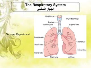
4. Consists of two parts:
(1) The upper respiratory system, which
includes the:
A- Nose األنف
B- Pharynx البلعوم
(2) The lower respiratory system, which
includes the:
A- Larynx الحنجرة
B- Trachea الهوائية القصبة
C- Bronchi الهوائية الشعب
D- Lungs الرئتين
4
Divisions of the Respiratory System
according to Structure
5. Functionally, the respiratory system consists of two parts:
(1) The conducting zone (
توصيلية منطقة
)
(2) The respiratory zone (
تنفسية منطقة
)
5
Divisions of the Respiratory System:
according to Function
6. 1. Provides surface for gas exchange:
Intake of O2 for delivery to body cells and removal of CO2
produced by body cells.
2. Helps regulate the blood pH.
3. Contains receptors for sense of smell,
4. filters inspired air,
5. produces vocal sounds (phonation), and
6. excretes small amounts of water and heat.
6
Functions of the Respiratory System
9. – The nose is a specialized organ at the entrance of the respiratory system
that consists of:
1. External nose
2. Internal nose (called the nasal cavity)
The external nose: It is the visible part on the face and consists of a
supporting framework of bone and hyaline cartilage covered with muscle
and skin and lined by a mucous membrane.
9
Nose
10. Internal nose (The nasal cavity):
• It is a large space in the anterior aspect of
the skull that lies inferior to the nasal bone
and superior to the oral cavity.
• It is lined with muscle and mucous
membrane.
The nasal septum divides the nasal cavity into
right and left sides and consists primarily of
hyaline cartilage.
The nasal cavity communicates with the
pharynx through two openings called the
internal nares.
10
cont.
11. The pharynx (or throat) is a funnel-shaped tube
about 13 cm (5 in.) long that starts at the internal
nares and extends to the level of the cricoid
cartilage.
• The pharynx lies just posterior to the nasal and
oral cavities, superior to the larynx.
• Its wall is composed of skeletal muscles.
• Relaxed skeletal muscles help keep the
pharynx patent and contraction assists in
swallowing.
Pharynx
11
12. • The pharynx functions as a passageway for air, food
and houses the tonsils, which participate in
immunological reactions against foreign invaders.
• The pharynx can be divided into three anatomical
regions:
1. Nasopharynx: The superior portion of the pharynx.
2. Oropharynx: The intermediate portion of the
pharynx.
3. Laryngopharynx: The inferior portion of the
pharynx.
cont.
12
14. Larynx
Larynx (or voice box), is a short passageway that connects the
laryngopharynx with the trachea.
• It lies in the midline of the neck anterior to the esophagus and the fourth
through sixth cervical vertebrae (C4–C6).
• The wall of the larynx is composed of nine pieces of cartilage.
Cartilages of the Larynx :
- 3 single cartilages:(Thyroid, Cricoid and Epiglottis Cartilage).
- 3 paired cartilages:- (Arytenoids , Corniculates and Cuneiforms
Cartilage).
14
16. Muscles of the Larynx:
1- Extrinsic laryngeal muscles
Action – Move the larynx as a whole
2. Intrinsic laryngeal muscles
Action – Move parts of the larynx
cont.
16
17. The trachea (or windpipe) is a tubular passageway
for air that is about 12 cm (5 in.) long and 2.5 cm (1
in.) in diameter.
• It is located anterior to the esophagus.
The layers of the tracheal wall, from deep to
superficial, are the:
(1) mucosa, (2) submucosa, (3) hyaline cartilage,
and (4) adventitia (composed of areolar connective
tissue).
Trachea
17
19. The trachea divides into:
a right main (primary) bronchus which
goes into the right lung, and
a left main (primary) bronchus, which
goes into the left lung.
The right main bronchus is more vertical,
shorter, and wider than the left .
Bronchi
19
20. On entering the lungs, the main
bronchi divide to form:
1- Smaller Bronchi –
Also called secondary bronchi
and lobar bronchi
Note: one for each lobe of the
lung.
2- Segmental Bronchi
Also called tertiary bronchi
3- Bronchioles.
4- Terminal Bronchioles. 20
21. Lungs
21
The lungs (= lightweights, because
they float) are paired cone-shaped
organs in the thoracic cavity.
• They are separated from each other
by the heart and other structures of
the mediastinum.
22. Each lung is enclosed and protected by a double-layered
serous membrane called the pleural membrane or pleura
divide to:
1. The superficial layer, called the parietal pleura (
غشاء
جداري
) .
It lines the wall of the thoracic cavity.
2. The deep layer, the visceral pleura (
حشوي غشاء
) .
It covers the lungs themselves.
cont.
22
23. Between the visceral and
parietal pleurae is a small
space, the pleural cavity,
which contains a small
amount of lubricating fluid
secreted by the membranes.
• This pleural fluid
reduces friction between
the membranes.
cont.
23
24. The lungs extend from the diaphragm to just slightly superior to the
clavicles and lie against the ribs anteriorly and posteriorly.
Each lung has:
1- Apex
2- Base
3- Costal Surface
4- Mediastinal Surface
5- Hilum
24
25. The two lungs in our body -
1- Right Lung
2- Left lung
cont.
25
26. Right lung
The Right Lung has:
Superior lobe
Middle lobe
Inferior lobe
– This lobe divided by Horizontal
Fissure and Oblique fissure
The Right Lung is
shorter than the left
lung
26
27. The Left Lung has:
Superior lobe
Inferior lobe
- This lobe divided by
oblique fissure
The Left Lung is 10%
smaller than the right lung.
Left lung
27
28. One or two fissures divide each lung into sections called lobes.
Both lungs have an oblique fissure, which extends inferiorly and
anteriorly; the right lung also has a horizontal fissure.
The oblique fissure in the left lung separates the superior lobe from
the inferior lobe.
Lobes (
فصوص
)
Fissures (
شقوق
)
28
29. Alveoli
(
هوائية حويصالت
)
Alveoli )analogous to individual grapes)
The wall of each alveolus (singular)
consists of two types of alveolar epithelial
cells:
1- Type I alveolar cells:
• The thin type I alveolar cells are the
main sites of gas exchange.
2- Type II alveolar cells (also called
septal cells):
• They are fewer in number and are
found between type I alveolar cells.
rounded or cuboidal epithelial cells with
free surfaces
29
30. Function of alveoli
• containing microvilli, secrete
alveolar fluid, which keeps the
surface between the cells and the air
moist.
• Exchange of O2 and CO2 between
the air spaces in the lungs and the
blood takes place by diffusion
across the alveolar and capillary
walls.
30
31. 1-The respiratory system can be divided according to
structure into -------
A. One part.
B. Two parts.
C. Three parts.
D. Four parts.
2- The upper respiratory system includes the
--------
A. Nose.
B. Mouth.
C. Larynx.
D. Trachea.
3- The lower respiratory system includes
the ----------
A. Pharynx.
B. Neck.
C. Nasal cavity.
D. Lungs.
4- The Left Lung is smaller than the right
lung by ---------
A. 10 %.
B. 20 %.
C. 30%.
D. 40 %. 31
System Review
Choose Correct answer?