
Recommended
More Related Content
What's hot
What's hot (20)
Viewers also liked
Similar to LPP_Poster_Wilms
Similar to LPP_Poster_Wilms (20)
LPP_Poster_Wilms
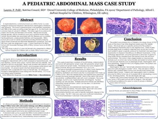
- 1. A PEDIATRIC ABDOMINAL MASS CASE STUDY Lauren. P. Polli1, Katrina Conard, MD2; 1Drexel University College of Medicine, Philadelphia, PA 19102 2Department of Pathology, Alfred I. duPont Hospital for Children, Wilmington, DE 19803 Acknowledgments Special thanks are extended to Steve Phillips, Heather Hardy, and Linda Krawczuk. Abstract A nephroblastoma, commonly known as a Wilms Tumor, is a rare malignant embryonal neoplasm derived from nephrogenic blastemal cells (Murphy). This tumor is predominantly in the pediatric population with 98% of the cases occurring under the age of 10. It is the fourth most common type of cancer in children. The mean age of occurrence for males is 36 months and females is 42 months. Risk factors include female gender, African American race and a positive family history. Certain abnormalities are associated with this disease such as aniridia, hemihypertrophy, undescended testicles and hypospadia. Wilms Tumors are a part of certain syndromes including WAGR syndrome, Denys-Drash syndrome and Beckwith-Wiedemann syndrome. Only 400- 500 new cases are diagnosed a year. Current treatments for Wilms Tumors with favorable histology (95% of Wilms Tumors) have an 85-90% cure rate. This case study is about a 3 year old female who presented to Alfred I. duPont Hospital for Children with a classic Wilms Tumor case. Introduction On April 8, 2012 a 3 year old female presented to the A.I. duPont Emergency Department with a chief complaint of abdominal pain. She had diarrhea, normal urine output, normal vitals and was afebrile. She refused to eat but would drink. Blood work showed mild anemia, LDH of 2731 mmol/L (normal 500-920 mmol/L), and increased white blood cells (neutrophils). On physical exam she had abdominal pain and distention with a palpable firm mass on the left side. An ultrasound showed a heterogeneous mass arising from the left kidney. A CT scan and biopsy were scheduled. Initial differential diagnoses included Wilms Tumor or Neuroblastoma. Methods An ultrasound guided needle biopsy was preformed and showed a Stage III Wilms Tumor with unfavorable histology and diffuse anaplasia. A port was placed and chemotherapy began on April 14, 2012. On July 16, 2012 a left nephrectomy was preformed to remove the left kidney, adrenal gland and tumor plus additional lymph nodes. A standard work up for a suspected Wilms Tumor patient include X- ray, ultrasound, CT scan, CBC and urinalysis. Treatment includes a combination of nephrectomy, chemotherapy and/or radiation. Results The surgical specimen consisted of the left kidney, adrenal gland and tumor; left perinephric lymph node; left paraaortic lymph node and 2 paracaval lymph nodes. The kidney and tumor weighed 692 grams (normal= 48.4 g) and measured 14.5 x 10 x 6 cm. The typical histological appearance of a Wilms Tumor displays the various stages of both normal and abnormal nephrogenesis. Blastema, epithelium and stroma are the 3 components that make up a Wilms Tumor. Both the needle biopsy and nephrectomy specimen showed the same histologic features. Most of the cells in the tumor are primitive, undifferentiated cells with scant cytoplasm. The nuclei are large, round to oval, hyperchromatic and irregular. Nuclear molding can also be seen. Multiple abnormal mitoses are identified. Attempted tubule formation, which can be seen, is a dominant feature of the tumor. Both intralobular and periolobular nephrogenic rests were identified within the kidney. The renal artery, renal vein and ureter were uninvolved by tumor on permanent sections. Immunohistochemistry was used to identify metastatic disease in the lymph nodes. The WT-1 stain was used and confirmed metastases and micrometastases in all lymph node specimens. Conclusion The patient was entered into a Children's Oncology Group (COG) study at the time of her initial diagnosis based upon the needle biopsy. This study laid out a specific protocol for the patient’s chemotherapy treatment prior to her nephrectomy. These studies include large pools of patients collected from across the country. Each patient is given a specific protocol for treatment and specimen examination to research the efficacy of different treatments. In the pathology department, once the specimen is removed 10 grams of snap frozen tissue is required from the primary tumor site and from normal kidney. The tumor is then staged based on macroscopic features such as tumor size, tumor focality and extent of tumor as well as microscopic features such as histological type, nephrogenic rests and metastases. Based on the COG staging system, this patient was classified as a Stage III. Nephrogenic rests are “abnormally retained embryonic kidney precursor cells arranged in clusters” (NCI). They are seen in 35% of kidneys with unilateral Wilms tumors and virtually every kidney with bilateral Wilms Tumors. Patients with nephrogenic rests in a kidney removed for Wilms Tumor are at increased risk for developing a tumor in the remaining kidney. This risk decreases with age. Patients with unfavorable histology (5% of Wilms Tumors) such as this one, have a 72% 4-year survival rate at Stage III. She will need continued surveillance for years to come to ensure a bilateral tumor does not occur. References A.D.A.M. Medical Encyclopedia. Wilms Tumor. 16 May 2012. 7 September 2012 <http://www.ncbi.nlm.nih.gov/pubmedhealth/PMH0002542/>. A.D.A.M. Medical Encyclopedia. Neuroblastoma. 7 February 2012. 7 September 2012 <http://www.ncbi.nlm.nih.gov/pubmedhealth/PMH0002381/>. American Associated for Clinical Chemistry. LDH. 11 July 2011. 7 September 2012 <http://labtestsonline.org/understanding/analytes/ldh/>. Mayo Clinic. Wilms’ Tumor. 2 September 2011. 7 September 2012 <http://www.mayoclinic.com/health/wilms-tumor/DS00436>. Murphy, William M, David J Gringnon and Elizabeth J Perlman. Tumors of the Kidney, Bladder, and Related Urinary Structures. Washington D.C.: American Registry of Pathology 2004 National Cancer Institute. Wilms Tumor and Other Childhood Kidney Tumors Treatment. 9 August 2012. 13 September 2012. <http://www.cancer.gov/cancertopics/pdq/treatment/wilms/HealthProfessional/page2>. Figure 1- Abdominal CT Scans showing large mass on left kidney. Figures 2 & 3- Gross images of nephrectomy specimen external surfaces. Figures 4 & 5- Gross images of nephrectomy specimen cut surfaces. Figures 6 & 7- Histologic appearance of Wilms Tumor Figures 8 & 9- Histologic appearance of Wilms Tumor