
Recommended
More Related Content
Similar to Identify Blood Components
Similar to Identify Blood Components (20)
Recently uploaded
Recently uploaded (20)
Identify Blood Components
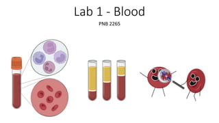
- 1. Lab 1 - Blood PNB 2265
- 2. Learning Objectives 1. Identify and evaluate various components of human blood 2. Identify blood under different disease states 3. Know how to conduct various blood tests including blood typing, Hemoglobin and Hematocrit
- 3. Stations 1. Histology Slides- demo slides on microscope (7) - Identify & draw different cell types - Differentiate and recognize Leukemia in general 2. Blood typing Station - Agglutination may take several minutes to occur - Clean test plates as quickly as possible, with soap and water DO NOT DISCARD 3. Hemoglobin Station : Each group does 1 test 4. Anatomage Table Station: view blood cell types
- 4. Introduction Function: • Transportation of oxygen and nutrients • Removing cellular waste Components: • Formed Elements • Red Blood Cells • White Blood Cells • Platelets • Extracellular Matrix • Plasma
- 5. Introduction- Cell types Learning Objectives: • Identify and describe various components of human blood. • Identify different disease states of blood
- 6. Introduction – Blood Diseases • Sickle Cell Anemia • Hereditary Condition • Abnormal hemoglobin is produced • Infectious Mononucleosis • Viral disease • Increased production of Monocytes and Lymphocytes • Polycythemia • Over production of RBCs • Result of Bone Marrow Cancer • Leukemia • Type of Cancer that involves the overproduction of leukocytes • Many different types
- 7. Blood Typing Learning Objectives • Learn about the antigen and antibodies that determine blood types • Learn the procedure to testing for blood type
- 8. Blood Types – Introduction: Antigens Blood travels all throughout the body to deliver nutrients and oxygen to all of your tissues. The presence of antigen on our cells allow for our immune cells to recognize foreign cells present in our bodies. When foreign cells are identified by our immune cells, the immune cells then act to destroy them. Our blood cells have specific antigens present on them that are different from the antibodies our immune cells carry. This helps are immune cells not to attack our own red blood cells.
- 9. Blood Type – Introduction: Antigens Patient's blood type is determined by the antigen that is present on the surface of their blood cells. Type A Type B Type AB
- 10. Introduction - Blood Type Test Unknown antigens on RBCs + + = = Unknown antigens on RBCs Anti-B antibodies Anti-A antibodies No Agglutination Agglutination Unknown Blood Type Type B Blood
- 11. Hemoglobin and Hematocrit Learning Objectives • Learn the procedure of measure the Hemoglobin levels and why its important • Learn the procedure and application of the Hematocrit
- 12. Introduction – Hemoglobin What is Hemoglobin? • Main Component of RBC • Four Folded Proteins called Globin • Red pigment called Heme Why is Hemoglobin important? • Each Iron molecule binds an oxygen How does Hemoglobin help the blood accomplish its function?
- 13. Introduction – Hematocrit • A diagnostic test to see the portion of RBCs in a blood sample • Multiple Different Disorders can be seen in a hematocrit • Polycythemia • Dehydration • Anemia
- 14. Question 1: What is the most abundant type of WBC found within normal blood? A. Basophil B. Monocyte C. Neutrophil D. Lymphocyte
- 15. Hematocrit Results! Normal Anemia Polycythemia Compensated Blood loss Dehydration Doping Leukemia
- 16. Question 2 What is the physiological reasons for the symptoms of Anemia? (Weakness, Shortness of Breath, Headaches, Poor Ability to Exercise) A. Lack of Hemoglobin to Bind Oxygen B. Increased amount of Hemoglobin to Bind more Oxygen C. Increased Plasma D. An Infection
- 17. Question 3 Does it lie within the normal range? If not determine a possible condition. A. This is within the normal range B. Infectious mononucleosis C. Polycythemia D. Sickle Cell Disease
- 18. If blood type “O” has anti-A and anti-B, antibodies, why is it a universal donor? Blood Type
- 19. Question 4 Determine the blood type. A. A- B. B- C. AB+ D. O-
- 20. Reminders • Make sure to upload all drawn images individually to Kura Cloud • Results and Discussion page is also to be done Alone! • Lab Report 1 is due before your next lab session starts
Editor's Notes
- Anemia – insufficient hemoglobin concentration Macrocytic anemia – Reasons / Folic acid + B12 deficiency also Gastric bypass prevents the absorption of B12 in blood (Intrinsic Factor released by parietal cells) / Pathology could cause stroke Microcytic anemia – Reasons/ Low Iron levels / Mutations in Hb / Pathology low 02 carrying capacity / Blue lips + cold extremities
- I would try to cut words out of this slide. The first sentence can be deleted bc you have already covered that
- Students will check with you if they did the blood typing correctly; answers in TA drawer
- C. Neutrophil
- Compensated blood loss: A hematocrit is frequently done to assess the extent of significant blood loss. A hematocrit that is done immediately after a hemorrhage usually does not show the extent of RBC loss because at the time of the hemorrhage, plasma and red blood cells are lost in equal proportions. However, within several hours after hemorrhage, plasma volume increases due to a shift of interstitial fluid into the vascular space. Red blood cells, however, cannot be replaced quickly, as the bone marrow takes approximately ten days to produce mature red blood cells. As a result, a hematocrit done several hours after a bleeding episode will show a more accurate picture - the hematocrit will be decreased because the plasma volume has compensated for fluid loss while the red blood cells that have been lost cannot be replaced for days. Blood doping mechanisms: 1. collect blood (preferably your own) a few weeks before competition, centrifuge and store RBCs, inject RBCs on competition day (old way) 2. Inject erythropoietin (EPO), a cytokine usually secreted in response to hypoxia that stimulates RBC production Risks of doping: Due to higher viscosity of blood: hypertension, decreased cardiac output, heart attack, stroke, embolism Leukemia results in significantly more WBCs. The main effect of leukemia on hematocrit is the diversion of resources from erythropoiesis (making RBCs) to making WBCs, resulting in a lower PCV. Leukemia can be diagnosed with blood smears
- C
- A. A-
- She is trying to increase her blood’s oxygen carrying capacity by increasing the amount of RBCs and therefore hemoglobin present. This would most closely resemble blood test results of polycythemia.