2. (Papenfuss 1942, Womersley 1967, Venegas et al.
1992, Tsutsui and Ohno 1993, Aruga et al. 1997)
but genera Egregia and Eisenia produce basal sporo-
phylls (Blanchette et al. 2002, Henkel and Murray
2007, Guiry and Guiry 2013). The kelp Aureophycus
aleuticus, which is not classified in a laminarean fam-
ily due to its exceptional features (e.g., sporophyte
morphology), forms sori on its semidiscoidal hold-
fast (Kawai et al. 2013).
Whether reproductive structures in the family
Alariaceae are limited to the sporophylls has been
examined. Alaria nana produced sori on the vegeta-
tive blade after sporophylls were experimentally
removed (Pfister 1991). In the field, natural popula-
tions of Undaria pinnatifida and Alaria crassifolia pro-
duced sori on their blades, usually toward the end
of their reproductive period (Stuart et al. 1999,
Kumura et al. 2006). Sori have also been observed
on the midrib of U. pinnatifida (Sanbosunga and
Hasegawa 1967), and on stipes of a Lessoniaceae
Lessonia nigrescens (Venegas et al. 1992). In
M. pyrifera (Linnaeus) C.Agardh, the only sporo-
phyll-bearing species in the family Laminariaceae,
different vegetative laminae (i.e., frond initials, sur-
face-canopy blades and apical scimitars) have been
reported to bear sori (Brandt 1923, Neushul 1963,
Lobban 1978, Graham et al. 2007). However, these
reports are vague, short, and descriptive. Here, we
detail the morphology (surface area) of sori from
different laminae (i.e., sporophylls, pneumatocyst-
bearing blades, and apical scimitars) and quantify
their reproductive output (numbers of meiospores
released) and the postsettlement viability (germina-
tion rate) of released meiospores. The ecological
implications of developing reproductive structures
on different parts of the sporophyte frond are
discussed.
MATERIAL AND METHODS
Seaweed collection. During low tide in spring 2012, ten adult
sporophytes of M. pyrifera (4.0 Æ 1.5 m; hereafter Macrocystis)
were collected in the upper sublittoral of a wave-sheltered site
in Hamilton Bay (45°47′ 51″ S; 170°38′ 39″ E), Otago Har-
bour, New Zealand. The specimens bore sori on different
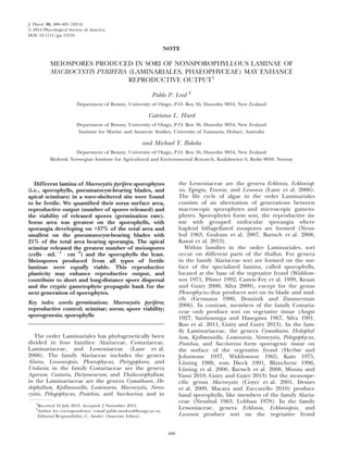
parts of their frond. Fertile laminae were classified according
to the general classification of Lobban (1978): sporophyll, SP
(Fig. 1a), specialized smooth lamina without a pneumatocyst;
blade, BL (Fig. 1b), lamina with a corrugated surface and
pneumatocyst; and apical scimitars, SC (Fig. 1c), lamina with
unilateral divisions.
Sorus area, meiospore culture, and germination. Sporophytes
were individually processed by separating and counting all
sorus-bearing laminae according to the three categories above
and photographing each lamina with a ruler. Sorus area
(Fig. 1) on different sorus-bearing laminae was measured
using the image-analysis software ImageJ (Schneider et al.
2012) and expressed as percentage of the total lamina area.
Fertile laminae were gently cleaned of epiphytes by brushing
them under filtered (0.2 lm) seawater. Samples were
wrapped in moist tissue paper and stored overnight at 4°C.
The next day, meiospores from different sorus-bearing
laminae (n = 6, from different individual sporophytes) were
separately released by immersing 2 cm2
sorus, excised with a
core sampler from the darkest sorus area (equivalent to Sorus
Class 2 as described by Bartsch et al. 2013), into 10 mL of
0.2 lm-filtered seawater for 15 min. The sorus was then
removed and the number of meiospores released was
counted using a hemocytometer (0.1 mm depth, bright-line,
Marienfeld, Germany). Meiospore densities were adjusted to
20,000–25,000 cell Á mLÀ1
and separately dispensed onto
each compartment of the six-well polystyrene tissue culture
vessels (Costar 3516; Corning Incorporated, New York, NY,
USA). Meiospores were cultivated in a temperature-controlled
room at 12°C under a 12:12 h light:dark photoperiod of
50 lmol photons Á mÀ2
Á sÀ1
of PAR (cool-white fluorescent;
Philips, Eindhoven, the Netherlands). The number of mei-
ospores that germinated was counted after 3 d. At least five
randomly chosen visual fields using a 109 objective of an
inverted microscope (Olympus CK2; Olympus Optical Co.
Ltd., Tokyo, Japan) were photographed using a video camera
(5.1M CMOS camera, UCMOS0510KPA). Photographs were
viewed using the digital camera software ToupView 3.5 where
350 meiospores were classified and counted to measure ger-
mination rates according to Roleda et al. (2012).
Statistical analysis. Percentage data (sorus area and meios-
pore germination) were logit transformed (Warton and Hui
2011) and meiospore release data were log transformed to
meet the ANOVA assumptions. The Kolgomorov-Smirnow test
was used to test Normality and the Levene’s test to test homo-
geneity of variances. One-way ANOVA (P < 0.05) was used to
test lamina-specific differences in sorus size, number of mei-
ospores released and the percentage germination. The post
hoc Tukey test was applied when significant differences were
encountered. All statistical analyses were run in SigmaStat
2.03 (SPSS Inc., Chicago, IL, USA).
RESULTS
Sorus area. Sori occurred in each of the three
types of lamina (Fig. 1). Sorus area was greatest on
the sporophylls (Fig. 2a) with sporangia developing
on >57% of the total area and smallest on the pneu-
matocyst-bearing blades with 21% of the total area
FIG. 1. Sorus-bearing lamina in Macrocystis pyrifera. (a) Sporo-
phyll, (b) pneumatocyst-bearing blade, and (c) apical scimitar;
scale bar = 2 cm. Corresponding line illustrations show the typi-
cal location where sori (S) are found in each lamina-type.
SORI ON NONSPOROPHYLLOUS LAMINAE 401
3. becoming fertile. This difference in sorus size was
statistically significant (ANOVA: F2,108 = 12.029,
P < 0.001) and a post hoc test (Tukey, P < 0.05)
revealed SP > SC = BL.
Meiospore release. Meiospores released from differ-
ent types of fertile laminae ranged from 5.15 9 103
to 6.35 9 104
cells Á mLÀ1
Á cmÀ2
. The number of
meiospores released varied between the fertile lami-
nae (ANOVA: F2,26 = 605.903, P < 0.001) with the
scimitar releasing the most meiospores and the spo-
rophylls the least (Tukey, P < 0.05; SP < BL < SC;
Fig. 2b).
Meiospore germination. Meiospore germination after
3 d post cultivation ranged from 39% to 66% (Fig. 2c)
and there was no significant difference in the develop-
ment of meiospores from the different laminae
(ANOVA: F2,17 = 1.547, P = 0.245).
DISCUSSION
It seems that reproduction in nonsporophyllous
tissue among species in the sporophyllous family
Alariaceae (Sanbosunga and Hasegawa 1967, Pfister
1991, Stuart et al. 1999, Kumura et al. 2006) and
for the sporophyllous Macrocystis (Brandt 1923,
Neushul 1963, Lobban 1978, this study) is not un-
usual. Results of our study show that meiospores
produced from sporophyllous and nonsporophyl-
lous (blade and apical scimitar) lamina are equally
viable, and germination rates are within the range
previously reported (Roleda et al. 2012).
The question of why sporophyll-bearing Laminari-
ales usually do not reproduce on vegetative parts of
the frond had been asked previously. Pfister (1992)
suggested three hypotheses: (i) confining reproduc-
tion to the sporophylls permits vegetative fronds to
remain a fast growing, “photosynthetic organ.” If
this hypothesis is correct, a decrease in vegetative
growth prior to reproduction is predicted; in sup-
port of this idea sporogenesis in the Laminariales
generally occurs when the growth rate decreases
(Kain 1975, Bartsch et al. 2008), although this idea
requires testing for Macrocystis. (ii) Sporogenesis on
nonsporophyllous laminae is selected against
because they are removed or damaged by waves in
wave-exposed sites. This idea may explain why we
encountered fertile apical scimitars only in
wave-sheltered sites. In a demographic survey of
Macrocystis in Southern New Zealand, all sporo-
phytes from wave-exposed sites have a torn apical
scimitar (P. Leal, unpublished data). (iii) “Physio-
logical constraints maintain reproduction on the
sporophylls” (Pfister 1992) whereby internal chemi-
cal cues mediate sporogenesis. Growth substances
have been related to sori formation in Laminaria dig-
itata (Hudson) J.V.Lamouroux, for which the pres-
ence of sporangium inhibitor substances keep the
young frond free of sori during the season of rapid
growth (Buchholz and L€uning 1999, L€uning et al.
2000). Similarly, the application of high external
concentrations of indole-acetic acid induced vegeta-
tive growth and delayed sori formation in Saccharina
japonica (Areschoug) C.E. Lane, C. Mayes, Druehl &
G.W. Saunders [= L. japonica; = L. ochotensis] (Kai
et al. 2006). The exposure to exogenous abscisic
acid produced an opposite reaction, at high concen-
tration it suppressed growth and promoted the spo-
rogenesis of the same species (Nimura and Mizuta
2002). Putative growth substances have been
detected in Macrocystis (de Nys et al. 1991) but any
role in controlling the onset of sporogenesis is
unknown (Stirk et al. 2003).
FIG. 2. Macrocystis pyrifera (a) sorus area, expressed as percent-
age of the total surface area of the bearing-sorus laminae, (b)
number of spores released per sorus area per individual sporo-
phyte, and (c) corresponding percentage germination. Sorus-
bearing laminae are sporophylls (SP), pneumatocyst-bearing
blades (BL), and apical scimitar (SC). Bars represent the
mean Æ SD. Different letters above the bar indicate a significant
difference (Tukey test, P = 0.05).
402 PABLO P. LEAL ET AL.
4. An alternative explanation is that the develop-
ment of sorus on blades and apical scimitars could
be a trait that enhances long distance meiospore
dispersal in area of slow flows and such a trait might
be inheritable. However, meiospores released into
the water column may take longer time to swim and
settle into the benthos and could therefore be
exposed to high irradiance and UVR compromising
their viability (i.e., germ tube, gametophyte, and
sporophyte developments; Edwards 1998, Roleda
et al. 2006, Cie and Edwards 2008). Therefore, meio-
spores originating from distal blades and apical
scimitars may play an integral role in successful
recruitment only if they are released and disperse
during periods of low irradiance, e.g., at night, dur-
ing cloudy or overcast days (Amsler and Neushul
1989, Cie and Edwards 2008) or during low tide
when the whole Macrocystis frond lay prostrate and
closer to the benthos (P. Leal, personal observation)
away from the holdfast where sporophylls are
located. Fertile scimitars and blades released more
cells per unit area compared to the sporophylls, but
the latter have more biomass per unit sporophyte.
Therefore, sporophylls may generate more meio-
spores settling proximate to the parental sporo-
phyte. The overall meiospore output from each
tissue type to the population’s banks of microscopic
spores (cf “seed banks”; Schiel and Foster 2006) and
the next generation of sporophytes requires further
study.
We know surprisingly little about the controls of
sporogenesis in Macrocystis compared to other mem-
bers of the Laminariales and the environmental
and/or endogenous triggers that control the devel-
opment of sori on nonsporophyllous lamina of Mac-
rocystis are currently unknown. Sorus production in
sporophylls of Macrocystis can continuously occur
with an appropriate translocation of photosynthates
from the surface canopy (Neushul 1963, Reed 1987,
Reed et al. 1996, Dayton et al. 1999, Graham 2002).
Control of reproduction in kelps has often been
attributed to physico-chemical factors such as photo-
period, temperature, and nutrients (tom Dieck
1991, Bartsch et al. 2008). For example, in S. latiss-
ima and Laminaria setchellii short photoperiod
induced cessation of blade growth followed by the
formation of sori (L€uning 1988, tom Dieck 1991).
Nutrient availability also strongly affects the repro-
duction of Laminariales. Nutrient-rich media (i.e.,
phosphorous and nitrogen) enhanced sorus forma-
tion in S. angustata (Mizuta et al. 1999, Nimura
et al. 2002), A. crassifolia and U. pinnatifida (Kumura
et al. 2006). All these species have a higher internal
content of phosphorous and nitrogen in sporoge-
nous tissue than in vegetative tissue, indicating a
strong influence of nutrients on reproduction of
Laminariales (Nimura et al. 2002, Kumura et al.
2006, Bartsch et al. 2008). Additionally in S. japon-
ica, the interaction of low water temperature, long
photoperiod, and low nutrients delayed the onset of
sorus formation (Mizuta et al. 1999). Accordingly,
the formation of sori in natural populations of Lam-
inariales seems to be restricted to periods of low or
no growth in autumn to winter, coinciding with
decreasing day-length and temperature and increas-
ing nutrient availability (Kain 1975, Bartsch et al.
2008). However, for Macrocystis sori may be present
throughout the year, as observed and reported in
the southeast and northeast Pacific coast of both
hemispheres (Reed et al. 1996, Buschmann et al.
2004, 2006), and southeastern coasts of New
Zealand (P. Leal, unpublished data).
One reason that we know so little about develop-
mental regulation in Macrocystis is its size – unlike
Laminaria listed above, it cannot be easily cultured to
a mature reproductive state in the laboratory,
thereby limiting our understanding of the factors
causing sporogenesis. If sporogenesis can be induced
on isolated discs of different Macrocystis laminae (i.e.,
sporophyll, pneumatocyst-bearing blade, and scimi-
tar) as in other Laminariaceae species (e.g., Buch-
holz and L€uning 1999, Gruber et al. 2011), some
outstanding questions may be answered on the envi-
ronmental (physico-chemical cues) and physiological
(e.g., age and size class, and growth substances) fac-
tors, that control of sporogenesis in this species.
Pablo P. Leal is supported by a scholarship from BECAS
CHILE-CONICYT. Catriona L. Hurd and Michael Y. Roleda
were supported by a Royal Society of New Zealand Marsden
grant (UOO0914). We are grateful to Pamela Fernandez and
Rocıo Suarez for assisting in field sampling. We also thank
the reviewers for the critical and helpful comments.
Amsler, C. D. Neushul, M. 1989. Diel periodicity of spore
release from kelp Nereocystis luetkeana (Mertens) Postels
et Ruprecht. J. Exp. Mar. Biol. Ecol. 134:117–27.
Angst, L. 1927. Gametophytes of Costaria costata. Publ. Puget Sd.
Biol. Sta. 5:293–307.
Aruga, Y., Kurashima, A. Yokohama, Y. 1997. Formation of
zoosporangial sori and photosynthetic activity in Ecklonia
cava Kjellman (Laminariales, Phaeophyta). J. Tokyo Univ.
Fish. 83:103–28.
Bartsch, I., Vogt, J., Pehlke, C. Hanelt, D. 2013. Prevailing sea-
surface temperatures inhibit summer reproduction of the
kelp Laminaria digitata at Helgoland (North Sea). J. Phycol.
49:1061–73.
Bartsch, I., Wiencke, C., Bischof, K., Buchholz, C. M., Buck, B.
H., Eggert, A., Feuerpfeil, P. et al. 2008. The genus Lami-
naria sensu lato: recent insights and developments. Eur. J. Phy-
col. 43:1–86.
Blanchette, C. A. 1996. Seasonal patterns of disturbance influence
recruitment of the sea palm, Postelsia palmaeformis. J. Exp.
Mar. Biol. Ecol. 197:1–14.
Blanchette, C. A., Miner, B. G. Gaines, S. D. 2002. Geographic
variability in form, size and survival of Egregia menziesii around
Point Conception, California. Mar. Ecol. Prog. Ser. 239:69–82.
Boo, G. H., Lindstrom, S. C., Klochkova, N. C., Yotsukura, N.,
Yang, E. C., Kim, H. G., Waaland, J. R., Cho, G. Y., Miller, K.
A. Boo, S. M. 2011. Taxonomy and biogeography of Aga-
rum and Thalassiophyllum (Laminariales, Phaeophyceae)
based on sequences of nuclear, mitochondrial, and plastid
markers. Taxon 60:831–40.
Brandt, R. P. 1923. Potash from kelp: early development and
growth of the giant kelp, Macrocystis pyrifera. U. S. Dept. Agric.
Bull. 1191:1–40.
SORI ON NONSPOROPHYLLOUS LAMINAE 403
5. Buchholz, C. L€uning, K. 1999. Isolated, distal blade discs of the
brown alga Laminaria digitata form sorus, but not discs,
near to the meristematic transition zone. J. Appl. Phycol.
16:579–84.
Buschmann, A. H., Moreno, C., Vasquez, J. A. Hernandez-
Gonzalez, M. C. 2006. Reproduction strategies of Macrocystis
pyrifera (Phaeophyta) in Southern Chile: the importance of
population dynamics. J. Appl. Phycol. 18:575–82.
Buschmann, A. H., Vasquez, J. A., Osorio, P., Reyes, E., Filun, L.,
Hernandez-Gonzalez, M. C. Vega, A. 2004. The effect of
water movement, temperature and salinity on abundance
and reproductive patterns of Macrocystis spp. (Phaeophyta) at
different latitudes in Chile. Mar. Biol. 145:849–62.
Castric-Fey, A., Beaupoil, C., Bouchain, J., Pradier, E. L’Hardy-
Halos, M. Th. 1999. The introduced alga Undaria pinnatifida
(Laminariales, Alariaceae) in the rocky shore ecosystem of
the St Malo area: morphology and growth of the sporophyte.
Bot. Mar. 42:71–82.
Cie, D. K. Edwards, M. S. 2008. The effects of high irradiance
on the settlement competency and viability of kelp zoosp-
ores. J. Phycol. 44:495–500.
Coyer, J. A., Smith, G. J. Andersen, R. A. 2001. Evolution of
Macrocystis spp. (Phaeophyceae) as determined by ITS1 and
ITS2 sequences. J. Phycol. 585:574–85.
Dayton, P. K., Tegner, M. J., Edwards, P. B. Riser, K. L. 1999.
Temporal and spatial scales of kelp demography: the role of
oceanographic climate. Ecol. Monogr. 69:219–50.
Demes, K. W., Graham, M. H. Suskiewicz, T. S. 2009. Pheno-
typic plasticity reconciles incongruous molecular and mor-
phological taxonomies: the giant kelp, Macrocystis
(Laminariales, Phaeophyceae), is a monospecific genus.
J. Phycol. 45:1266–9.
tom Dieck, I. 1991. Circannual growth rhythm and photoperiodic
sorus induction in the kelp Laminaria setchellii (Phaeophyta).
J. Phycol. 27:341–50.
Dominik, C. M. Zimmerman, R. C. 2006. Dynamics of carbon
allocation in a deep-water population of the deciduous kelp
Pleurophycus gardneri (Laminariales). Mar. Ecol. Prog. Ser.
309:143–57.
Edwards, M. S. 1998. Effects of long-term kelp canopy exclusion
on the abundance of the annual alga Desmarestia ligulata
(Light F). J. Exp. Mar. Biol. Ecol. 228:309–26.
Germann, I. 1986. Growth phenology of Pleurophycus gardneri
(Phaeophyceae, Laminariales), a deciduous kelp of the
northeast Pacific. Can. J. Bot. 64:2538–47.
Graham, M. H. 2002. Prolonged reproductive consequences of
short-term biomass loss in seaweeds. Mar. Biol. 140:901–11.
Graham, M. H., Vasquez, J. A. Buschmann, A. H. 2007. Global
ecology of the giant kelp Macrocystis: from ecotypes to ecosys-
tems. Oceanogr. Mar. Biol. Annu. Rev. 45:39–88.
Gruber, A., Roleda, M. Y., Bartsch, I., Hanelt, D. Wiencke, C.
2011. Sporogenesis under ultraviolet radiation in Laminaria
digitata (Phaeophyceae) reveals protection of photosensitive
meiospores within soral tissue: physiological and anatomical
evidence. J. Phycol. 47:603–14.
Guiry, M.D. Guiry, G.M. 2013. AlgaeBase. World-wide electronic
publication, National University of Ireland, Galway. Available
at: http://www.algaebase.org (searched on 11 October
2013).
Henkel, S. K. Murray, S. N. 2007. Reproduction and morpho-
logical variation in Southern California populations of the
lower intertidal kelp Egregia menziesii (Laminariales). J. Phycol.
43:242–55.
Herbst, C. C. Johnstone, G. R. 1937. Life history of Pelagophycus
porra. Bot. Gaz. 99:339–54.
Kai, T., Nimura, K., Yasui, H. Mizuta, H. 2006. Regulation of
sorus formation by auxin in Laminariales sporophyte. J. Appl.
Phycol. 18:95–101.
Kain, J. M. 1975. The biology of Laminaria hyperborea VII. Repro-
duction of the sporophyte. J. Mar. Biol. Ass. U.K. 55:567–82.
Kawai, H., Hanyuda, T., Ridgway, L. M. Holser, K. 2013. Ances-
tral reproductive structure in basal kelp Aureophycus aleuticus.
Sci. Rep. 3:2491.
Kraan, S. Guiry, M. D. 2000. Molecular and morphological
character inheritance in hybrids of Alaria esculenta and
A. praelonga (Alariaceae, Phaeophyceae). Phycologia 39:554–9.
Kumura, T., Yasui, H. Mizuta, H. 2006. Nutrient requirement
for zoospore formation in two alariaceous plants Undaria pin-
natifida (Harvey) Suringar and Alaria crassifolia Kjellman
(Phaeophyceae: Laminariales). Fish. Sci. 72:860–9.
Lane, C. E., Mayes, C., Druehl, L. D. Saunders, G. W. 2006. A
multi-gene molecular investigation of the kelp (Laminariales,
Phaeophyceae) supports substantial taxonomic re-organiza-
tion. J. Phycol. 42:493–512.
Lobban, C. S. 1978. The growth and death of the Macrocystis spo-
rophyte (Phaeophyceae, Laminariales). Phycologia 17:196–212.
L€uning, K. 1988. Photoperiodic control of sorus formation in the
brown alga Laminaria saccharina. Mar. Eco. Prog. Ser. 45:137–44.
L€uning, K., Wagner, A. Buchholz, C. M. 2000. Evidence for
inhibitors of sporangium formation in Laminaria digitata
(Phaeophyceae) during the season of rapid growth. J. Phycol.
36:1129–34.
Macaya, E. C. Zuccarello, G. C. 2010. DNA barcoding and
genetic divergence in the giant kelp Macrocystis (Laminari-
ales). J. Phycol. 46:736–42.
Mizuta, H., Nimura, K. Yamamoto, H. 1999. Inducible condi-
tions for sorus formation of the sporophyte discs of Lami-
naria japonica Areschoug of the Sporophyte (Phaeophyceae).
Fish. Sci. 65:104–8.
Mizuta, H. Yasui, H. 2010. Significance of radical oxygen pro-
duction in sorus development and zoospore germination in
Saccharina japonica (Phaeophyceae). Bot. Mar. 53:409–16.
Neushul, M. 1963. Studies on the giant kelp, Macrocystis II. Repro-
duction. Am. J. Bot. 50:354–9.
Nimura, K. Mizuta, H. 2002. Inducible effects of abscisic acid
on sporophyte discs from Laminaria japonica Areschoug
(Laminariales, Phaeophyceae). J. Appl. Phycol. 14:159–63.
Nimura, K., Mizuta, H. Yamamoto, H. 2002. Critical contents of
nitrogen and phosphorus for sorus formation in four Lami-
naria species. Bot. Mar. 45:184–8.
de Nys, R., Jameson, P. E. Brown, M. T. 1991. The influence of
cytokinins on the growth of Macrocystis pyrifera. Bot. Mar.
34:465–7.
Papenfuss, G. F. 1942. Studies of South African Phaeophyceae. I.
Ecklonia maxima, Laminaria pallida, Macrocystis pyrifera. Am.
J. Bot. 29:15–24.
Pfister, C. A. 1991. Reproductive plasticity in the kelp Alaria nana
(Phaeophyceae). J. Phycol. 27:763–6.
Pfister, C. A. 1992. Costs of reproduction in an intertidal kelp:
patterns of allocation and life history consequences. Ecology
73:1586–96.
Reed, D. C. 1987. Factors affecting the production of sporophylls
in the giant kelp Macrocystis pyrifera (L.) C.Ag. J. Exp. Mar.
Biol. Ecol. 113:61–9.
Reed, D. C., Ebeling, A. W., Anderson, T. W. Anghera, M.
1996. Differential reproductive responses to fluctuating
resources in two seaweeds with different reproductive strate-
gies. Ecology 77:300–16.
Roleda, M. Y., Hanelt, D. Wiencke, C. 2006. Exposure to ultra-
violet radiation delays photosynthetic recovery in Arctic kelp
zoospores. Photosynth. Res. 88:311–22.
Roleda, M. Y., Morris, J. N., McGraw, C. M. Hurd, C. L. 2012.
Ocean acidification and seaweed reproduction: increased
CO2 ameliorates the negative effect of lowered pH on meios-
pore germination in the giant kelp Macrocystis pyrifera (Lami-
nariales, Phaeophyceae). Glob. Change Biol. 18:854–64.
Sanbosunga, Y. Hasegawa, Y. 1967. Studies on Laminariales in
culture. I. On the formation of zoosporangia on the thalli
of Undaria pinnatifida and Costaria costata in culture. Bull.
Hokkaido Reg. Fish. Res. Lab. 32:41–8.
Schiel, D. R. Foster, M. S. 2006. The population biology of
large brown seaweeds: ecological consequences of multi-
phase life histories in dynamic coastal environments. Annu.
Rev. Ecol. Evol. Syst. 37:343–72.
Schneider, C. A., Rasband, W. S. Eliceiri, K. W. 2012. NIH Image
to ImageJ: 25 years of image analysis. Nat. Methods 9:671–5.
404 PABLO P. LEAL ET AL.
6. Silva, P. C. 1991. Nomenclatural remarks on Agarum (Laminaria-
ceae, Phaeophyceae). Jpn. J. Phycol. 39:217–21.
Silva, P. C. 2009. Historical, nomenclatural, and distributional
notes on two Pacific coast kelps: Lessoniopsis littoralis and Pleu-
rophycus gradneri (Phaeophyceae, Laminariales, Alariaceae).
Madro~no 56:112–7.
Stirk, W. A., Novak, O., Strnad, M. van Staden, J. 2003. Cytoki-
nins in macroalgae. Plant Growth Regul. 41:13–24.
Stuart, M. D., Hurd, C. L. Brown, M. T. 1999. Effect of seasonal
growth rate on morphological variation of Undaria pinnatifida
(Alariaceae, Phaeophyceae). Hydrobiologia 398/399:191–9.
Tsutsui, I. Ohno, M. 1993. Growth and maturation of Undaria
pinnatifida, U. undarioides, and Eckloniopsis radicosa at Susaki
Bay of Kochi in Japan. Aquac. Sci. 41:55–60.
Venegas, M., Tala, F. B. Vasquez, J. A. 1992. Sporangial sori on
stipes of Lessonia nigrescens Bory (Laminariales, Phaeophyta):
a high frequency phenomenon in intertidal populations of
northern Chile. Bot. Mar. 35:573–8.
Warton, D. I. Hui, F. K. C. 2011. The arcsine is asinine: the
analysis of proportions in ecology. Ecology 92:3–11.
Widdowson, T. B. 1965. A taxonomic study of the genus Hedophyl-
lum setchell. Can. J. Bot. 43:1409–20.
Widdowson, T. B. 1971. A taxonomic revision of the genus Alaria
Greville. Syesis 4:11–49.
Womersley, H. B. S. 1967. A critical survey of the marine algae of
southern Australia. Aust. J. Bot. 15:189–270.
SORI ON NONSPOROPHYLLOUS LAMINAE 405