1. The Role of Neuronal Chaperone ProSAAS in Blocking 𝛼-synuclein Cytotoxicity
Nevin Varghese, Timothy S. Jarvela, Iris Lindberg
Department of Anatomy and Neurobiology, University of Maryland-BalEmore, BalEmore MD
In Parkinson’s Disease, abnormal aggrega3on of 𝛼-synuclein is toxic to dopaminergic
neurons and results in serious neuronal dysfunc3on, impairing motor skills. Recent
research suggests that molecular chaperone proteins can be cytoprotec3ve against
neurodegenera3ve biochemical processes including protein aggrega3on. ProSAAS is a
widely expressed secretory chaperone in the brain. We hypothesize that proSAAS is
cytoprotec3ve against the aggrega3on and fibrilla3on of 𝛼-synuclein in Parkinson’s
Disease. The purpose of this research is to determine the ability of proSAAS to decrease 𝛼-
synuclein cytotoxicity. Full length and trunca3on constructs of proSAAS were affinity
purified and tested for cytoprotec3ve effects. Addi3on of recombinant proSAAS to SH-SY5Y
neuroblastoma cells overexpressing 𝛼-synuclein rescued the synuclein-mediated cytotoxic
effects. To see if protec3on was specific for 𝛼-synuclein cytotoxicity, proSAAS was tested
against other forms of cytotoxicity. ProSAAS had no effect on cytotoxicity due to oxida3ve
stress (H2O2) or to endoplasmic re3culum stress (Tunicamycin). We conclude that proSAAS
cytoprotec3on is specific for synuclein-mediated cytotoxicity. Ongoing research aims to
determine the specific protein sequence responsible for cytoprotec3on, using purified
truncated constructs of proSAAS, as highly conserved amino acid residues 160-180 have
been shown to be cri3cal in blocking in vitro fibrilla3on.
Parkinson’s Disease is the second most common neurodegenera3ve disease in the United
States. It primarily affects the motor system, and symptoms result from dopamine loss and
neuronal cell death in the substan3a nigra. A key characteris3c of Parkinson’s Disease is
abnormal 𝛼-synuclein protein aggrega3on into Lewy Bodies in neurons. 𝛼-synuclein is a 140
amino acid na3vely unfolded protein believed to have a func3onal role in synap3c
transmission regula3on. Proteostasis is a molecular process by which chaperones prepare
or fix unfolded and misfolded proteins for transla3on or degrada3on. However, through
environment stressors, accumula3on of errors, and muta3ons, proteosta3c mechanisms
begin to fail and proteins like 𝛼-synuclein are more prone to misfolding, aggrega3ng, and
fibrilla3ng. When the protein forms fibrils, it adapts toxic behavior which can cause
proteasome disrup3on, mitochondrial dysfunc3on, and ER-Golgi traffic blockage. Probable
therapeu3c targets are secretory chaperones that block 𝛼-synuclein aggrega3on. The focus
of this project is on the widely expressed neuronal secretory chaperone proSAAS.
Preliminary data shows that proSAAS colocalizes with 𝛼-synuclein in Lewy Bodies in the
substan3a nigra. Further, proSAAS can block 𝛼-synuclein fibrilla3on in vitro. In this project,
we produced proSAAS dele3on mutants to test for efficacy at blocking 𝛼-synuclein toxicity
in cell culture.
Is the neuronal chaperone proSAAS capable of decreasing 𝛼-synuclein-mediated
cytotoxicity?
Furin site
Purified proSAAS dele.on constructs
(21kDa)
Amino acid #
Pep.des
Endogenous
proSAAS
1-180
1-224
A: Structural representa3on of endogenous proSAAS (21 kDa and 27 kDa). Alpha helices
are red, furin cleavage site is yellow, and known pep.des are pink. B: N- or C- terminal
dele.on constructs of proSAAS were purified and used in cytotoxicity and fibrilla.on
assays.
In order to prepare purified protein, recombinant proSAAS expression vectors were grown
in bacteria. Aaer inducing for protein expression with IPTG, the bacteria was incubated for
18 hours at 27°C and then centrifuged. Cell pellets were disrupted by addi3on of Bug
Buster and lysozyme. Benzonase nuclease and PMSF were then added to break down DNA
and inhibit proteases, respec3vely. The lysate was incubated while rocking for 15 minutes,
and then centrifuged at 12.5k RPM for 20 minutes. The new sample supernatant was then
affinity purified using fast protein liquid chromatography (FPLC), and collected frac3ons
were analyzed by SDS-PAGE. Frac3ons with bands matching our desired proSAAS construct
molecular weight were then dialyzed into 0.1 M ace3c acid, concentrated by 10 kDa cutoff
centrifugal filters, and diluted in 5 mM ace3c acid for use in experiments.
0
500
1000
1500
2000
2500
3000
0 8 16 24 32 40 48 56 64 72
Absorbance 280 nm (mAU)
A: Proteostasis network. Proteins are
synthesized in the endoplasmic re.culum.
Molecular chaperones fold proteins to
form stable proteins. When proteins are
misfolded or destabilized from
aggrega.on, they are sent to the
ubiqui.n-proteasome system for
degrada.on.
B: Fibrilla3on pathway. Na.ve form
monomers nucleate, forming poten.ally
toxic annular or linear protofibrils. These
protofibrils then elongate to form toxic
fibrils. 𝛼-synuclein is thought to follow this
unidirec.onal fibrilla.on pathway.
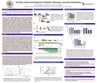
ProSAAS blocks 𝛼-synuclein mediated cytotoxicity. SH-
SY5Y neuroblastoma cells were transfected with 𝛼-
synuclein expression and LacZ control vectors. AZer 24
hours, they were split into a 96 well plate and treated
with proSAAS, ace.c acid buffer, and ovalbumin. AZer 48
hours, the percentage of surviving cells was measured
through the WST-1 assay. Cell survival increased from
75% to 85% aZer proSAAS addi.on. Each point is
represented by the mean ± SEM (*p<0.05, **p<0.01, two-
way t-test).
ProSAAS cytoprotec3on is specific for 𝛼-synuclein toxicity. A: SH-SY5Y neuroblastoma
cells were seeded into a 96 well plate. ProSAAS (PS) and two concentra.ons (5.0 μg/mL
and 10.0 μg/mL) of ER stressor Tunicamycin (TM) were added. AZer 48 hours, cells were
measured for percentage survival using the WST-1 assay. B: Cells were treated with
proSAAS and oxida.ve stressor 100.0 μg/mL hydrogen peroxide (H2O2). C: Cells were
treated with proSAAS, two concentra.ons (100.0 μg/mL and 250.0 μg/mL) of hydrogen
peroxide, and 5.0 μg/mL Tunicamycin. Variability in toxicity between experiments is due
to concentra.on of cells per well, but not in the rela.ve effects of (+/-) proSAAS. Each
point is represented by the mean ± SEM (n.s. p>0.05, ***p<0.001, two-way t-test).
• ProSAAS residues 160-180 are essen3al to its an3-aggregant func3on.
• ProSAAS is effec3ve at reducing 𝛼-synuclein cytotoxicity in SH-SY5Y cells.
• ProSAAS cytoprotec3on is specific for 𝛼-synuclein toxicity in SH-SY5Y cells, as it has no
effect on toxicity from ER or oxida3ve stress.
Time (minutes)
Research Ques3on
1 2 3 4 5 6 7 8 9 10 11 12
1. Residues 160-180 are Necessary for Op3mal An3-Aggregant Behavior
ProSAAS residues 160-180 are cri3cal to preven3ng 𝛼-synuclein aggrega3on. Experiment
of 𝛼-synuclein fibrilla.on inhibi.on by proSAAS 21 kDa, dele.on constructs, carbonic
anhydrase (control), and alpha-crystallin (posi.ve control). Each point is represented by
the mean ± SEM, n=6. (*p<0.05, **p<0.01, ***p<0.001, two-way ANOVA, vs. carbonic
anhydrase control).
ProSAAS protein purifica3on. A: ProSAAS protein frac.ons were collected and run on a
SDS polyacrylamide gel. Frac.ons 4-12 show intense bands at 21 kDa, the molecular
weight of “full length” proSAAS. B: Chromatogram of .me versus absorbance following
FPLC. Binding, wash, and elu.on buffer regions are shown for 21 kDa proSAAS sample.
B A
Aggrega3on
Stable
protein
FoldingSynthesis
Degrada3on
Disaggrega3on
A
A
B
0 50 100 150 200
0
5000
10000
proSAAS 1-159
proSAAS 97-180
proSAAS 1-180
alpha-crystallin
carbonic anhydrase
proSAAS 62-180
Time (h)
ThTFluorescence(au)
***
***
***
***
0
50
100
Cell Viability (%)
0
50
100
Cell Viability (%)
0
50
100
Cell Viability (%)
Future Studies
• Does proSAAS interact with 𝛼-synuclein in the extracellular space to prevent
cytotoxicity?
• Does proSAAS block 𝛼-synuclein oligomeriza3on intra- and/or extracellularly?
• Does proSAAS have an effect on an increased fibrilla3on 𝛼-synuclein A53T mutant?
• Create a finer map of proSAAS func3onal domains.
A B
C
Elu3on Buffer
B
Abstract
Background
Methods
Proteostasis Network and Fibrilla3on Pathway
ProSAAS Secondary Structure
3. ProSAAS Protec3on Against α-synuclein Cytotoxicity
2. ProSAAS Blocks α-synuclein Cytotoxicity
Conclusions
Acknowledgements
This research was supported by the grant NIA NIH R21AG045741-02. Thanks to the McLean
Lab for split-Venus 𝛼-synuclein vectors.
n.s. n.s.
n.s.
n.s.
n.s.
n.s.
+Buffer
+Ovalbumin
+ProSAAS
+Buffer
+Ovalbumin
+proSAAS
0
50
100
α-synucleinLacZ
**
**
%Survival
***
***
***
Adapted from Roberts, H.L. and Brown, D.R., 2015. Seeking a
mechanism for the toxicity of oligomeric α-
synuclein. Biomolecules, 5(2), pp.282-305.
Wash Buffer
Binding Buffer