Rectum & Anal Canal.pptx

Rectum means straight as if ruled. This is a misnorma,for it is curved in conformity with the hollow of the sacrum. Rectum is continuous with the sigmoid colon and there is no change of structure at the junction. The distinction is a matter of peritoneal attachment; where there is a mesocolon, the gut is called sigmoid colon and where there is no mesentery, it is called rectum . Where the muscle coats are replaced by sphincters it becomes the anal canal. The rectum begins in the hollow of the sacrum at the level of its 3rd. Piece and it curves forwards over coccyx and ano-coccygeal raphe. It is 15 cm long. The 3 tinea of the sigmoid colon come together over the rectum invest it in a complete outer layer of the longitudinal muscle. The upper and lower ends of the rectum lie in the midline but the ampulla is convex to the left. Rectal valves of Houston,2 on the left and one on the Right are produced by circular muscles of the gut.

Recommended
More Related Content
What's hot
What's hot (20)
Similar to Rectum & Anal Canal.pptx
Similar to Rectum & Anal Canal.pptx (20)
More from Dr Ndayisaba Corneille
More from Dr Ndayisaba Corneille (20)
Recently uploaded
Recently uploaded (20)
Rectum & Anal Canal.pptx
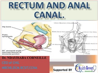
- 1. Dr. NDAYISABA CORNEILLE CEO of CHG MBChB,DCM,BCSIT,CCNA Supported BY
- 2. RECTUM • Rectum means straight as if ruled. This is a misnorma,for it is curved in conformity with the hollow of the sacrum. • Rectum is continuous with the sigmoid colon and there is no change of structure at the junction. The distinction is a matter of peritoneal attachment; where there is a mesocolon, the gut is called sigmoid colon and where there is no mesentery, it is called rectum . Where the muscle coats are replaced by sphincters it becomes the anal canal. • The rectum begins in the hollow of the sacrum at the level of its 3rd. Piece and it curves forwards over coccyx and ano-coccygeal raphe. Dr Ndayisaba Corneille 2
- 3. The sigmoid colon and Rectum. Dr Ndayisaba Corneille 3
- 4. Cont. Rectum • It is 15 cm long. • The 3 tinea of the sigmoid colon come together over the rectum invest it in a complete outer layer of the longitudinal muscle. • The upper and lower ends of the rectum lie in the midline but the ampulla is convex to the left. • Rectal valves of Houston,2 on the left and one on the Right are produced by circular muscles of the gut. Dr Ndayisaba Corneille 4
- 5. RECTAL VALVES OF HAUSTON Dr Ndayisaba Corneille 5
- 6. Cont. At the sigmoid-rectal junction, the 3 tinea coli spread out to form the outer longitudinal layer and the fatty appendices epiploicae are discontinued. The rectum posses no mesentery, but the pelvic peritoneum of the posterior wall of the pouch of Douglas is dapped over it in the upper part . The upper 1/3 is clad in front and on both sides, the middle 1/3 in front only and the lower 1/3 not at all by peritoneum. The upper part of the rectum is usually empty, but the lower part ,resting on the pelvic floor ,is distended into the ampulla and contains resting flatus and faeces. The upper and lower part of the rectum lie in the midline, but the ampulla is convex to the left. 3 lateral curves are produced, each being marked on the anterior of the ampulla by horizontal shelf (RECTAL VALVES of HOUSTON). THEY LIE 2 ON THE LEFT AND ONE ON THE RIGHT .THE Valves ARE PRODUCED BY CIRCULAR MUSCLE OF THE GUT. THE PURPOSE OF THE VALVES IS NOT CLEAR, BUT THEY MAY BE CONCERNED WITH THE SEPARATION OF MUCUS FROM THE FAECES, Holding UP FAECES WHILE ALLOWINNG FLATUS TO PASS. Dr Ndayisaba Corneille 6
- 7. Blood supply to the Rectum. • Derived principally from the artery of the hind gut, the inferior mesenteric artery, whose superior rectal branch ,supplies the mucus membrane as far down as the mucocutaneous junction of the anal canal (Hilton's line ). The muscle wall of the rectum receives a reinforcement from the middle rectal artery, a branch of the internal Iliac artery. • The inferior rectal artery, a branch of the internal pudendal artery, anastomoses with the middle rectal artery. • The superior rectal artery divides into right and left branches and the former into anterior and posterior branches. Dr Ndayisaba Corneille 7
- 8. Blood supply to the Rectum and Anal canal Dr Ndayisaba Corneille 8
- 9. Cont. of blood supply to the RECTUM • These 3 branches sink into the muscle walls ,in line with the 3 primary hemorrhoids( 4,7 and 11 o'clock). • The muscles of the wall of the ampulla and upper part of the anal canal receives branches from the middle and inferior rectal arteries and supply the blood to the mucus membrane but little anastomoses with branches of the superior rectal artery. Dr Ndayisaba Corneille 9
- 10. Venous return. • Veins correspond with arteries . • Superior rectal vein is a tributary of the portal circulation and drains into the inferior mesenteric vein. The middle and inferior rectal veins drain into the internal iliac and internal pudendal veins respectively. The anastomosis between the rectal veins form an important portal systemic anastomosis. Dr Ndayisaba Corneille 10
- 11. Lymphatic drainage. • Correspond with the 3 main sources of arterial supply. • Lymph from the upper part of the rectum to the inferior mesenteric lymph nodes to the para aortic lymph nodes. • Lymph from the lower part of the rectum follow the middle rectal artery to the internal iliac group of lymph nodes. Dr Ndayisaba Corneille 11
- 12. Nerve supply. • derived from both parts of the autonomic nervous system. • Sympathetics –from pelvic plexuses and by fibers which accompany inferior mesenteric and superior rectal arteries from the coeliac plexus. • Parasympathetic supply – from S2 ,3 or S3,4 by the nervi erigentes. • The rectum is only sensitive to stretch. Dr Ndayisaba Corneille 12
- 13. Fascia of the rectum. • Posteriorly-a sheet of fascia , suspends the lower part of the ampulla to the hollow of the sacrum. It encloses the superior rectal vessels (artery, veins and lymphatics). It is known as fascia Waldeyer. • Laterary – the middle rectal artery and branches and branches of the pelvic is enclosed in slight condensation of areolar tissue – the lateral ligament of the rectum. • The fascia of Waldeyer, the lateral ligaments, the pelvic peritoneum and vessels and most of all the pelvic floor combine to hold the rectum stable and in position. Dr Ndayisaba Corneille 13
- 14. Cont. • Anteriorly, some muscle fibers leave the lower part of the ampulla and pass forwards towards the apex of the prostate and the commencement of the membranous urethra. They form the recto urethralis muscle. Dr Ndayisaba Corneille 14
- 15. RECTUM AND ANAL CANAL Dr Ndayisaba Corneille 15
- 16. Relations of the rectum. • POSTERIORLY: 3 INFERIOR sacral vertebra and coccyx and the anal coccygeal ligament. Pyriformis, levetor ani muscles the sacral plexus and the sympathetic trunk. • Anteriorly : Males- upper 2/3 of the rectum is covered by peritoneum and is related sigmoid colon and coils of the ileum which occupy the retro vesical pouch. • The lower 1/3 of the rectum which is devoid of peritoneum, is covered by the posterior surface of the bladder, to the termination of the vas deferens and the seminal vesicles on each side of the prostate. Dr Ndayisaba Corneille 16
- 17. Female relations. • Relations of the upper 2/3 of the rectum are similar to those of the males. • Lower 1/3 is related to the posterior surface of the vagina. Dr Ndayisaba Corneille 17
- 18. Relationship of the rectum in a female. Dr Ndayisaba Corneille 18
- 19. The anal canal. Dr Ndayisaba Corneille 19
- 20. The anal canal. – The junction of the rectum and the anal canal is at the pelvic floor i.e. the level where the pubo rectalis muscle clasps the gut and angles it forwards. – Anal canal extends from this level to the skin of the perineum‘. It is 1.5” long. – The upper 2/3 is derived from the cloaca(endoderm) the lower 1/3 from the anal pit (ectoderm) – The junction of the 2 is at Hilton's white line. This is a water shed dividing the upper and lower zones arterial supply and venous and lymphatic drainage. – In the upper (cloacal ) there is a dentate ring produced by folds mucus membrane termed anal “valves”. Dr Ndayisaba Corneille 20
- 21. JUNCTION BETWEEN THE RECTUM AND THE ANAL CANAL . Dr Ndayisaba Corneille 21
- 22. Anal Canal & Right ischiorectal fossa The anal canal columns' are found in the upper 2/3 of the anal canal(folds of the mucosa). Dr Ndayisaba Corneille 22
- 23. Cloacal part of the rectum. • It is endodermal in origin. • The mucus membrane is typical large gut with columnar epithelium. • There are mucus glands in the submucosa • Arterial supply; It is from the superior rectal artery as far as the white line. The sphincters outside the mucus membrane are supplied by the middle or inferior rectal arteries. • The veins drain upward in the in the sub mucus plexus of the ampulla • Lymphatics pass upwards to the inferior mesenteric of pre aortic lymph nodes. • The nerves are from the pelvic plexus (autonomic) Dr Ndayisaba Corneille 23
- 24. Anal canal cont. • Above the anal valves , the longitudinal mucus folds extend upwards in the anal canal and reach the lower limit of the rectum. • The contain sub mucus veins and are named anal columns. Dr Ndayisaba Corneille 24
- 25. .Anal part • It is ectodermal in origin with thin hairless skin. It is covered with stratified squamous epithelium. • Arterial supply inferior rectal artery. • Venous drainage inferior rectal vein (systemic) • LYMPHATICS drain into the medial group of superficial inguinal lymph nodes. • Nerves inferior rectal nerve (somatic) Dr Ndayisaba Corneille 25
- 26. Blood supply to the rectum and anal canal. Dr Ndayisaba Corneille 26
- 27. ANAL SPHINCTERS • AT the beginning of the anal canal, the inner circular layer becomes greatly thickened forming the internal sphincter, and this stops at the white line of Hilton. • The longitudinal layer thins out, and from it arise inter muscular septa ( fibro elastic fibers) • The external sphincter lines the entire length of the anal canal. It is striated muscle. It is made up of 3 parts 1)subcutaneous external. sphincter-lies in the perianal fat 2)superficial external sphincter- it is elliptical. Its anterior part is attached to the perineal body and the posterior part attached to the anal coccygeal body. 3)deep external sphincter Dr Ndayisaba Corneille 27
- 28. Anal sphincters • The anal canal is always closed except during the passing of stool and flatus. • The whole extent of the canal (1.5”) is enclosed in a sphincteric tube of muscle. The internal and external sphincter. Each occupies 2/3 of the anal canal so that they overlap in the middle 1/3. • The internal sphincter is smooth muscle (visceral) and lies around the upper 2/3 of the anal canal. Dr Ndayisaba Corneille 28
- 29. External anal sphincters Dr Ndayisaba Corneille 29
- 30. Internal sphincter • Occupies the upper 2/3s of the anal canal i.e. down to the Hiltons line. It is the thickened lower end of (inner) circular layer of the gut. • The sphincter is not competent when acting alone. Dr Ndayisaba Corneille 30
- 31. • Pubo rectalis is thickened lower border of levetor ani and when it contracts ,it increases the angle between the rectum and the anal canal. • FUNCTIONALLY THE EXTERNAL SPHINCTER is the most important. Dr Ndayisaba Corneille 31
- 32. THE ANAL CANAL Dr Ndayisaba Corneille 32
- 33. VEINS OF THE ANAL CANAL Dr Ndayisaba Corneille 33
- 34. VEINS OF THE ANAL CANAL • 3 OF THE VEINS SITUATED AT 4,7,11 OCLOCK are apt to become varicose as the 3 primary hemorrhoids. • Superior and middle rectal veins take blood to the inferior rectal vein. Dr Ndayisaba Corneille 34
- 35. Mucus membrane of the anal canal. • Cloacal part ,endodermal in origin, covered with large gut mucus membrane, columnar epithelium. Dr Ndayisaba Corneille 35
- 36. Dr Ndayisaba Corneille 36
- 37. Fissure in ano Dr Ndayisaba Corneille 37
- 38. Internal haemorroid Dr Ndayisaba Corneille 38
- 39. Rectal piles Dr Ndayisaba Corneille 39
- 40. Dr Ndayisaba Corneille 40
- 41. Dr Ndayisaba Corneille 41
- 42. END Dr Ndayisaba Corneille THANKS FOR LISTENING By DR NDAYISABA CORNEILLE MBChB,DCM,BCSIT,CCNA Contact us: amentalhealths@gmail.com/ ndayicoll@gmail.com whatsaps :+256772497591 /+250788958241 42