SlideShare a Scribd company logo
1 of 7
Download to read offline
Fisheries Research 161 (2015) 1–7
Contents lists available at ScienceDirect
Fisheries Research
journal homepage: www.elsevier.com/locate/fishres
X-ray imaging as a time-saving, non-invasive technique for
diet analysis
Melanie C. Beckmann∗
, James F. Gilliam, R. Brian Langerhans
Department of Biological Sciences and W.M. Keck Center for Behavioral Biology, North Carolina State University, Raleigh, NC 27695-7617, USA
a r t i c l e i n f o
Article history:
Received 4 February 2014
Received in revised form 20 May 2014
Accepted 25 May 2014
Handling Editor George A. Rose
Keywords:
Dietary patterns
Digital radiography
Fish
Nonlethal
Stomach-content analysis
a b s t r a c t
Dietary patterns of animals have a long-recognized importance in ecology and evolution, with numerous
and diverse applications. While many methods of diet assessment exist, the most common method of
direct diet examination for most small vertebrates is stomach-content analysis, using labor-intensive
surgical removal of the gut following death. Methods that can reduce the time required to collect diet
information without necessarily sacrificing specimens could prove invaluable for a range of applications.
We evaluated digital X-ray imaging as a non-invasive method for examination of stomach contents of
small fishes. Based on both a feeding experiment and examination of field-collected preserved specimens,
we found that digital radiography consistently revealed the presence of moderate- to high-density prey
items in the stomach, such as small arthropods. Moreover, X-ray imaging allowed for rapid identification
of some particular prey items such as detritus, dipteran larvae, ostracods, hard-shelled molluscs, and small
fish. However, this method failed to detect some low-density prey items present in some stomachs, and
could not be used for precise taxonomic identifications in most cases. Overall, we found that digital X-ray
images can be quickly acquired from anesthetized or preserved animals, permit rapid identification of
certain prey items, and facilitate digital data archives. Future studies that employ this method should first
“ground-truth” the radiological signatures of prey items observed within a given study using stomach-
content analysis, which then permits rapid data collection strictly using X-ray images. This method can
provide information useful for determining the inclusion of certain prey items in diets, even quantifying
particular taxonomic groups of prey (% occurrence, % by number). Thus our results indicate that for certain
study goals, X-ray radiography may provide a time reducing, non-invasive technique for diet analysis of
small vertebrates.
Published by Elsevier B.V.
1. Introduction
The measurement and understanding of dietary patterns in
animals has central importance in ecology, evolutionary biol-
ogy, conservation, and management. Diet analysis comprises a
long-standing tool for addressing a range of questions, such as
community assembly, trophic relationships among species, habitat
use, management of threatened, game, or commercially harvested
species, and resource competition’s role in driving major ecolog-
ical and evolutionary patterns (e.g. Bolnick et al., 2003; Collar
et al., 2009; Morin, 2011; Odum, 1983; Polis and Winemiller, 1996;
Schluter, 2000; Schoener, 1971). A number of methods exist for
assessing animal diets, such as visual observations of feeding, mor-
phological and molecular identification of prey taxa in feces and
∗ Corresponding author. Tel.: +1 919 515 3514.
E-mail addresses: mcbeckma@ncsu.edu (M.C. Beckmann), langerhans@ncsu.edu
(R. Brian Langerhans).
stomachs, stable isotope analysis, and lipid analysis (e.g. Hyslop,
1980; Peterson and Fry, 1987; Valentini et al., 2009). For small ver-
tebrates, especially fishes, amphibians, and reptiles, morphological
examination of stomach contents is the most commonly employed
technique for direct diet analysis. Nonlethal techniques, such as
stomach flushing using tubes or gastric lavage, is sometimes pos-
sible for larger individuals (e.g. Giles, 1980; Light et al., 1983), but
post-mortem dissection represents the most common approach. In
fisheries research and management, stomach dissections are regu-
larly used for the analysis of diet.
Typically, the stomach/intestines are surgically removed from
freshly-killed or preserved specimens, partially digested prey items
extracted, and taxonomic identification of prey accomplished using
microscopic examination. Once prey items have been identi-
fied, a range of approaches can be used for statistical analysis
of diet (review of methods are beyond the scope of this paper,
see Cortes, 1997; Hyslop, 1980). This method requires the death
of the specimen and is time and labor intensive, and requires
specialized training to process and identify the contents. Thus,
http://dx.doi.org/10.1016/j.fishres.2014.05.015
0165-7836/Published by Elsevier B.V.
2 M.C. Beckmann et al. / Fisheries Research 161 (2015) 1–7
alternative methods that can utilize live specimens or reduce the
time required to collect diet data would be advantageous, as this
would streamline the collection of diet information without sacri-
ficing specimens.
Here we examined digital X-ray radiography as a rapid, non-
invasive method for assessing diet of small fishes. A number
of recent advances in digital radiography make this assessment
timely: e.g. increases in resolution and magnification permit the
detection of very small objects of low density, prices of digital radio-
graphy equipment has recently dropped considerably, portability
of X-ray units has increased substantially, and many universities
already have digital X-ray machines capable of at least moder-
ate resolution of small vertebrates. While previous studies have
employed X-ray radiography in the context of animal diets (e.g.
rates of feed intake and gastric evacuation; Talbot and Higgins,
1983; McCarthy et al., 1992; Jobling et al., 1993, 2001), no previous
study has examined the utility of this technique for identifying diet
items of small vertebrates.
In this study, we investigated gut/stomach contents using X-
ray imaging in four small fish species (15–80 mm standard length):
Eastern mosquitofish (Gambusia holbrooki, Girard 1859), Bahamas
mosquitofish (G. hubbsi, Breeder 1934), Trinidadian guppy (Poecilia
reticulata, Peters 1859), and Hart’s killifish (Anablepsoides hartii;
formerly Rivulus hartii, Boulenger 1890). Collectively, these omniv-
orous species are known to exhibit a broad diet, including detritus,
algae, aquatic and terrestrial insects, crustaceans, molluscs, and
even juvenile fishes. We hypothesized that X-ray images would
reveal the presence of prey contents in the stomach, and permit
the detection and identification of some broad groups of prey taxa
based on their dense body parts (e.g. shells, exoskeletons, bones),
such as molluscs (e.g. gastropods, bivalves), crustaceans (e.g. ostra-
cods, shrimp), insects (e.g. chironomids, beetles), and vertebrates
(e.g. fish, tadpoles).
2. Materials and methods
Our goal was to determine whether X-ray imaging could reveal
the presence or absence of prey in stomachs of small fishes, and
allow the identification of 5 different types of prey items that vary
in density (mass per unit volume): (1) soft homogeneous prey (e.g.
algae, detritus), (2) weakly shelled arthropods (e.g. small shrimp,
ants), (3) moderate-density arthropods (e.g. ostracods, beetles),
(4) hard-bodied prey (e.g. shelled molluscs, crabs) and (5) verte-
brates (e.g. small fish, anurans). We took a two-pronged approach
to accomplish this: we conducted a feeding experiment with live
fish to directly assess the accuracy of diet identification using X-ray
imaging, and we examined preserved, wild-caught fish specimens
to evaluate the utility of the approach for the examination of natural
dietary patterns.
2.1. Feeding experiment
We performed a feeding experiment using 21 live individuals of
G. holbrooki and 3 individuals of A. hartii. All fish were collected from
the wild (G. holbrooki: North Carolina, USA; A. hartii: Trinidad) and
housed in 38-L aquaria in common laboratory conditions. Prior to
the feeding experiment, fish were placed individually into separate
8-L tanks and starved for 48 h. For G. holbrooki, we assigned three
adult females at random to each of seven diet treatments: (1) no
prey: starved, (2) soft, low-density homogeneous prey: TetraMin
Pro flakes, (3) low-density crustaceans: live Artemia sp. nauplii,
(4) low-density insects: live ants, (5) moderate-density insects:
thawed bloodworms (Chironomus tetans), (6) hard-shelled prey:
live snails (Physa acuta), and (7) vertebrate: one live G. holbrooki
Table 1
Collection and sample size information for wild-caught adult specimens examined
in this study.
Species Collection location N
Gambusia holbrooki Melbourne, Florida, USA 60
James Island Park, South Carolina, USA 60
Gambusia hubbsi East Twin blue hole, Andros Island, Bahamas 40
West Twin blue hole, Andros Island, Bahamas 40
Hubcap blue hole, Andros Island, Bahamas 40
Poecilia reticulata Kahala, Oahu, Hawaii, USA 120
Anablepsoides hartii Arima Valley, Trinidad 23
juvenile. We fed two live P. reticulata juveniles (3–4 mm SL) to A.
hartii to test for detection of vertebrate consumption in this species.
Within 1 h of feeding, we X-rayed each individual and saved
a digital image. We placed each live fish into a small, moist plastic
bag, laid the fish on its side within a petri dish, and set the dish in the
X-ray machine to capture a lateral image. We used a custom-built
digital X-ray unit comprising a micro-focus X-ray source (Hama-
matsu L6731-01) and a digital X-ray detector (PaxScan 2520E)
housed in a lead-shielded cabinet, set to 45 kV and 40 ␮A. Radi-
ation exposure to each fish was low, approximately 25–50 mrem –
roughly equal to a human dental X-ray for comparison. Fish were
then immediately placed into a recovery tank. Removal from water,
X-ray imaging, and return to recovery tank typically only required
approximately 30 s. Identification of all stomach contents based on
digital X-ray images was conducted blind of fish ID.
2.2. Preserved specimens
We examined digital X-ray images (using method and equip-
ment described above) of wild-caught specimens preserved in 70%
ethanol to assess the ability to detect natural dietary patterns of
preserved small fish with X-ray images. We examined 120 G. hol-
brooki, 120 G. hubbsi, 120 P. reticulata, and 23 A. hartii (see Table 1
for collection and sample size details).
For each preserved specimen, we attempted to determine
contents of the stomach based on the X-ray image. After view-
ing a number of images, six natural categories emerged from our
identifications: (1) no prey contents, (2) soft homogeneous prey
(e.g. algae, detritus), (3) low-density prey (e.g. branchiopods, ants),
(4) moderate-density prey (e.g. ostracods, dipterans), (5) shelled
mollusc prey, and (6) vertebrate (fish) prey. To determine the accu-
racy of diet identification, we dissected three randomly selected
specimens of each species from each diet category using the tra-
ditional method of surgically removing the gut and identifying the
contents under a microscope (Leica S8 APO stereoscope). We then
compared the diet classification from X-ray images to that from
direct morphological identification.
3. Results
3.1. Feeding experiment
The guts of all six starved fish appeared empty in X-ray images
(Fig. 1a and b). We could not detect flakes or Artemia sp. nauplii
with our X-ray images (Fig. 1c). In two out of the three fish fed
ants, small hard parts of prey were visible in their guts in the X-
ray Images – presumably reflecting broken pieces of the ants – but
these were difficult to identify as ants or even as insects (Fig. 1d).
Based on X-ray images, we accurately detected prey items in all
fish fed bloodworms (Fig. 1e), P. acuta snails (Fig. 1f), newborn G.
holbrooki (Fig. 1g), and juvenile P. reticulata (Fig. 1h). Thus, we con-
sistently could not detect the two lowest density prey types, but
could detect the three highest density prey types; ants appeared
M.C. Beckmann et al. / Fisheries Research 161 (2015) 1–7 3
Fig. 1. Representative X-ray images from each diet treatment in the feeding experiment showing only the intestinal region of each image (anterior to the left). (a) Starved
female G. holbrooki, depicting no apparent object signatures within the gut (only yolked eggs, spherical objects visible in the image, were observed in the relevant body
region). (b) Starved female A. hartii, depicting no apparent object signatures within the gut. (c) Female G. holbrooki fed TetraMin Pro flakes, depicting no apparent object
signatures within the gut. (d) Female G. holbrooki fed ants, depicting several small low-density items within the gut (yolked eggs also visible in image). (e) Pregnant female
G. holbrooki fed bloodworms, clearly depicting the shape of the Chironomus tetans larvae (embryo otoliths appear as paired black spots in the image). (f) Female G. holbrooki
fed Physa acuta, depicting the consumed snail shells. (g) Female G. holbrooki fed a newborn G. holbrooki, depicting the otoliths and body outline of the fish prey. (h) Female
A. hartii fed two juvenile P. reticulata, depicting the otoliths, vertebrae, and parts of the skulls of the fish prey.
to represent prey densities around the threshold of detectability
using this method.
3.2. Preserved specimens
First, we found that stomachs we identified as empty based
on X-ray images actually contained small, low-density prey items,
such as soft-bodied prey, weakly shelled prey, or filamentous
plant material (Fig. 2a). We further frequently encountered these
prey items in dissected stomachs that we identified to contain
other prey types, but these prey were typically not visible in
X-ray images. Second, guts that we identified as containing soft,
homogeneous prey or low- to moderate-density arthropod prey
were filled with a variety of prey items, including ants, dipteran
larvae, small adult dipterans, and filamentous plant material
(Figs. 2b and c and 3a and b). Guts of P. reticulata in particu-
lar appeared to be filled homogeneously with low-density prey,
which we confirmed to comprise detritus and algal material; inter-
spersed higher-density items within these guts represented small
arthropods (Fig. 2c). Third, among the moderate-density items vis-
ible in radiographs, dipteran larvae were occasionally identifiable
based on their characteristic size and elongate shape (Fig. 3b), and
4 M.C. Beckmann et al. / Fisheries Research 161 (2015) 1–7
Fig. 2. Representative X-ray images of the intestinal regions of adult wild-caught specimens (left) and the low- to moderate-density prey removed from their stomachs
(right). (a) Female G. hubbsi incorrectly identified to have an empty stomach, while it actually contained a soft-bodied insect larva and an ant. (b) Male A. hartii identified
as containing soft-bodied prey and low- to moderate-density arthropods according to the X-ray, and dissected contents revealed Chironomidae and Simuliidae larvae and
filamentous plant material. (c) Female P. reticulata identified as containing homogeneous low-density items, such as detritus, and several higher-density items, such as
arthropods; dissected contents revealed detritus and homogeneous algal material, along with several small adult hymenopterans.
ostracods were always easily identified (Fig. 3a and b). Fourth,
snail shells were among the most easily identifiable prey item in
X-ray images, as the shells appear highly defined in radiographs
and all dissected stomachs confirmed our identifications via radio-
graphs (Fig. 3c). Finally, fish prey were generally readily identifiable
(Fig. 3d), with the exception that pregnant live-bearing fish carry-
ing one or a few late-stage embryos can appear on an X-ray image
to have instead consumed small fish (we encountered one instance
here, where a pregnant G. hubbsi female contained a single embryo
at the final stage of development, which would generally be difficult
to distinguish from consumed fish prey).
4. Discussion
Diet analysis holds central importance in a range of ecological
and evolutionary applications. Traditional methods for diet analy-
sis of small vertebrates are time and labor intensive, and typically
involve the death of the animal. This situation can prove problem-
atic in the face of time constraints, or when the animals of interest
need to remain alive, such as in the cases of endangered species,
captive breeding programs, the need to minimize impacts on popu-
lations, and to avoid negative public perceptions of management
or conservation programs (Crossman and Hamilton, 1978; Hartleb
and Moring, 1995; Light et al., 1983). Small vertebrates pose spe-
cial challenges to nonlethal diet analysis, as techniques commonly
applied to larger animals are typically not feasible with small indi-
viduals (Hyslop, 1980; Strange and Kennedy, 1981; Waters et al.,
2004). Thus, a need exists for methods of diet analysis amenable to
small vertebrates that reduce the required time and labor, and can
be applied to live animals.
We found that X-ray imaging can be used to quickly identify
some particular moderate- to high-density prey taxa within stom-
ach contents of both live and preserved small fishes. Although
identification of these prey taxa with X-ray images is quite rapid,
the taxonomic resolution will generally be much lower than that
obtained via microscopic examination of stomach contents. How-
ever, the method can detect very small prey items within small
organisms – the entire body cavity length for the fish examined
in this study typically only spanned 8–15 mm, with prey parts as
small as 0.1 mm long identified in images.
In our feeding experiment, we found that X-ray imaging could
be employed to accurately identify chironomids, snails, and small
fish prey, as we correctly identified these prey in all cases. More-
over, small ants were often detectable as small, low-density items
within the stomach; although, these items could not be identi-
fied as hymenoptera or even insects without examination of the
M.C. Beckmann et al. / Fisheries Research 161 (2015) 1–7 5
Fig. 3. Representative X-ray images of the intestinal regions of adult wild-caught specimens found to contain moderate- to high-density prey items (left) and the prey
removed from their stomachs (right). (a) Female G. holbrooki identified as containing many ostracods, and several low- to moderate-density items in its stomach (yolked
eggs also visible); dissected contents revealed many ostracods, along with plant material. (b) Female G. holbrooki identified as containing several clumps of ostracods and an
elongate moderate-density item indicative of a dipteran larva in its stomach; dissected contents revealed ostracods, a Brachycera larva, a small annelid, and plant material.
(c) Female G. holbrooki identified as containing four snail shells in the gut (embryos with otoliths also visible); dissected contents revealed four snail shells and filamentous
plant material. (d) Female G. hubbsi identified as containing one small fish prey (the pairs of otoliths are the darkest spots, while the jaw and vertebrae are also evident);
dissected contents revealed a single small fish prey.
stomach contents. Our analysis of preserved wild-caught speci-
mens confirmed that we could use X-ray imaging to accurately
identify homogeneous algae/detritus, ostracods, snails, and fish,
while we could typically also identify the presence of dipteran
larvae. Moreover, the presence of some other prey, such as small
arthropods, could often be determined from X-ray images, although
the taxonomic status could not be determined without stomach-
content analysis.
Because species-specific, morphological diagnostic characters
are often not visible in X-ray images, identification to species
level was not reliable in most cases in this study. In some simple
natural systems, there may only be a single species of a partic-
ular prey type, in which species-level identification is possible.
If a study requires detailed information regarding the taxonomic
identity of a variety of food items consumed by an animal, or
requires volumetric or weight information, then X-ray imaging
will not be suitable to accomplish the study goals. Thus, X-ray
imaging can provide rapid information useful for certain types of
diet analysis, but not others. For instance, the method can rapidly
acquire presence/absence of particular prey taxa in stomachs of
6 M.C. Beckmann et al. / Fisheries Research 161 (2015) 1–7
Fig. 4. (a) X-ray image of TetraMin Pro flakes when examined alone (not within fish stomach). (b) X-ray image of three ants when examined in isolation.
individuals, and presence/absence of broad groupings of prey types
where taxonomic identities are difficult to discern with X-ray
images and are also less important for the study. In some cases,
numerical information may also be obtained from X-ray images.
Thus, digital radiography might often allow for collection of data
for two commonly employed dietary metrics, percent frequency
of occurrence and percent by number of prey taxa in stomachs
(Cortes, 1997). While a range of prey taxa were detectable using
digital radiography, our study also revealed that not all types of
prey items were visible in X-ray images. For instance, we could
not detect flake food within fish stomachs in our feeding experi-
ment, and sometimes could not detect ants. These prey items, when
imaged in isolation, are clearly evident in X-ray images taken with
our equipment (Fig. 4). However, when contained within surround-
ing fish tissue, these low-density food items become very difficult
to detect. Further, our examination of wild-caught preserved spec-
imens confirmed that guts appearing empty on X-ray images could
actually contain soft-bodied, low-density food items such as ants,
soft-bodied insect larvae, and plant material. Consequently, this
method cannot accurately distinguish empty stomachs from stom-
achs that contain certain types of low-density prey.
In addition to the limitations of this method discussed already
regarding detection and identification of particular prey items,
this method may also be affected by variable processing (e.g. prey
crushing and tearing) and digestion rates of different prey within
different species (e.g. Macdonald et al., 1982). Prey densities can
decline during digestion, making the method more amenable to
foregut analyses and prey items that are swallowed whole where
moderate-density parts are passed in feces.
Compared to more labor-intensive methods, such as gut dis-
section and stomach-content analysis using identification with
a microscope, taking and analyzing X-ray images provides a
dramatic improvement in the time required for analysis, and per-
mits the examination of live animals. For instance, digital X-ray
images usually take about 1 min per specimen to acquire, with
images immediately available for inspection (no film development
required), and digital files ready for archiving for long-term stor-
age and future reference. After initial training for identification
of the relevant prey categories from radiographs, scoring stom-
ach contents from images usually only requires a few minutes
per specimen. Meanwhile, surgical extraction of a stomach and
microscopic analysis of diet typically require much more time
per specimen. Microscopic identification of prey taxa extracted
from stomachs demands considerable skills that require substan-
tial training for the identification of a wide range of possible prey
taxa, while identification of a smaller set of prey categories on
X-ray images necessitates much less training. The use of X-ray
imaging versus direct stomach-content analysis for diet studies
of small vertebrates largely represents a tradeoff between time,
training, precision, and lethality: X-ray imaging allows the rapid
extraction of broad dietary information requiring minimal train-
ing where the specimens can remain alive after examination,
while direct stomach-content analysis represents a more time-
consuming extraction of detailed dietary information requiring
substantial training where the individuals must typically die prior
to examination. Thus, a researcher or manager must weigh these
four primary factors when deciding whether X-ray imaging might
represent the most appropriate method for their study.
The methods described here should apply to small vertebrates
other than fish, such as reptiles, amphibians, and small birds
and mammals. Indeed, during the course of other projects that
involved X-ray imaging of small lizards and frogs, we have wit-
nessed a number of obvious diet items in the images, such as
terrestrial arthropods and vertebrates (RBL, unpublished data). Fur-
ther, portable X-ray applications are becoming more commonplace
today, and this implies that diets of live animals could be exam-
ined directly in the field. Because the method requires little time
and handling, and can be employed with extremely low levels of
radiation exposure, this method could represent a suitable solu-
tion for diet studies of threatened species, or in situations where
it is important not to impact the study population, including small
populations, breeding programs, zoo animals, or lab animals.
We envision other uses of digital radiography for ecologists and
evolutionary biologists as well, such as the use in reproductive
and developmental studies of viviparous fishes. For instance, X-ray
images provide highly accurate information regarding whether a
fish is pregnant or not, and can often permit the counting of the
number of developing embryos within some species. Thus, studies
examining the fecundity of females might employ X-ray imaging to
rapidly collect information on the frequency of pregnancy, number
of offspring, and perhaps estimates of the size and developmental
stage of embryos.
5. Conclusion
Overall, our study revealed that X-ray imaging can provide an
appropriate tool for diet analysis of small vertebrates, depending
on the goals of the study. The two most obvious advantages of the
approach are the reductions in time required for data collection and
the ability to non-invasively examine live animals. This method
is probably most useful for studies that seek rapid collection of
broad dietary information, where detailed taxonomic information
or prey-size information is less important for the goals. This non-
lethal method can rapidly and accurately provide information on
certain types of prey items contained within the stomachs of spec-
imens, and should thus receive increased use in diet studies in the
future.
M.C. Beckmann et al. / Fisheries Research 161 (2015) 1–7 7
Acknowledgements
We would like to thank J. Warrillow for assistance with the
feeding experiment; B. Lamphere and E. Hain for valuable input
during early stages of the project; R. Martin, R. Riesch, S. Dia-
mond, and J. Heinen-Kay for assistance collecting specimens; and
a grant from the National Science Foundation to RBL for funding
(DEB-0842364).
References
Bolnick, D.I., Svanback, R., Fordyce, J.A., Yang, L.H., Davis, J.M., Hulsey, C.D., Forister,
M.L., 2003. The ecology of individuals: incidence and implications of individual
specialization. Am. Nat. 161, 1–28.
Collar, D.C., O’Meara, B.C., Wainwright, P.C., Near, T.J., 2009. Piscivory limits
diversification of feeding morphology in centrarchid fishes. Evolution 63,
1557–1573.
Cortes, E., 1997. A critical review of methods of studying fish feeding based on anal-
ysis of stomach contents: application to elasmobranch fishes. Can. J. Fish. Aquat.
Sci. 54, 726–738.
Crossman, E.J., Hamilton, J.G., 1978. An apparatus for sampling gut contents of large,
living fishes. Environ. Biol. Fish. 3, 297–300.
Giles, N., 1980. A stomach sampler for use on live fish. J. Fish Biol. 16, 441–444.
Hartleb, C.F., Moring, J.R., 1995. An improved gastric lavage device for removing
stomach contents from live fish. Fish. Res. 24, 261–265.
Hyslop, E.J., 1980. Stomach contents analysis-a review of methods and their appli-
cation. J. Fish Biol. 17, 411–429.
Jobling, M., Christiansen, J.S., Jørgensen, E.H., Arnesen, A.M., 1993. The application
of X-radiography in feeding and growth studies with fish: a summary of exper-
iments conducted on arctic charr. Rev. Fish. Sci. 1 (3), 223–237.
Jobling, M., Coves, D., Damsgard, B., Kristiansen, H.R., Koskela, J., Petursdottir, T.E.,
Kadri, S., Gudmundsson, O., 2001. Techniques for Measuring Feed Intake. Food
Intake in Fish. New York, Wiley.
Light, R.W., Adler, P.H., Arnold, D.E., 1983. Evaluation of gastric lavage for stomach
analyses. North Am. J. Fish. Manage. 3, 81–85.
Macdonald, J.S., Waiwood, K.G., Green, R.H., 1982. Rates of digestion of different prey
in Atlantic Cod (Gadus morhua), Ocean Pout (Macrozoarces americanus), Winter
Flounder (Pseudopleuronectes amerkanus), and American Plaice (Hippoglossoides
platessoides). Can. J. Fish. Aquat. Sci. 39, 651–659.
McCarthy, I.D., Carter, C.G., Houliha, D.F., 1992. The effect of feeding hierarchy on
individual variability in daily feeding of rainbow trout, Oncorhynchus mykiss
(Walbaum). J. Fish Biol. 41, 257–263.
Morin, P.J., 2011. Community Ecology. Wiley-Blackwell, Malden, MA.
Odum, H.T., 1983. Systems Ecology: An Introduction. Wiley, New York.
Peterson, B.J., Fry, B., 1987. Stable isotopes in ecosystem studies. Annu. Rev. Ecol.
Syst. 18, 293–320.
Polis, G.A., Winemiller, K.O., 1996. Food Webs: Integration of Patterns & Dynamics.
Chapman & Hall, New York.
Schluter, D., 2000. The Ecology of Adaptive Radiation. Oxford University Press,
Oxford.
Schoener, T.W., 1971. Theory of feeding strategies. Annu. Rev. Ecol. Syst. 2, 369–404.
Strange, C.D., Kennedy, G.J.A., 1981. Stomach flushing of salmonids: a simple and
effective technique for the removal of the stomach contents. Fish. Manage. 12,
9–15.
Talbot, C., Higgins, P.J., 1983. A radiographic method for feeding studies on fish using
metallic iron powder as marker. J. Fish Biol. 23, 211–220.
Valentini, A., Miquel, C., Nawaz, M.A., Bellemain, E., Coissac, E., Pompanon, F., Gielly,
L., Cruaud, C., Nascetti, G., Wincker, P., Swenson, J.E., Taberlet, P., 2009. New per-
spectives in diet analysis based on DNA barcoding and parallel pyrosequencing:
the trnL approach. Mol. Ecol. Res. 9, 51–60.
Waters, D.S., Kwak, T.J., Arnott, J.B., Pine, W.E., 2004. Evaluation of stomach tubes and
gastric lavage for sampling diets from blue catfish and flathead catfish. North
Am. J. Fish. Manage. 24, 258–261.

More Related Content

Featured

How to Prepare For a Successful Job Search for 2024
How to Prepare For a Successful Job Search for 2024How to Prepare For a Successful Job Search for 2024
How to Prepare For a Successful Job Search for 2024Albert Qian
 
Social Media Marketing Trends 2024 // The Global Indie Insights
Social Media Marketing Trends 2024 // The Global Indie InsightsSocial Media Marketing Trends 2024 // The Global Indie Insights
Social Media Marketing Trends 2024 // The Global Indie InsightsKurio // The Social Media Age(ncy)
 
Trends In Paid Search: Navigating The Digital Landscape In 2024
Trends In Paid Search: Navigating The Digital Landscape In 2024Trends In Paid Search: Navigating The Digital Landscape In 2024
Trends In Paid Search: Navigating The Digital Landscape In 2024Search Engine Journal
 
5 Public speaking tips from TED - Visualized summary
5 Public speaking tips from TED - Visualized summary5 Public speaking tips from TED - Visualized summary
5 Public speaking tips from TED - Visualized summarySpeakerHub
 
ChatGPT and the Future of Work - Clark Boyd
ChatGPT and the Future of Work - Clark Boyd ChatGPT and the Future of Work - Clark Boyd
ChatGPT and the Future of Work - Clark Boyd Clark Boyd
 
Getting into the tech field. what next
Getting into the tech field. what next Getting into the tech field. what next
Getting into the tech field. what next Tessa Mero
 
Google's Just Not That Into You: Understanding Core Updates & Search Intent
Google's Just Not That Into You: Understanding Core Updates & Search IntentGoogle's Just Not That Into You: Understanding Core Updates & Search Intent
Google's Just Not That Into You: Understanding Core Updates & Search IntentLily Ray
 
Time Management & Productivity - Best Practices
Time Management & Productivity -  Best PracticesTime Management & Productivity -  Best Practices
Time Management & Productivity - Best PracticesVit Horky
 
The six step guide to practical project management
The six step guide to practical project managementThe six step guide to practical project management
The six step guide to practical project managementMindGenius
 
Beginners Guide to TikTok for Search - Rachel Pearson - We are Tilt __ Bright...
Beginners Guide to TikTok for Search - Rachel Pearson - We are Tilt __ Bright...Beginners Guide to TikTok for Search - Rachel Pearson - We are Tilt __ Bright...
Beginners Guide to TikTok for Search - Rachel Pearson - We are Tilt __ Bright...RachelPearson36
 
Unlocking the Power of ChatGPT and AI in Testing - A Real-World Look, present...
Unlocking the Power of ChatGPT and AI in Testing - A Real-World Look, present...Unlocking the Power of ChatGPT and AI in Testing - A Real-World Look, present...
Unlocking the Power of ChatGPT and AI in Testing - A Real-World Look, present...Applitools
 
12 Ways to Increase Your Influence at Work
12 Ways to Increase Your Influence at Work12 Ways to Increase Your Influence at Work
12 Ways to Increase Your Influence at WorkGetSmarter
 
Ride the Storm: Navigating Through Unstable Periods / Katerina Rudko (Belka G...
Ride the Storm: Navigating Through Unstable Periods / Katerina Rudko (Belka G...Ride the Storm: Navigating Through Unstable Periods / Katerina Rudko (Belka G...
Ride the Storm: Navigating Through Unstable Periods / Katerina Rudko (Belka G...DevGAMM Conference
 
Barbie - Brand Strategy Presentation
Barbie - Brand Strategy PresentationBarbie - Brand Strategy Presentation
Barbie - Brand Strategy PresentationErica Santiago
 
Good Stuff Happens in 1:1 Meetings: Why you need them and how to do them well
Good Stuff Happens in 1:1 Meetings: Why you need them and how to do them wellGood Stuff Happens in 1:1 Meetings: Why you need them and how to do them well
Good Stuff Happens in 1:1 Meetings: Why you need them and how to do them wellSaba Software
 
Introduction to C Programming Language
Introduction to C Programming LanguageIntroduction to C Programming Language
Introduction to C Programming LanguageSimplilearn
 

Featured (20)

How to Prepare For a Successful Job Search for 2024
How to Prepare For a Successful Job Search for 2024How to Prepare For a Successful Job Search for 2024
How to Prepare For a Successful Job Search for 2024
 
Social Media Marketing Trends 2024 // The Global Indie Insights
Social Media Marketing Trends 2024 // The Global Indie InsightsSocial Media Marketing Trends 2024 // The Global Indie Insights
Social Media Marketing Trends 2024 // The Global Indie Insights
 
Trends In Paid Search: Navigating The Digital Landscape In 2024
Trends In Paid Search: Navigating The Digital Landscape In 2024Trends In Paid Search: Navigating The Digital Landscape In 2024
Trends In Paid Search: Navigating The Digital Landscape In 2024
 
5 Public speaking tips from TED - Visualized summary
5 Public speaking tips from TED - Visualized summary5 Public speaking tips from TED - Visualized summary
5 Public speaking tips from TED - Visualized summary
 
ChatGPT and the Future of Work - Clark Boyd
ChatGPT and the Future of Work - Clark Boyd ChatGPT and the Future of Work - Clark Boyd
ChatGPT and the Future of Work - Clark Boyd
 
Getting into the tech field. what next
Getting into the tech field. what next Getting into the tech field. what next
Getting into the tech field. what next
 
Google's Just Not That Into You: Understanding Core Updates & Search Intent
Google's Just Not That Into You: Understanding Core Updates & Search IntentGoogle's Just Not That Into You: Understanding Core Updates & Search Intent
Google's Just Not That Into You: Understanding Core Updates & Search Intent
 
How to have difficult conversations
How to have difficult conversations How to have difficult conversations
How to have difficult conversations
 
Introduction to Data Science
Introduction to Data ScienceIntroduction to Data Science
Introduction to Data Science
 
Time Management & Productivity - Best Practices
Time Management & Productivity -  Best PracticesTime Management & Productivity -  Best Practices
Time Management & Productivity - Best Practices
 
The six step guide to practical project management
The six step guide to practical project managementThe six step guide to practical project management
The six step guide to practical project management
 
Beginners Guide to TikTok for Search - Rachel Pearson - We are Tilt __ Bright...
Beginners Guide to TikTok for Search - Rachel Pearson - We are Tilt __ Bright...Beginners Guide to TikTok for Search - Rachel Pearson - We are Tilt __ Bright...
Beginners Guide to TikTok for Search - Rachel Pearson - We are Tilt __ Bright...
 
Unlocking the Power of ChatGPT and AI in Testing - A Real-World Look, present...
Unlocking the Power of ChatGPT and AI in Testing - A Real-World Look, present...Unlocking the Power of ChatGPT and AI in Testing - A Real-World Look, present...
Unlocking the Power of ChatGPT and AI in Testing - A Real-World Look, present...
 
12 Ways to Increase Your Influence at Work
12 Ways to Increase Your Influence at Work12 Ways to Increase Your Influence at Work
12 Ways to Increase Your Influence at Work
 
ChatGPT webinar slides
ChatGPT webinar slidesChatGPT webinar slides
ChatGPT webinar slides
 
More than Just Lines on a Map: Best Practices for U.S Bike Routes
More than Just Lines on a Map: Best Practices for U.S Bike RoutesMore than Just Lines on a Map: Best Practices for U.S Bike Routes
More than Just Lines on a Map: Best Practices for U.S Bike Routes
 
Ride the Storm: Navigating Through Unstable Periods / Katerina Rudko (Belka G...
Ride the Storm: Navigating Through Unstable Periods / Katerina Rudko (Belka G...Ride the Storm: Navigating Through Unstable Periods / Katerina Rudko (Belka G...
Ride the Storm: Navigating Through Unstable Periods / Katerina Rudko (Belka G...
 
Barbie - Brand Strategy Presentation
Barbie - Brand Strategy PresentationBarbie - Brand Strategy Presentation
Barbie - Brand Strategy Presentation
 
Good Stuff Happens in 1:1 Meetings: Why you need them and how to do them well
Good Stuff Happens in 1:1 Meetings: Why you need them and how to do them wellGood Stuff Happens in 1:1 Meetings: Why you need them and how to do them well
Good Stuff Happens in 1:1 Meetings: Why you need them and how to do them well
 
Introduction to C Programming Language
Introduction to C Programming LanguageIntroduction to C Programming Language
Introduction to C Programming Language
 

FisheriesResearch2015_X-Ray

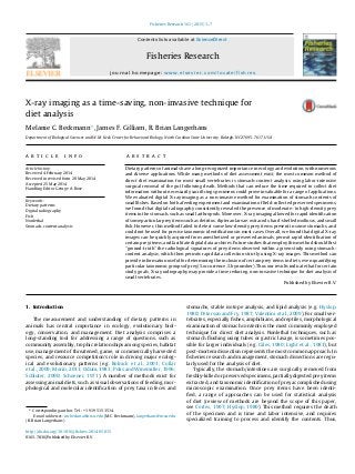
  • 1. Fisheries Research 161 (2015) 1–7 Contents lists available at ScienceDirect Fisheries Research journal homepage: www.elsevier.com/locate/fishres X-ray imaging as a time-saving, non-invasive technique for diet analysis Melanie C. Beckmann∗ , James F. Gilliam, R. Brian Langerhans Department of Biological Sciences and W.M. Keck Center for Behavioral Biology, North Carolina State University, Raleigh, NC 27695-7617, USA a r t i c l e i n f o Article history: Received 4 February 2014 Received in revised form 20 May 2014 Accepted 25 May 2014 Handling Editor George A. Rose Keywords: Dietary patterns Digital radiography Fish Nonlethal Stomach-content analysis a b s t r a c t Dietary patterns of animals have a long-recognized importance in ecology and evolution, with numerous and diverse applications. While many methods of diet assessment exist, the most common method of direct diet examination for most small vertebrates is stomach-content analysis, using labor-intensive surgical removal of the gut following death. Methods that can reduce the time required to collect diet information without necessarily sacrificing specimens could prove invaluable for a range of applications. We evaluated digital X-ray imaging as a non-invasive method for examination of stomach contents of small fishes. Based on both a feeding experiment and examination of field-collected preserved specimens, we found that digital radiography consistently revealed the presence of moderate- to high-density prey items in the stomach, such as small arthropods. Moreover, X-ray imaging allowed for rapid identification of some particular prey items such as detritus, dipteran larvae, ostracods, hard-shelled molluscs, and small fish. However, this method failed to detect some low-density prey items present in some stomachs, and could not be used for precise taxonomic identifications in most cases. Overall, we found that digital X-ray images can be quickly acquired from anesthetized or preserved animals, permit rapid identification of certain prey items, and facilitate digital data archives. Future studies that employ this method should first “ground-truth” the radiological signatures of prey items observed within a given study using stomach- content analysis, which then permits rapid data collection strictly using X-ray images. This method can provide information useful for determining the inclusion of certain prey items in diets, even quantifying particular taxonomic groups of prey (% occurrence, % by number). Thus our results indicate that for certain study goals, X-ray radiography may provide a time reducing, non-invasive technique for diet analysis of small vertebrates. Published by Elsevier B.V. 1. Introduction The measurement and understanding of dietary patterns in animals has central importance in ecology, evolutionary biol- ogy, conservation, and management. Diet analysis comprises a long-standing tool for addressing a range of questions, such as community assembly, trophic relationships among species, habitat use, management of threatened, game, or commercially harvested species, and resource competition’s role in driving major ecolog- ical and evolutionary patterns (e.g. Bolnick et al., 2003; Collar et al., 2009; Morin, 2011; Odum, 1983; Polis and Winemiller, 1996; Schluter, 2000; Schoener, 1971). A number of methods exist for assessing animal diets, such as visual observations of feeding, mor- phological and molecular identification of prey taxa in feces and ∗ Corresponding author. Tel.: +1 919 515 3514. E-mail addresses: mcbeckma@ncsu.edu (M.C. Beckmann), langerhans@ncsu.edu (R. Brian Langerhans). stomachs, stable isotope analysis, and lipid analysis (e.g. Hyslop, 1980; Peterson and Fry, 1987; Valentini et al., 2009). For small ver- tebrates, especially fishes, amphibians, and reptiles, morphological examination of stomach contents is the most commonly employed technique for direct diet analysis. Nonlethal techniques, such as stomach flushing using tubes or gastric lavage, is sometimes pos- sible for larger individuals (e.g. Giles, 1980; Light et al., 1983), but post-mortem dissection represents the most common approach. In fisheries research and management, stomach dissections are regu- larly used for the analysis of diet. Typically, the stomach/intestines are surgically removed from freshly-killed or preserved specimens, partially digested prey items extracted, and taxonomic identification of prey accomplished using microscopic examination. Once prey items have been identi- fied, a range of approaches can be used for statistical analysis of diet (review of methods are beyond the scope of this paper, see Cortes, 1997; Hyslop, 1980). This method requires the death of the specimen and is time and labor intensive, and requires specialized training to process and identify the contents. Thus, http://dx.doi.org/10.1016/j.fishres.2014.05.015 0165-7836/Published by Elsevier B.V.
  • 2. 2 M.C. Beckmann et al. / Fisheries Research 161 (2015) 1–7 alternative methods that can utilize live specimens or reduce the time required to collect diet data would be advantageous, as this would streamline the collection of diet information without sacri- ficing specimens. Here we examined digital X-ray radiography as a rapid, non- invasive method for assessing diet of small fishes. A number of recent advances in digital radiography make this assessment timely: e.g. increases in resolution and magnification permit the detection of very small objects of low density, prices of digital radio- graphy equipment has recently dropped considerably, portability of X-ray units has increased substantially, and many universities already have digital X-ray machines capable of at least moder- ate resolution of small vertebrates. While previous studies have employed X-ray radiography in the context of animal diets (e.g. rates of feed intake and gastric evacuation; Talbot and Higgins, 1983; McCarthy et al., 1992; Jobling et al., 1993, 2001), no previous study has examined the utility of this technique for identifying diet items of small vertebrates. In this study, we investigated gut/stomach contents using X- ray imaging in four small fish species (15–80 mm standard length): Eastern mosquitofish (Gambusia holbrooki, Girard 1859), Bahamas mosquitofish (G. hubbsi, Breeder 1934), Trinidadian guppy (Poecilia reticulata, Peters 1859), and Hart’s killifish (Anablepsoides hartii; formerly Rivulus hartii, Boulenger 1890). Collectively, these omniv- orous species are known to exhibit a broad diet, including detritus, algae, aquatic and terrestrial insects, crustaceans, molluscs, and even juvenile fishes. We hypothesized that X-ray images would reveal the presence of prey contents in the stomach, and permit the detection and identification of some broad groups of prey taxa based on their dense body parts (e.g. shells, exoskeletons, bones), such as molluscs (e.g. gastropods, bivalves), crustaceans (e.g. ostra- cods, shrimp), insects (e.g. chironomids, beetles), and vertebrates (e.g. fish, tadpoles). 2. Materials and methods Our goal was to determine whether X-ray imaging could reveal the presence or absence of prey in stomachs of small fishes, and allow the identification of 5 different types of prey items that vary in density (mass per unit volume): (1) soft homogeneous prey (e.g. algae, detritus), (2) weakly shelled arthropods (e.g. small shrimp, ants), (3) moderate-density arthropods (e.g. ostracods, beetles), (4) hard-bodied prey (e.g. shelled molluscs, crabs) and (5) verte- brates (e.g. small fish, anurans). We took a two-pronged approach to accomplish this: we conducted a feeding experiment with live fish to directly assess the accuracy of diet identification using X-ray imaging, and we examined preserved, wild-caught fish specimens to evaluate the utility of the approach for the examination of natural dietary patterns. 2.1. Feeding experiment We performed a feeding experiment using 21 live individuals of G. holbrooki and 3 individuals of A. hartii. All fish were collected from the wild (G. holbrooki: North Carolina, USA; A. hartii: Trinidad) and housed in 38-L aquaria in common laboratory conditions. Prior to the feeding experiment, fish were placed individually into separate 8-L tanks and starved for 48 h. For G. holbrooki, we assigned three adult females at random to each of seven diet treatments: (1) no prey: starved, (2) soft, low-density homogeneous prey: TetraMin Pro flakes, (3) low-density crustaceans: live Artemia sp. nauplii, (4) low-density insects: live ants, (5) moderate-density insects: thawed bloodworms (Chironomus tetans), (6) hard-shelled prey: live snails (Physa acuta), and (7) vertebrate: one live G. holbrooki Table 1 Collection and sample size information for wild-caught adult specimens examined in this study. Species Collection location N Gambusia holbrooki Melbourne, Florida, USA 60 James Island Park, South Carolina, USA 60 Gambusia hubbsi East Twin blue hole, Andros Island, Bahamas 40 West Twin blue hole, Andros Island, Bahamas 40 Hubcap blue hole, Andros Island, Bahamas 40 Poecilia reticulata Kahala, Oahu, Hawaii, USA 120 Anablepsoides hartii Arima Valley, Trinidad 23 juvenile. We fed two live P. reticulata juveniles (3–4 mm SL) to A. hartii to test for detection of vertebrate consumption in this species. Within 1 h of feeding, we X-rayed each individual and saved a digital image. We placed each live fish into a small, moist plastic bag, laid the fish on its side within a petri dish, and set the dish in the X-ray machine to capture a lateral image. We used a custom-built digital X-ray unit comprising a micro-focus X-ray source (Hama- matsu L6731-01) and a digital X-ray detector (PaxScan 2520E) housed in a lead-shielded cabinet, set to 45 kV and 40 ␮A. Radi- ation exposure to each fish was low, approximately 25–50 mrem – roughly equal to a human dental X-ray for comparison. Fish were then immediately placed into a recovery tank. Removal from water, X-ray imaging, and return to recovery tank typically only required approximately 30 s. Identification of all stomach contents based on digital X-ray images was conducted blind of fish ID. 2.2. Preserved specimens We examined digital X-ray images (using method and equip- ment described above) of wild-caught specimens preserved in 70% ethanol to assess the ability to detect natural dietary patterns of preserved small fish with X-ray images. We examined 120 G. hol- brooki, 120 G. hubbsi, 120 P. reticulata, and 23 A. hartii (see Table 1 for collection and sample size details). For each preserved specimen, we attempted to determine contents of the stomach based on the X-ray image. After view- ing a number of images, six natural categories emerged from our identifications: (1) no prey contents, (2) soft homogeneous prey (e.g. algae, detritus), (3) low-density prey (e.g. branchiopods, ants), (4) moderate-density prey (e.g. ostracods, dipterans), (5) shelled mollusc prey, and (6) vertebrate (fish) prey. To determine the accu- racy of diet identification, we dissected three randomly selected specimens of each species from each diet category using the tra- ditional method of surgically removing the gut and identifying the contents under a microscope (Leica S8 APO stereoscope). We then compared the diet classification from X-ray images to that from direct morphological identification. 3. Results 3.1. Feeding experiment The guts of all six starved fish appeared empty in X-ray images (Fig. 1a and b). We could not detect flakes or Artemia sp. nauplii with our X-ray images (Fig. 1c). In two out of the three fish fed ants, small hard parts of prey were visible in their guts in the X- ray Images – presumably reflecting broken pieces of the ants – but these were difficult to identify as ants or even as insects (Fig. 1d). Based on X-ray images, we accurately detected prey items in all fish fed bloodworms (Fig. 1e), P. acuta snails (Fig. 1f), newborn G. holbrooki (Fig. 1g), and juvenile P. reticulata (Fig. 1h). Thus, we con- sistently could not detect the two lowest density prey types, but could detect the three highest density prey types; ants appeared
  • 3. M.C. Beckmann et al. / Fisheries Research 161 (2015) 1–7 3 Fig. 1. Representative X-ray images from each diet treatment in the feeding experiment showing only the intestinal region of each image (anterior to the left). (a) Starved female G. holbrooki, depicting no apparent object signatures within the gut (only yolked eggs, spherical objects visible in the image, were observed in the relevant body region). (b) Starved female A. hartii, depicting no apparent object signatures within the gut. (c) Female G. holbrooki fed TetraMin Pro flakes, depicting no apparent object signatures within the gut. (d) Female G. holbrooki fed ants, depicting several small low-density items within the gut (yolked eggs also visible in image). (e) Pregnant female G. holbrooki fed bloodworms, clearly depicting the shape of the Chironomus tetans larvae (embryo otoliths appear as paired black spots in the image). (f) Female G. holbrooki fed Physa acuta, depicting the consumed snail shells. (g) Female G. holbrooki fed a newborn G. holbrooki, depicting the otoliths and body outline of the fish prey. (h) Female A. hartii fed two juvenile P. reticulata, depicting the otoliths, vertebrae, and parts of the skulls of the fish prey. to represent prey densities around the threshold of detectability using this method. 3.2. Preserved specimens First, we found that stomachs we identified as empty based on X-ray images actually contained small, low-density prey items, such as soft-bodied prey, weakly shelled prey, or filamentous plant material (Fig. 2a). We further frequently encountered these prey items in dissected stomachs that we identified to contain other prey types, but these prey were typically not visible in X-ray images. Second, guts that we identified as containing soft, homogeneous prey or low- to moderate-density arthropod prey were filled with a variety of prey items, including ants, dipteran larvae, small adult dipterans, and filamentous plant material (Figs. 2b and c and 3a and b). Guts of P. reticulata in particu- lar appeared to be filled homogeneously with low-density prey, which we confirmed to comprise detritus and algal material; inter- spersed higher-density items within these guts represented small arthropods (Fig. 2c). Third, among the moderate-density items vis- ible in radiographs, dipteran larvae were occasionally identifiable based on their characteristic size and elongate shape (Fig. 3b), and
  • 4. 4 M.C. Beckmann et al. / Fisheries Research 161 (2015) 1–7 Fig. 2. Representative X-ray images of the intestinal regions of adult wild-caught specimens (left) and the low- to moderate-density prey removed from their stomachs (right). (a) Female G. hubbsi incorrectly identified to have an empty stomach, while it actually contained a soft-bodied insect larva and an ant. (b) Male A. hartii identified as containing soft-bodied prey and low- to moderate-density arthropods according to the X-ray, and dissected contents revealed Chironomidae and Simuliidae larvae and filamentous plant material. (c) Female P. reticulata identified as containing homogeneous low-density items, such as detritus, and several higher-density items, such as arthropods; dissected contents revealed detritus and homogeneous algal material, along with several small adult hymenopterans. ostracods were always easily identified (Fig. 3a and b). Fourth, snail shells were among the most easily identifiable prey item in X-ray images, as the shells appear highly defined in radiographs and all dissected stomachs confirmed our identifications via radio- graphs (Fig. 3c). Finally, fish prey were generally readily identifiable (Fig. 3d), with the exception that pregnant live-bearing fish carry- ing one or a few late-stage embryos can appear on an X-ray image to have instead consumed small fish (we encountered one instance here, where a pregnant G. hubbsi female contained a single embryo at the final stage of development, which would generally be difficult to distinguish from consumed fish prey). 4. Discussion Diet analysis holds central importance in a range of ecological and evolutionary applications. Traditional methods for diet analy- sis of small vertebrates are time and labor intensive, and typically involve the death of the animal. This situation can prove problem- atic in the face of time constraints, or when the animals of interest need to remain alive, such as in the cases of endangered species, captive breeding programs, the need to minimize impacts on popu- lations, and to avoid negative public perceptions of management or conservation programs (Crossman and Hamilton, 1978; Hartleb and Moring, 1995; Light et al., 1983). Small vertebrates pose spe- cial challenges to nonlethal diet analysis, as techniques commonly applied to larger animals are typically not feasible with small indi- viduals (Hyslop, 1980; Strange and Kennedy, 1981; Waters et al., 2004). Thus, a need exists for methods of diet analysis amenable to small vertebrates that reduce the required time and labor, and can be applied to live animals. We found that X-ray imaging can be used to quickly identify some particular moderate- to high-density prey taxa within stom- ach contents of both live and preserved small fishes. Although identification of these prey taxa with X-ray images is quite rapid, the taxonomic resolution will generally be much lower than that obtained via microscopic examination of stomach contents. How- ever, the method can detect very small prey items within small organisms – the entire body cavity length for the fish examined in this study typically only spanned 8–15 mm, with prey parts as small as 0.1 mm long identified in images. In our feeding experiment, we found that X-ray imaging could be employed to accurately identify chironomids, snails, and small fish prey, as we correctly identified these prey in all cases. More- over, small ants were often detectable as small, low-density items within the stomach; although, these items could not be identi- fied as hymenoptera or even insects without examination of the
  • 5. M.C. Beckmann et al. / Fisheries Research 161 (2015) 1–7 5 Fig. 3. Representative X-ray images of the intestinal regions of adult wild-caught specimens found to contain moderate- to high-density prey items (left) and the prey removed from their stomachs (right). (a) Female G. holbrooki identified as containing many ostracods, and several low- to moderate-density items in its stomach (yolked eggs also visible); dissected contents revealed many ostracods, along with plant material. (b) Female G. holbrooki identified as containing several clumps of ostracods and an elongate moderate-density item indicative of a dipteran larva in its stomach; dissected contents revealed ostracods, a Brachycera larva, a small annelid, and plant material. (c) Female G. holbrooki identified as containing four snail shells in the gut (embryos with otoliths also visible); dissected contents revealed four snail shells and filamentous plant material. (d) Female G. hubbsi identified as containing one small fish prey (the pairs of otoliths are the darkest spots, while the jaw and vertebrae are also evident); dissected contents revealed a single small fish prey. stomach contents. Our analysis of preserved wild-caught speci- mens confirmed that we could use X-ray imaging to accurately identify homogeneous algae/detritus, ostracods, snails, and fish, while we could typically also identify the presence of dipteran larvae. Moreover, the presence of some other prey, such as small arthropods, could often be determined from X-ray images, although the taxonomic status could not be determined without stomach- content analysis. Because species-specific, morphological diagnostic characters are often not visible in X-ray images, identification to species level was not reliable in most cases in this study. In some simple natural systems, there may only be a single species of a partic- ular prey type, in which species-level identification is possible. If a study requires detailed information regarding the taxonomic identity of a variety of food items consumed by an animal, or requires volumetric or weight information, then X-ray imaging will not be suitable to accomplish the study goals. Thus, X-ray imaging can provide rapid information useful for certain types of diet analysis, but not others. For instance, the method can rapidly acquire presence/absence of particular prey taxa in stomachs of
  • 6. 6 M.C. Beckmann et al. / Fisheries Research 161 (2015) 1–7 Fig. 4. (a) X-ray image of TetraMin Pro flakes when examined alone (not within fish stomach). (b) X-ray image of three ants when examined in isolation. individuals, and presence/absence of broad groupings of prey types where taxonomic identities are difficult to discern with X-ray images and are also less important for the study. In some cases, numerical information may also be obtained from X-ray images. Thus, digital radiography might often allow for collection of data for two commonly employed dietary metrics, percent frequency of occurrence and percent by number of prey taxa in stomachs (Cortes, 1997). While a range of prey taxa were detectable using digital radiography, our study also revealed that not all types of prey items were visible in X-ray images. For instance, we could not detect flake food within fish stomachs in our feeding experi- ment, and sometimes could not detect ants. These prey items, when imaged in isolation, are clearly evident in X-ray images taken with our equipment (Fig. 4). However, when contained within surround- ing fish tissue, these low-density food items become very difficult to detect. Further, our examination of wild-caught preserved spec- imens confirmed that guts appearing empty on X-ray images could actually contain soft-bodied, low-density food items such as ants, soft-bodied insect larvae, and plant material. Consequently, this method cannot accurately distinguish empty stomachs from stom- achs that contain certain types of low-density prey. In addition to the limitations of this method discussed already regarding detection and identification of particular prey items, this method may also be affected by variable processing (e.g. prey crushing and tearing) and digestion rates of different prey within different species (e.g. Macdonald et al., 1982). Prey densities can decline during digestion, making the method more amenable to foregut analyses and prey items that are swallowed whole where moderate-density parts are passed in feces. Compared to more labor-intensive methods, such as gut dis- section and stomach-content analysis using identification with a microscope, taking and analyzing X-ray images provides a dramatic improvement in the time required for analysis, and per- mits the examination of live animals. For instance, digital X-ray images usually take about 1 min per specimen to acquire, with images immediately available for inspection (no film development required), and digital files ready for archiving for long-term stor- age and future reference. After initial training for identification of the relevant prey categories from radiographs, scoring stom- ach contents from images usually only requires a few minutes per specimen. Meanwhile, surgical extraction of a stomach and microscopic analysis of diet typically require much more time per specimen. Microscopic identification of prey taxa extracted from stomachs demands considerable skills that require substan- tial training for the identification of a wide range of possible prey taxa, while identification of a smaller set of prey categories on X-ray images necessitates much less training. The use of X-ray imaging versus direct stomach-content analysis for diet studies of small vertebrates largely represents a tradeoff between time, training, precision, and lethality: X-ray imaging allows the rapid extraction of broad dietary information requiring minimal train- ing where the specimens can remain alive after examination, while direct stomach-content analysis represents a more time- consuming extraction of detailed dietary information requiring substantial training where the individuals must typically die prior to examination. Thus, a researcher or manager must weigh these four primary factors when deciding whether X-ray imaging might represent the most appropriate method for their study. The methods described here should apply to small vertebrates other than fish, such as reptiles, amphibians, and small birds and mammals. Indeed, during the course of other projects that involved X-ray imaging of small lizards and frogs, we have wit- nessed a number of obvious diet items in the images, such as terrestrial arthropods and vertebrates (RBL, unpublished data). Fur- ther, portable X-ray applications are becoming more commonplace today, and this implies that diets of live animals could be exam- ined directly in the field. Because the method requires little time and handling, and can be employed with extremely low levels of radiation exposure, this method could represent a suitable solu- tion for diet studies of threatened species, or in situations where it is important not to impact the study population, including small populations, breeding programs, zoo animals, or lab animals. We envision other uses of digital radiography for ecologists and evolutionary biologists as well, such as the use in reproductive and developmental studies of viviparous fishes. For instance, X-ray images provide highly accurate information regarding whether a fish is pregnant or not, and can often permit the counting of the number of developing embryos within some species. Thus, studies examining the fecundity of females might employ X-ray imaging to rapidly collect information on the frequency of pregnancy, number of offspring, and perhaps estimates of the size and developmental stage of embryos. 5. Conclusion Overall, our study revealed that X-ray imaging can provide an appropriate tool for diet analysis of small vertebrates, depending on the goals of the study. The two most obvious advantages of the approach are the reductions in time required for data collection and the ability to non-invasively examine live animals. This method is probably most useful for studies that seek rapid collection of broad dietary information, where detailed taxonomic information or prey-size information is less important for the goals. This non- lethal method can rapidly and accurately provide information on certain types of prey items contained within the stomachs of spec- imens, and should thus receive increased use in diet studies in the future.
  • 7. M.C. Beckmann et al. / Fisheries Research 161 (2015) 1–7 7 Acknowledgements We would like to thank J. Warrillow for assistance with the feeding experiment; B. Lamphere and E. Hain for valuable input during early stages of the project; R. Martin, R. Riesch, S. Dia- mond, and J. Heinen-Kay for assistance collecting specimens; and a grant from the National Science Foundation to RBL for funding (DEB-0842364). References Bolnick, D.I., Svanback, R., Fordyce, J.A., Yang, L.H., Davis, J.M., Hulsey, C.D., Forister, M.L., 2003. The ecology of individuals: incidence and implications of individual specialization. Am. Nat. 161, 1–28. Collar, D.C., O’Meara, B.C., Wainwright, P.C., Near, T.J., 2009. Piscivory limits diversification of feeding morphology in centrarchid fishes. Evolution 63, 1557–1573. Cortes, E., 1997. A critical review of methods of studying fish feeding based on anal- ysis of stomach contents: application to elasmobranch fishes. Can. J. Fish. Aquat. Sci. 54, 726–738. Crossman, E.J., Hamilton, J.G., 1978. An apparatus for sampling gut contents of large, living fishes. Environ. Biol. Fish. 3, 297–300. Giles, N., 1980. A stomach sampler for use on live fish. J. Fish Biol. 16, 441–444. Hartleb, C.F., Moring, J.R., 1995. An improved gastric lavage device for removing stomach contents from live fish. Fish. Res. 24, 261–265. Hyslop, E.J., 1980. Stomach contents analysis-a review of methods and their appli- cation. J. Fish Biol. 17, 411–429. Jobling, M., Christiansen, J.S., Jørgensen, E.H., Arnesen, A.M., 1993. The application of X-radiography in feeding and growth studies with fish: a summary of exper- iments conducted on arctic charr. Rev. Fish. Sci. 1 (3), 223–237. Jobling, M., Coves, D., Damsgard, B., Kristiansen, H.R., Koskela, J., Petursdottir, T.E., Kadri, S., Gudmundsson, O., 2001. Techniques for Measuring Feed Intake. Food Intake in Fish. New York, Wiley. Light, R.W., Adler, P.H., Arnold, D.E., 1983. Evaluation of gastric lavage for stomach analyses. North Am. J. Fish. Manage. 3, 81–85. Macdonald, J.S., Waiwood, K.G., Green, R.H., 1982. Rates of digestion of different prey in Atlantic Cod (Gadus morhua), Ocean Pout (Macrozoarces americanus), Winter Flounder (Pseudopleuronectes amerkanus), and American Plaice (Hippoglossoides platessoides). Can. J. Fish. Aquat. Sci. 39, 651–659. McCarthy, I.D., Carter, C.G., Houliha, D.F., 1992. The effect of feeding hierarchy on individual variability in daily feeding of rainbow trout, Oncorhynchus mykiss (Walbaum). J. Fish Biol. 41, 257–263. Morin, P.J., 2011. Community Ecology. Wiley-Blackwell, Malden, MA. Odum, H.T., 1983. Systems Ecology: An Introduction. Wiley, New York. Peterson, B.J., Fry, B., 1987. Stable isotopes in ecosystem studies. Annu. Rev. Ecol. Syst. 18, 293–320. Polis, G.A., Winemiller, K.O., 1996. Food Webs: Integration of Patterns & Dynamics. Chapman & Hall, New York. Schluter, D., 2000. The Ecology of Adaptive Radiation. Oxford University Press, Oxford. Schoener, T.W., 1971. Theory of feeding strategies. Annu. Rev. Ecol. Syst. 2, 369–404. Strange, C.D., Kennedy, G.J.A., 1981. Stomach flushing of salmonids: a simple and effective technique for the removal of the stomach contents. Fish. Manage. 12, 9–15. Talbot, C., Higgins, P.J., 1983. A radiographic method for feeding studies on fish using metallic iron powder as marker. J. Fish Biol. 23, 211–220. Valentini, A., Miquel, C., Nawaz, M.A., Bellemain, E., Coissac, E., Pompanon, F., Gielly, L., Cruaud, C., Nascetti, G., Wincker, P., Swenson, J.E., Taberlet, P., 2009. New per- spectives in diet analysis based on DNA barcoding and parallel pyrosequencing: the trnL approach. Mol. Ecol. Res. 9, 51–60. Waters, D.S., Kwak, T.J., Arnott, J.B., Pine, W.E., 2004. Evaluation of stomach tubes and gastric lavage for sampling diets from blue catfish and flathead catfish. North Am. J. Fish. Manage. 24, 258–261.