1. Maryam Rahmani1
, Melanie Pepin1,2
, Dr. Peter H. Byers1,2
1
University of Washington, 2
University of Washington Department of Pathology
Introduction:
Does ultrasound identification of in-utero fractures predict Osteogensis Imperfecta?
University of Washington GenOM Project 2012
Background:
Methods:
Acknowledgements:
Results:
Conclusion:References:
Abstract:
Osteogenesis imperfecta (OI) is an inherited brittle bone disorder that varies in clinical severity from mild to lethal forms. The mutation generally occurs in type one collagen genes (COL1A1 and COL1A2), and
it can be confirmed by molecular testing. The goal of this project is to discover which combinations of clinical signs of osteogenesis imperfecta are most likely to yield positive results for a mutation in COL1A1 or
COL1A2, and to develop guidelines which could be used by physicians when diagnosing OI with in utero fetuses. The data for this study was collected from in utero cases submitted to the University of Washington
Collagen Diagnostic Lab from around the world. In this study, I tabulated the multiple signs of OI from patient data organized by the signs shown on ultrasounds previous to genetic testing (ex: shortened long
bones, bowed bones, multiple fractures, etc.), and the results of genetic testing. With this information, it will be easier for doctors and genetic counselors to recognize cases of OI in fetuses and determine when
the appropriate genetic testing is required. The findings in this study will make it easier for care to be administered to the fetuses and family members dealing with osteogenesis imperfecta.
Osteogenesis Imperfecta (OI) is one of the more
common results of type one collagen mutations.
Many research facilities and medical agencies
recognize cases of OI in fetuses via ultrasound
imaging, by noticing multiple fractures, bowed
bones, and shortened long bones in the fetus.
In the University of Washington Collagen
Diagnostic Lab, doctors and genetic counselors from
around the world send in samples from fetuses
suspected to have OI based on clinical signs seen in
ultrasound images. The DNA is screened for
mutations in the type one collagen genes, and it is
then determined if the fetus has OI or not. We are
searching for patterns between the clinical signs of
fetuses, the results of genetic screenings, and
eventually the clinical diagnosis of osteogenesis
imperfecta. Patient information from about forty
cases of fetuses suspected to have OI were
compared to see which signs were most present in a
fetus with a collagen mutation.
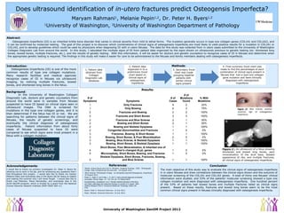
Figure 11
: An ultrasound of a fetus showing
shortened and bowed long bones, poor
mineralization of the skull base, beaded
appearance of ribs, and multiple fractures;
all clinical signs of osteogenesis imperfecta
1
Neish, Ariane Staub and Carl S. Winalski. Imaging Findings. 1995 . Photograph
Image. http://brighamrad.harvard.edu. 10 August 1012.
Figure 22
: Blue sclerae, another
common sign of osteogenesis
imperfecta
2
Blue Sclerae. Photograph Image. en.wikipedia.org/wiki/Osteogensis_imperfecta.
10 August 2012.
# of
Symptoms Symptoms
# of
Cases
# of
Mutations
found
% With
Mutations
1 Only Fractures 9 2 22%
1 Only Bowing 4 3 75%
2 Fractures and Bowing 2 2 100%
2 Fractures and Short Bones 3 3 100%
2 Fractures and Blue Sclerae 2 1 50%
2 Bowing and Short Bones 2 1 50%
2 Bowing and Skeletal Dysplasia 1 1 100%
2 Congenital Abnormalities and Fractures 1 0 0%
3 Fractures, Bowing, & Short Bones 1 1 100%
3 Bowing, Short Bones, & Poor Mineralization 2 0 0%
3 Bowing, Blue Sclerae, & Skeletal Dysplasia 1 1 100%
3 Bowing, Short Bones, & Skeletal Dysplasia 1 1 100%
4
Short Bones, Poor Mineralization, & Inherited one of
Mother's changed ALpL genes 1 0 0%
5 Microcephaly, Short Bones, Bowing, and Fractures 1 0 0%
5
Skeletal Dysplasia, Short Bones, Fractures, Bowing,
and Blue Sclerae 1 1 100%
The main objective of this study was to evaluate the clinical signs of osteogenesis imperfecta
in in utero fetuses and draw correlations between the clinical signs shown and the outcome of
molecular screening of the COL1A1 and COL1A2 genes. A total of thirty one fetuses’ clinical
information were studied, and 53% of the patients’ molecular screening showed a type one
collagen mutation and were diagnosed with osteogenesis imperfecta. One hundred percent
of the 53% of patients had bowed bones and multiple fractures listed as clinical signs
present. Based on these results, fractures and bowed long bones seem to be the most
common clinical signs present in fetuses clinically diagnosed with osteogenesis imperfecta.
1. Patient data
obtained from
Collagen
Diagnostic Lab
database
2. Patient data
organized in to a
preliminary Excel
chart based on
clinical signs of
osteogenesis
imperfecta
3. Secondary Excel
chart was made
grouping together
patients with
similar clinical
signs present
4. Final summary Excel chart was
made to find the percentages of each
combination of clinical signs present in
fetuses that had a type one collagen
gene mutation and were clinically
diagnosed with osteogenesis
imperfecta
Khalil, A., Pajkrt, E. and Chitty, L. S. (2011), Early prenatal diagnosis of skeletal
anomalies. Prenat. Diagn., 31: 115–124. doi: 10.1002/pd.2676
Konstantinidou, Anastasia E, et al. "Genetic skeletal disorders of the fetus
and infant: pathologic and molecular findings in a series of 41 cases." Birth
Defects Research Part A: Clinical and Molecular Teratology (2009): NA.
Print.
Pepin, Melanie. Personal Interview. 10 July 2012.
Byers, Peter H. Personal Interview. 10 July 2012.
I would like to thank my primary investigator Dr. Peter H. Byers for
allowing me to work in his lab, and for answering any questions that I
had throughout this project. I would also like to thank my mentor
Melanie Pepin and everybody else in the Byers lab for being so helpful
and making this summer one I will never forget. I would also like to
thank Lisa Peterson, Elena Hernandez, and everybody else in the
ALVA GenOM program, which is funded by a grant from the National
Human Genome research Institute (NHH HG02 360-11)