2. Astana Medical University Kazakhstan (NJSC)
SPECIALTY: GENERALMEDICINE
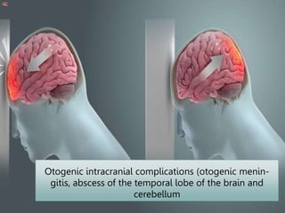
SUBJECT: Basics of otorhinolaryngology
CHECKEDBY- Yersakhanova B.К.
PreparedBy : MANDEEPSINGH
COURSE : 4RD year
Group : 478
SESSION– 2022-2023
IWS
3. The main forms of intracranial
complications
1. Otogenic meningitis
2. Otogenic abscesses of the brain and
cerebellum
3. Otogenic sepsis
4. Diffuse purulent
inflammation of the soft
and arachnoid
membranes of the brain,
developing as a result of
bacterial infection from
the cavities of the middle
ear (secondary
meningitis).
Otogenic meningitis
5. Classification of otogenic meningitis:
Depending on the severity of the
current, there are forms meningitis:
- acute,
- subacute,
- lightning fast
- recurrent.
6. Clinic of otogenic meningitis
General symptomsinfectious disease:
- rise in temperature to 38-40°FROM,type ofcontinua,
- severe general condition, tachycardia,heart sounds are muffled; breathing
quickened,
Meningeal symptoms:
- headache, vomiting,
-neck stiffness,
- symptoms of Kernig, Brudzinsky, zygomatic symptom
ankylosing spondylitis
- general hyperesthesia
Focal symptoms:
- pyramidal symptoms of Babinsky, Oppenheim,
Rossolimo, Gordon,
- damage to the cranial nerves - more often abducent (VInerve),
less often oculomotor (IIInerve), even less often block (IV
nerve).
7. Differential Diagnosis
Tuberculous meningitis-more often in children, a sluggish and slow
course is characteristic, other organs are often affected, primarily the
lungs. The cerebrospinal fluid is often clear, flows out under pressure;
cytosis is not pronounced (150-300), however, the cerebrospinal fluid
contains a large number of lymphocytes (up to 80%) and protein (3.3-6.6
g/l)
Epidemic cerebrospinal meningitis-proceeds rapidly, its onset is
often preceded by catarrh of the upper respiratory tract, petechiae on the
skin, herpetic eruptions on the lips are observed. taken into account
epidemiological situation. The diagnosis is confirmed by the detection of
meningococci in the cerebrospinal fluid.
Serous meningitis-often develops against the background of acute
otitis media caused by a viral infection. Often this happens during an
epidemic of influenza, mass diseases of SARS. Changes in the
cerebrospinal fluid are less pronounced than with a purulent process,
cytosis (usually lymphocytic) does not exceed 200-300 cells per 1 μl, the
sugar content is normal.
8. Treatment of otogenic meningitis:
Its basis issurgical debridement of the source of
infection in the ear(extended debridement surgery
on the ear).
Conservative treatment:
-massive antibiotic therapy with the appointment of high
doses of broad-spectrum antibiotics,
- dehydration (mannitol, lasix, magnesium sulfate solution,
etc.),
- detoxification (hemodez, Ringer-Locke solution, vitamins
B1, B6, ascorbic acid),
- orally or parenterally potassium preparations,
- symptomatic therapy (cardiac glycosides, analeptics,
analgesics)
9. Otogenic brain abscesses
a -open extradural
abscess;
b – closed extradural
abscess;
c - subdural abscess;
d - intracerebral
abscess.
10. Abscess of the brain and
cerebellum
Intracerebral abscesses -a limited
accumulation of pus in the substance
of the brain, which occurs a second
time in the presence of a source of
infection in the cavities of the middle
ear.
11. Etiology of otogenic intracranial
complications
From the primary focus in
the middle ear, mixed
flora is often sown:
Staphylococci
streptococci
Pseudomonas
aeruginosa
Proteus
12. Pathogenesis of otogenic
intracranial complications
Routes of infection:
1. Contact
2. Hematogenous
3. Lymphogenic
4. labyrinthogenic
5. Along preformed paths
6. Through digestion
13. General information:
Abscesses of the temporal lobe of the brainmeet4 times
more often, how cerebellar abscesses, and are usually
round in shape, while in the cerebellum they are slit-like.
Intracerebral abscesses are more likely to develop with
chronic suppurative otitis media, infection in the substance
of the brain penetrates by contact. They usually occur in
the immediate vicinity of the focus of infection and are
located rather superficially - at a depth of 2-4 cm.
At acute otitis mediapossible hematogenous or
lymphogenous spread of infection; in this case, an abscess
can form in areas of the brain remote from the primary
focus.
14. 4 stages of brain abscess:
I-initial (encephalitic) stageusually lasts 1-2 weeks and is
accompanied by a mild headache, lethargy, fever up to 37.2 -
37.5C, nausea and vomiting.
II-latentstage (imaginary well-being) lasts from 2 to 6 weeks.
There may be lethargy, pallor of the patient, lack of appetite,
periodically headache. The temperature is usually normal, the
blood formula without deviations from the norm.
The first two stages often go unnoticed or their symptoms are
interpreted as an exacerbation of otitis media.
III-explicit (manifest) stage.The duration is about 2 weeks, there
is a picture of a serious illness with a tendency to a rapid and
steady deterioration of the patient's condition. Symptoms of this
stage - see below.
IV-terminal stage.Occurs when an abscess breaks into the
ventricles of the brain or into the subarachnoid space.
16. Symptoms of an explicit abscess
stage
Three groups of symptoms:
-common signs of severe infectious disease(weakness,
weakness, lack of appetite, the patient is lethargic, drowsy,
the skin is pale, sometimes with an earthy or icteric tint, the
facial expression is suffering, sometimes the temperature
rises to 38-39°FROM, inflammatory changes in the blood).
-cerebral symptoms(severe headache, vomiting not
associated with food intake, forced head position with
cerebellar abscess, sometimes bradycardia up to 45 in 1
min, often meningeal symptoms)
-focal neurological symptomsdepending on the location of
the abscess.
17. Focal symptoms
with an abscess of the
temporal lobe of the brain:
- Aphasia(with damage to the left temporal lobe in right-handers)
observed in 75-80% of cases.
Amnestic aphasia- if the patient is shown an object and asked what it is
called, he cannot answer and describes this object (for example: a
pencil is what they write with, a spoon is what they eat, etc.)
Sensory aphasia- the patient loses the meaning of words, as if they are
speaking to him in a language he does not understand. With intact
hearing, he does not understand what he is told; his speech becomes
incomprehensible and turns into a meaningless set of words.
- Hemianopia- loss of visual fields on both sides, due to the involvement
in the process of the visual path passing through the temporal lobe to
the occipital lobe. An important symptom in the defeat of the right
temporal lobe in right-handers.
- epileptiform syndrome- one of the focal signs in the defeat of the
temporal lobe of the brain. Sometimes this symptom is the first
indication of an abscess that is forming.
- Temporal ataxia- with right-sided localization of the abscess of the
temporal lobe of the brain in right-handers, temporal ataxia is
manifested by the fall of the patient to the left
18. Symptoms
with cerebellar abscess:
-Violation of muscle tone (missing with one hand on the
side of the lesion when performing finger-nose, finger-
finger tests).
- In the Romberg pose and when walking in a straight
line deviation towards the affected lobe of the
cerebellum.
- Impossibility of execution flanking gait,fall in the
direction of defeat.
- Difficulty in doing heel test.
- Adiadochokinesis - the inability to quickly and
smoothly produce pronation and supination, there is a
sharp lag of the hand on the affected side.
-Cerebellar nystagmus usually directed towards the
affected side he rude, coarse, accompanied by other
cerebellar symptoms.
19. Diagnosis of intracerebral abscesses:
Clinical signs are taken into account, among which the
leading role for the localization of the pathological
process isfocal symptoms.
Radiography of the skull and temporal bones, X-ray
and magnetic resonance computed tomography of the
brain.
Echoencephalography (M-echo shift),
Lumbar puncture.
If necessary, encephalography, angiography, pneumo-
and ventriculography, radioisotope scintigraphy.
The patient is consulted by a neurologist, an
ophthalmologist, an otoneurologist, an
otolaryngologist.
20. Treatment for otogenic intracerebral
abscesses
Emergency, according to vital indications, an
extended radical operation is performed with
opening of the cranial fossae.
The substance of the brain is punctured to a depth of no more
than 4 cm.
When an abscess is detected, the needle is left in place and an
incision is made through it in the dura mater, most often
crosswise.
The ear forceps are inserted through the needle in a closed
state until the abscess of the brain. Having opened the forceps
by 1.5-2 cm, they are removed from the brain and thereby form
a passage into the substance of the brain to the abscess.
A strip of glove rubber is inserted into the abscess cavity.
Along with the operation, active antibacterial, dehydration,
detoxification therapy is carried out, as in purulent meningitis.
21. Thrombosis of the sigmoid sinus and
otogenic sepsis.
sinus thrombosis- formation and subsequent
infection thrombus in the lumen of the venous
sinus up to its complete occlusion, accompanied
by inflammation of the vascular wall and the
development of otogenic sepsis.
Thrombosis can spread retrogradely into the
transverse sinus, and down to the bulb of the
jugular vein and the jugular vein.
22. Limited pachymeningitis,
periphlebitis
Perisinus abscess
formation
Purulent fusion of a
thrombus Obturating thrombus
Spread of purulent emboli
by blood flow, sepsis
Phlebitis, the formation of a
parietal thrombus
Pathogenesis of sigmoid sinus
thrombosis and otogenic sepsis
Destruction by a carious
or cholesteatoma process
of the posterior wall of the
mastoid process
23. Conclusion:
Intracranial complications arising from chronic foci
of inflammation in the middle ear or in the
paranasal sinuses are among the life-threatening.
Without surgical sanitation, performed as early as
possible, a patient with such a complication has
practically no chance of recovery.
Therefore, a doctor of any profile must have
sufficient knowledge to suspect the presence of a
complication and promptly refer the patient to a
specialized medical institution.
Editor's Notes
Influenza and other ARIs account for 60-70% of all childhood infectious diseases