
Recommended
More Related Content
Similar to BMS1 K16 INFECTIONS OF THE CENTRAL NERVOUS SYSTEM Radang 1.ppt
Similar to BMS1 K16 INFECTIONS OF THE CENTRAL NERVOUS SYSTEM Radang 1.ppt (20)
Recently uploaded
Recently uploaded (20)
BMS1 K16 INFECTIONS OF THE CENTRAL NERVOUS SYSTEM Radang 1.ppt
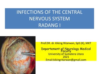
- 1. INFECTIONS OF THE CENTRAL NERVOUS SYSTEM RADANG I Prof.DR. dr. Kiking Ritarwan, SpS (K), MKT Departement of Neurology Medical Faculty University of Sumatera Utara 2021 Email:kikingritarwan@gmail.com
- 2. CNS INFECTION • MENINGITIS: TBM, Bacterial, Viral, Jamur - Inflamation of the meningeal covering of Brain and spinal cord. - LEPTOMENINGITIS (arachnoid + pia) PACHYMENINGITIS (duramater) - TYPE OF MENINGITIS : Bacterial, TB, Viral, fungal. • ENCEPHALITIS VIRAL • MYELITIS • ABSCESS CEREBRI
- 4. ANATOMY OF MENINGES DURAMATER PACHYMENIX Dura mater otak digambarkan terdiri dari dua lapis, yaitu lapisan endosteal dan lapisan meningeal. Lapisan tersebut bersatu dengan erat, kecuali pada garis-garis tertentu, tempat mereka berpisah untuk membentuk sinus venosus. ARACHNOID LEPTOMENINX Arachnoidea mater merupakan membran yang halus dan bersifat impermeabel, yang menutupi otak dan medulla spinalis dan terletak di antara pia mater di bagian dalamnya dan dura mater di bagian luar. Arachnoidea mater dipisahkan dari dura mater oleh ruang potensial (ruang subdural) yang terisi oleh selapis cairan; dipisahkan dari pia mater oleh ruang subarachnoid yang berisi cairan serebrospinal. PIAMATER LEPTOMENINX Pia mater adalah membran vaskular yang diliputi oleh sel-sel mesotelial yang gepeng. Struktur ini melekat erat pada otak dan medulla spinalis.
- 5. 1. Type of Meningitis Bacterial Meningitis (ICD X: G00.9) • Is an inflamatory response to bacterial infections involving the pia and arachnoid membrane covering the brain and spinal cord. • Many org. can produce pyogenic meningitis • It can be categorised into: a. Spontaneous community acquired meningitis b. Post traumatic meningitis following neurosurgery or fx of the skull. c. Device associated meningitis particularly in assoc. With CSF Shunts and drain. Tuberculous Meningitis (ICD X: A17.0). • Tuberculosis of the central nervous system (CNS) is the most serious complication of tuberculosis, especially in children. • TB hematogenous spread infection to the brain parenchyma or meninges. • TBM is an infection of the meninges caused by the acid- fast bacillus Mycobacterium tuberculosis.
- 6. Type of Meningitis 1.1. Bacterial Meningitis • The causative org. of meningitis can be predicted based on the patient’s age, exposure to an epidemic, vacc. Against common agents (eg. H. Influenza, Streptococcus pneumonie, N. meningitidis) and Immune state. • Pathology is characterized by inflammation of the meninges and cortical blood vessels 1.2. TBM • Mycobacterium tuberculosis gol ordo Actinomycetales, famili Mycobacteriaceae, genus Mycobacterium • Sifat : aerob, spora (-), motil (-), berkembang biak lambat • Mati dgn pemanasan & sinar UV • Bakteri batang tahan asam dgn pewarnaan Ziehl–Neelsen /Auramin leading to nickname “ red snapper”.
- 7. 1.1.Etiology of Bacterial meningitis Age Microorg. - Neonate ( 0-2 bln) Streptococ group B, E coli, list. Stap. Aureus, Enterobacter, Pseudomonas, Haemofilus - Child S. pneumonie, N. meningitidis, H. influenzae. - Youth ( 6-20th) N. meningitidis, S. pneumonie,H. infl. - Adult ( > 20 thn) S. pneu, N. meningi, Streptococ,Staph.
- 8. 1.1.Empiric antimicrobial Therapy for Bacterial Meningitis AGE GROUP ANTIMICROBIAL AGENTS Neonates Ampicillin + Cefotaxime or cefipime Infants and Children Ceftriaxone, cefotaxime or cefipime plus vancomycin Adults (15-50 yo) Community acquired Ceftriaxone, cefotaxime or cefipime + Vancomycin Postneurosurg. Ceftazideime + Vancomycin Immunocompromised Ceftazidime + Ampicilin Other adults Ceftriaxone, cefotaxime or cefepime +Vancomycin + Ampicillin Roos, K.L. Tormoehlen L.M. Central Nervous System Infection in Practical Neurology
- 9. 1.1.Clinical Picture Bacterial Meningitis • The conditions occurs equally in both sexes • Children aged 6 month to 1 year are at the greatest risk and children under 15 years of age comprise 75% of all cases. Patients aged 60 and older may be atypical. • Symptoms and signs I. early infection: fever, headache, malaise,vomite II. Higher ICP: vomite, headache, seizure, alteration of consciousness, papiledema III. Meningeal irritation: nuchal rigidity, Kernig and Brudzinski + IV. CSF:neutrophilic(PMN) pleocytosis, low glucose level (< 40 mg/dl), elevated protein concentration
- 10. MENINGEAL SIGN 1. NECK STIFFNESS 2. BRUDZINSKI I 3. KERNIG SIGN ( N > 1350 )
- 11. MENINGEAL SIGN 4. BRUDZINSKI II Resistance to neck flexion causes of the nuchal rigidity most commonly Inflammation (Meningitis ) Subarachnoid hemorrhage POSITIVE Meningeal sign
- 12. PEMERIKSAAN TANDA PERANGSANGAN MENINGEAL Pemeriksaan Kaku Kuduk (Nuchal/ Neck Rigidity) 1. Letakkan tangan kiri pemeriksa di bawah kepala pasien yang sedang berbaring. Rotasikan kepala ke kanan dan ke kiri untuk menyingkirkan adanya proses lokal. 2. Fleksikan kepala pasien dan diusahakan agar dagu dapat menyentuh dada. 3. Perhatikan ada / tidaknya tahanan Pemeriksaan Brudzinski I 1. Letakkan tangan kiri pemeriksa di bawah kepala pasien yang sedang berbaring. 2. Fleksikan kepala pasien dan diusahakan agar dagu dapat menyentuh dada. 3, Perhatikan ada / tidaknya fleksi kedua tungkai. Dikatakan positif , jika terjadi fleksi kedua tungkai. Pemeriksaan Brudzinski II 1. Penderita disuruh berbaring, dengan kedua tungkai ekstensi. 2. Fleksikan salah satu sendi panggul sampai membuat sudut 900, sementara sendi lutut difleksikan maksimal. 3. Ekstensikan sendi lutut hingga mencapai 1350 antara tungkai bawah dan tungkai atas 4. Perhatikan ada / tidaknya fleksi tungkai kontralateral. Dikatakan positif, jika terjadi fleksi tungkai kontralateral.
- 13. 1.1. CSF Findings CSF Parameter Bacterial meningitis NORMAL • WBC Count > 1000/ ul, < 5 • Cell types >60% PMN • Glucose < 40 mg/ dl 45 - 60 • Protein > 200 mg/ dl 20 - 45 • Gram stain + 80% • Culture + > 90%
- 14. 1.1.Diagnostic Prosedure • Lumbal Puncture • Blood should be drawn for blood culture before administration of antibiotic. • Bacterial antigen • Chest, skull mastoid and paranasal sinus x rays • MRI or CT Neuroimaging shoul be performed before LP in the following settings:60 yo or older, Depressed LOC, Focal neurologic signs, papilledema, Patients is immunocompromised.
- 15. Lumbal Puncture Peralatan Lumbal Pungsi Peralatan yang diperlukan untuk tindakan lumbal pungsi adalah sebagai berikut. 1. Sarung tangan steril 2. Iodine solusio 3. Alkohol 4. Kassa steril 5. Duk 6. Lidocaine (1%) 7. Syringe 5 ml 8. Jarum spinal (22G) 9. Manometer 10. Tabung LCS 11. Reagen Nonne dan Pandy 12. Plester
- 16. 1.1. Treatment 1.Antibiotic therapy should be administrated. A minimum of 2 weeks of therapy is recommended. Age Antibiotic 0 – 4 mgg Cefotaxim + Ampicillin 4-12 mgg Gen III. Cephalos+ Ampi 3 bln- 18 thn Gen III. Ceph + Ampi atau Ampi + chloramph. 18 thn – 50 thn Gen III. Ceph + Ampi >50 thn (adults) Gen III. Cephalosporin+ ampicillin 2. When possible etiologies for meningitis include H. Influenza or S Pneumoniae in child, or S Pneumoniae in adults, give dexamethasone 0,15 mg/kg (IV) every 6 hours for 2-4 days in child and 10 mg IV every 6 hours for 4 days in adults.
- 17. 1.1.Complication Bacterial meningitis • Cerebral abscess • Empyema subdural • Convulsie • Shock septic • Cerebral edema • Infarck serebral • Herniation
- 18. 1.1.Sequele bacterial meningitis • Mental retardation • Hydrocephalus • Convulsie, psikose • Parese, deafness, blind.
- 19. HYDROCEPHALUS • Complication or manifestation of Bacterial and TB Meningitis. • Definition: Hydrocephalus is an excessive accumulation of CSF within the cranial cavity. • Two main functional subdivisions of Hydrocephalus: 1. Obstructive 2. Communicating
- 20. Etiology • Hydrocephalus may occur under the following conditions: 1. Cerebral malformation 2. Increased production of CSF 3. Obstruction of CSF circulation a. Tumor of the lateral ventricles b. Obstruction of the third ventricles by tumors, colloid cysts, or parasitic cyst. c. Pressure on the third ventricles
- 21. Etiology ….. 3.d. Aqueductal narrowing by congenital stenosis e. Obstruction in the fourth ventricle by tumors or parasityc cyst f. Occlusions of the foramina of Luscha and Magendie by cerebellopontine angle tumors by fibrosis following meningitis or subarachnoid hemorrhage. g. Impaired circulation of CSF in the subarachnoid space, ex SAH, chronic meningitis, brain injury, intramedullary spinal cord
- 22. Etiology…. 4. Reduced absorption of the CSF. ex: meningitis, SAH, Thrombosis of the mayor venous sinuses may also decrease CSF absorption. 5. Compensation for cerebral atrophy Hydrocephalus ex vacuo.
- 23. Cerebrospinal Fluid (CSF) • Sebagian besar CSS (dua pertiga atau lebih) diproduksi di pleksus choroideus ventrikel serebri (utamanya ventrikel lateralis). Sejumlah kecil dibentuk oleh sel ependim yang membatasi ventrikel dan membran arakhnoid dan sejumlah kecil terbentuk dari cairan yang bocor ke ruangan perivaskuler disekitar pembuluh darah otak (kebocoran sawar darah otak). • Pada orang dewasa, produksi total CSS yang normal adalah sekitar 21 mL/jam (500 mL/ hari), volume CSS total hanya sekitar 150 mL. • CSS mengalir dari ventrikel lateralis melalui foramen intraventrikular (foramen Monroe) ke venrikel ketiga, lalu melewati cerebral aquaductus (aquaductus sylvii) ke venrikel keempat, dan melalui apertura medialis (foramen Magendi) dan apertura lateral (foramen Luschka) menuju ke sisterna cerebelomedular (sisterna magna). Dari sisterna cerebelomedular, CSS memasuki ruang subarakhnoid, bersirkulasi disekitar otak dan medula spinalis sebelum diabsorpsi pada granulasi arachnoid yang terdapat pada hemisfer serebri
- 26. HASIL FOTO HEAD CT SCAN HYDROCEPHALUS
- 27. SIGN AND SYMPTOMS In young children 1. Cranium enlarges at a rate > facial growth 2. Irritability 3. Fontanella full and bulging 4. Enlargement and engorgement of scalp veins; due to reversal of the flow from the intracerebral sinuses due to increased ICP In older child/ adults 1. Increased ICP - Headache - nausea - vomiting - visual impairement - Ataxia - papil edema
- 28. Oedema serebri • Def: increasing of intra/extra brain cellulair cause by local or diffuse brain process • Type • Vasogenic • Citotoxic • Osmotic • hydrostatic 28 Kiking Ritarwan
- 29. Edema cerebri 29 Kiking ritarwan Types of Edema Neurology in clinical practice Vasogenic edema = It is an extracellular edema which mainly affects the white matter via leakage of fluid from capillaries. Tr capitis, stroke, meningitis, encephalitis, SOL, malignant hypertension Cytotoxic edema =Imaging wise cytotoxic edema seen as hypodensity involving cortical grey matter as well as white matter result in loss of normal grey white matter interphase. Cytotoxic edema is seen with various intoxications and early ischemia. asphyxia, cardiac arrest, toxic subst Osmotic edema water intoxication, hemodyalisis Hydrostatic edema Hydrocephalus Available from : www. Neuroradiologycases.com/2011
- 30. Diagnostic Procedure 1. MRI and CT Scan 2. LP : should be delayed or avoided
- 31. Treatment of Hydrocephalus • Tumor or cyst causing hydrocephalus shoud be removed surgically if possible • Ventriculostomy and ventricular drainage may be required in emergency situations • In less emergent situations, a ventricular peritoneal shunts: - most commonly - lateral ventricle is the usual proximal location - intraperitoneal pressure, normal is near atmospheric
- 32. 1.2. PATHOLOGY TBM Aerosol transmission of Tuberculosis Tuberculosis is spread by droplet nuclei which are expelled when a person with infectious TB coughs, sneezes, speaks, or sings
- 33. Classification of neurotuberculosis • Intracranial - Tuberculous meningitis - Tuberculoma - Tuberculous abscess - Tuberculous encephalopathy - Tuberculous vasculopathy • Spinal - Pott’s spine and Pott’s paraplegia - Tuberculous arachnoiditis - Spinal tuberculoma - Spinal meningitis
- 34. 1.2.Clinical staging of patients with TBM (terminus/ advance)
- 35. 1.2.Diagnosis Meningitis TB • Kepastian diagnosis sulit • Algoritme diagnostik • Pada anak • Pada orang dewasa • Sistem skoring klinik • Kategori diagnostik (definite atau bukan) – Menggunakan pola klinis, adanya abnormalitas LCS dan adanya TB ekstraneural (nodus lymph,sumsum tulang, hati, pleura).
- 36. 1.2.Diagnosis Meningitis TB • Algoritme diagnostik – Pasien anak • Hal yang berhubungan dengan diagnosis MTB: – Riwayat sakit > 7 hari – Atrofi papil – Defisit neurologi fokal – Gangguan gerak ekstrapiramidal – Persentase netrofil di CSS < 50% • Sensitivitas 98%, spesifisitas 98% (jika didapatkan > 3 kriteria) Kumar et al., Arch. Dis. Child. 1999; 81; 221-4
- 37. 1.2.Diagnosis Meningitis TB • Algoritme diagnostik – Pasien dewasa • Hal yang berhubungan dengan diagnosis MTB: – Usia < 36 tahun – Leukosit darah perifer < 15.000 – Riwayat sakit > 6 hari – Leukosit di CSS < 760 – Netrofil di CSS < 75% • Sensitivitas 86%, spesifisitas 79% Thwaites 2002, Lancet; 360: 1287-92)
- 38. 1.2.Sistem Skoring Klinik Sensitivitas untuk diagnosis TBM 99% Belum diujikan terhadap kelompok HIV+
- 39. 1.2.Diagnosis Meningitis TB • Kategori diagnostik – Thwaites • MTB definite: – Gejala klinis meningitis dan – Gambaran LCS abnormal dan – BTA di LCS (mikroskopi) dan/atau kultur TB positif (PCR?)
- 40. 1.2.Diagnosis Meningitis TB • Kategori diagnostik – Thwaites • MTB probable: – Gejala klinis meningitis dan – Gambaran LCS abnormal dan – Didapatkan setidaknya satu dari 2 hal berikut: » Kecurigaan TB paru aktif (thorax foto) » Didapatkan BTA dari sampel lain selain LCS
- 41. 1.2.Diagnosis Meningitis TB • Kategori diagnostik – Thwaites • MTB possible: – Gejala klinis meningitis dan – Gambaran LCS abnormal dan – Didapatkan setidaknya 4 dari 7 hal berikut: » Riwayat menderita TB » Predominansi MN di LCS » Lama sakit > 5 hari » Rasio glukosa LCS: darah < 0.5 » Penurunan kesadaran » Warna LCS kuning / xanthochrom » Didapatkan defisit neurologi fokal
- 42. 1.2.Kategori diagnosis Ogawa • Definite - bila kultur positi - otopsi positip, atau keduanya • Probable - likuor pleiositosis (>5/mm3), kultur bak- teri dan jamur negatip + salah satu: 1. test tuberkulin positip 2. TB diluar SSP atau TB aktip sebelumnya 3. glukosa likuor < 40 mg/dl 4. protein likuor > 60 mg/dl
- 43. 1.2.Grading Meningitis TB (MIRC) • Grade I – Sadar penuh, tanpa defisit neurologis fokal • Grade II – Grade 2a: GCS 15 dengan defisit neurologi fokal – Grade 2b: GCS 10 – 14 dengan atau tanpa defisit • Grade III – GCS < 10 dengan atau tanpa defisit neuro fokal
- 44. Complication TBM • Arteritis thrombosis of a major artery cerebral infarction. • Hydrocephalus • Seizures • Focal motor deficits and impaired cognitive • Hypopituitarism in childhood.
- 45. 1.2. Differential DX TBM • Viral encephalitis • Partially treated pyogenic meningitis • Fungal infection • Other inflammatory disorders • The presence of active TB elsewhere, and the results of CSF examination are usually sufficient to establish the dx.
- 46. 1.2. Diagnostic Prosedures TBM 1. Lumbal Puncture CSF Parameter TB meningitis • WBC Count < 500/ ul, MN • Gluco moderate or marked decrease • Protein marked increase • Gram stain + +.- • CSF lactic acid > 35 mg/dl.
- 47. 1.2. Diagnostic Prosedure TBM 2. Laju endap Darah 3. Radiologic 3a. Chest x ray: detect pulmonary involvement 3b. Head CT scan enhancement of the basal cistern. 3b. MRI are more sensitive than CT scans in detecting basal meningitis infarction owing to arteritis hydrocephalus and parenchymal tuberculomas often in combination in AIDS patient. 4.Mikrobiologi: BTA + KULTUR 5. Arteriografi
- 48. 1.2. Images of CT Scans • Contrast-enhanced computed tomography (CT) scan in a patient with tuberculous meningitis demonstrating marked enhancement in the basal cistern and meninges, with dilatation of the ventricles.
- 49. 1.2. Investigations TBM • CSF examination • CSF Smear examination: Zeihl Nelson’s, Gram’s and India Ink stain. • CSF culture on solid media: Egg or agar based BACTEC systems. • Adjunctive tests CSF tuberculostearic acid, adenosine deaminase, radiolabelled bromide partition test. • Molecular diagnosis : Nucleic acid amplification, DNA finger printing, PCR.
- 50. • Petechial hemorrhages in the subcortical white matter of the brain as a result of tuberculous meningitis–associated vasculitis.
- 51. • Extensive infarcts of the right basal ganglia and internal capsule after the appearance of vasculitis in the thalamoperforating arteries in a child treated for tuberculous meningitis.
- 52. 1.2.Treatment TBM • 1. Combination of antituberculous drug Therapy WHO GILROY ATS - Initial INH+R+PZA+E INH+R+PZA INH+R+PZA atau S atau R+ PZA+S -2MO - 2 MO - 2 MO - Continued INH+R INH+R INH+R -7 MO - 9 MO - 9 MO Pyridoxine 50 mg/ hr • 2. Spinal arachnoiditis and arteritis may show improvement when terated with corticosteroid. • 3. Seizure anticonvulsant • 4. ventriculoperitoneal shunt.
- 53. Adjunctive steroid therapy • A recent Cochrane review and meta-analysis of 7 randomised controlled trials involving 1140 participants (with 411 deaths) concluded that corticosteroids improved outcome in HIV- negative children and adults with TBM, but the benefit in HIV infected individuals remains uncertain. Prasad K, Singh MB. Corticosteroids for managing tuberculous meningitis. Cochrane Database Syst Rev 2008;(1):CD002244.
- 54. Differentiated Corticosteroid Regimen in TBM Thwaites et al, J Infect 2009; 59: 167-187
- 57. • CSF Consentration of certain antituberculosis drugs Anti Tuberculosis Drug DRUGS Daily Dose Mg/ kg Serum Ug/dl Normal Meningens Ug/ms Inflammated menigens ug/ms Isoniazid 5 - 10 3 - 5 0,6- 1,6 2,0 – 3,2 Rifampicine 10 - 20 0,4 – 1,2 0 0,4-1,0 Ethambutol 15 - 25 1,0 -7,7 0 0,5-2,5 Pyrazinamide 25 - 30 15 - 50 10 30 – 50 Streptomycine 15 - 40 25 – 50 trace 2.0 – 9.0 Misra ,UK. Tuberculous meningitis. XVII World Congress of neurology, London,(2001)
- 58. • Good penetration in CSF Treatment: Isoniazid, Rifampicine,Pyrazinamide,prothianamide/ethion amide and cycloserine. • Only in the presence of meningeal inflamation: kanamycin, amikacin and capreomycin • Poor or no penetration: PAS and ethambutol PRINCIPLES THERAPHY TBM
- 59. Prognosis TBM • Mortality 10 & 20% • The prognosis is poor in infants, the elderly, when treatment is delayed, and in patients with poor nutrition or debilation from HIV infection or other chronic disease. • The outcome is clearly associated with the stage of the disease at dx and the introduction of early treatment. Those who are conscious and without neurological deficits have a good prognosis; those in coma at the beginning of treatment have 20% mortality and only 20 oercent make complete recovery.
- 60. 1.3. Viral meningitis • Viral meningitis shares clinical features with bacterial meningitis, but patients appear less ill and the disease follows a more benign course. • Headache, often meningismus and photophobia, is often the presenting symptoms. • The most pathogens include herpes simplex-1 (HSV1), mumps, enterovirus, herpes zoster, adenoviruses and Epstein barr virus.
- 61. Herpes Zoster • Reactivasi infeksi virus dari ganglion radiks dorsalis – Lesi kulit muncul 1-5 hari – Sepanjang Dematom (sesuai lesi kulit pada varicella) – Nyeri dan gatal • Durasi lesi bergantung pada – Usia. Muda = 2-3minggu, geriatri = 5-6 minggu – Tingkat keparahan – Immunosuppression • Insiden meningkat dengan bertambahnya umur dan imunosupresan
- 62. Dx procedure Viral meningitis • Lumbal Puncture Cells Glucose Protein Smear CSF lactic < 500 Normal Mild incr No org < 35 mg/dl MN /mm3 PCR MRI predominant temporal lobe and insular changes in HSE-1 and basal ganglia lesion in japanese encephalitis.
- 63. Treatment 1. Antiviral Activity. Acyclovir 10 mg/ kg iv every 8 hours for 10-14 days. Intravenous Acyclovir can cause transient renal insufficiency secondary or crystallization of the drug in renal epithelial cells. 2. Anticonvulsant therapy 3. Therapy for increased ICP.
- 64. 1.4.FUNGAL MENINGITIS • ETIOLOGY Fungi invade of CNS producing meningitis in a small fraction of patients with systemic fungal infection (mycoses) The most pathogens are Cryptococcus neoformans, Coccidiodes immitis, Candida albicans, Aspergillus, H. Capsulatum, Blastomyces, and Mucor Mucormycosis and aspergillosis usually spreads to the CNS from infected sinuses and generally cause local inflamation and necrosis rather than a diffuse meningitis
- 65. • Fungi can cause infection in patients with: 1. Cancer 2. Receiving corticosteroids 3. Other immunosuppressive drugs (Diabetes, malignancy, immunosuppressive th., or AIDS) 4. IV drug abuse. • Route of entry A. Haematogenous: from the heart, lung, GIT and skin B. Direct: from the orbit and paranasal sinuses.
- 66. Clinical Picture of Fungal Meningitis • Symptoms progress over days, sometimes 2-4 weeks, with headache, nausea, vomiting and mild encephalopathy. • Neurologic examination: 1. meningeal irritation (+) 5, Visual loss 2. papilledema 6. Confusional state 3. Cranial nerve palsies 7. Focal paralysis 4. Ptosis
- 67. 1.4. Investigations • Lab investigations: 1. Blood culture 2. Serum glucose 3.Arterial blood gases 4. Electrolyte 5. Liver function test 6. Urinalysis • CSF Examinations: LCS tinta India dan kultur • Imaging: CT brain dengan kontras
- 68. Invest….. • CSF Exam: - Pressure: Increased - Appearance: varies with organism - White Blood cells: 50 – 10.000 (mixed or lymphocytic). - Glucose :Normal - Protein: increased - Cryptoccal antigen is more sensitive - Fungal culture of CSF(+)
- 69. Invest…. • Chest X-ray : Hilar lymphadenopathy, cavitation, effusion. • CT or MRI: mass lesion (Cryptococcus)
- 70. 1.4.Treatment Meningitis Cyptococcus • Amphotericin B - Protocol, starting with 0,7-1 mg/ Body weight /day - doubling the dose daily until reaching 16 mg per day, than increasing at increments of 10 mg until reaching full therapeutic dose of 0,5 to 1,5 mg/ kg per day IV. * Fluconazole: 800-1200mg/ hari (PO) selama 2 minggu.
- 72. 2. Myelitis • Inflamation of the spinal cord • I. Transverse Myelitis, II. Disseminata, III. Difussa • Transverse myelitis (MYELOPATHY) is a syndrome characterized by acute spinal cord dysfunction both halves the cord in transverse section.
- 73. • Myelitis transversalis – inflamasi akut atau sub akut – mengenai suatu area fokal di medula spinalis – karakteristik klinis disfungsi neurologis pada saraf motorik, sensorik dan otonom dan traktus saraf di medula spinalis
- 74. ACUTE TRANSVERSE MYELITIS • IS USUALLY BILATERAL AND TENDS TO CAUSE MORE SEVERE WEAKNESS THAN THE TYPICAL ATTACKS OF PARTIAL MYELITIS. • The condition may be peri infectious or postinfectious process and has been associated with many viral infection, including poliovirus, echovirus and coxsackieviruses.
- 75. Etiologie Transverse myelitis • 1. Congenital – vascular malformation • 2. Infectious – viral infection • 3. Autoimune- peri or post infection or vaccinial myelitis. • 4. Multiple sclerosis • 5. Neoplastic • 6. Toxic- secondary to heroin injection • 7. Vascular • 8. Degenerative- irradiation • 9. Idiopathic.
- 76. PATOLOGI • JHTMC (John Hopkins Transverse Myelitis Center) kondisi inflamasi yang berhubungan dengan mekanisme immune- mediated • Pasien myelitis transversalis perubahan inflamasi pada medula spinalisnya • Abnormalitas patologi ( bervariasi ) – infiltrasi lokal oleh limfosit dan monosit dalam segmen medula spinalis dan daerah perivaskuler – adanya aktifitas yang bervariasi dari mikroglia dan astroglia
- 77. • Besar dan luasnya gambaran inflamasi faktor etiologi dan profile perubahan myelopati : – Myelitis post infeksius perubahan white matter, demielinasi, gangguan aksonal – myelitis transversalis gambaran yang melibatkan keduanya secara bersamaan baik white maupun grey matter
- 78. Viral causes of acute myelitis • Herpesvirus: HSV2, Varicella Zoster, HSV1, Epstein barr, Cytomegalo, human herpes6. • Enterovirus: Poliovirus, Enterovirus 70, Echovirus, Coxsackievirus. • Arbovirus: west nile virus • Other: Mumps, HIV, Dengue.
- 79. Affinities virus in myelitis • Enterovirus anterior horn or nuclei of the brain stem • Herpes zoster dorsal root ganglion
- 80. Clinical manifestation • Acute paraplegic or Quadriplegic. • Urinary retention. • Sensory disturbances
- 81. Diagnostic prosedure • CSF examination: - mild to moderate lymphocytic pleocytosis (10-1000 cell/mm3), elevated protein (100-500 mg/dl), and normal or mildly depressed glucose level. • PCR- virus spesific PCR and antibody titer should be performed. • MRI-T2 weighted shows increased signal intensity involving gray matter and surronding white matter.
- 82. DIAGNOSIS BANDING : • Multiple sclerosis • Penyakit sistemik (SLE, Sjorgen disease) • Venous infarct • Malformasi vaskuler (fistula AV, AVM, angioma kavernosa) • Fibrocartilagenous embolism • Myelopati radiasi
- 83. Treatment Viral myelitis • Antiviral treatment: • Glucocorticoid • Spasticity: baclofen (lioresal) 10 mg q6h, benzodiazepin and tizanidine.
- 84. 3. BRAIN ABSCESS (ICD X: G06.0) • Brain abscess is a focal intracerebral infection that begin as a localized area of cerebritis and develops into a collection of pus surrounded by a weil- vascularized capsule. • Multiple brain abscesses most frequently in : CHD, lung abscess, empyema, bronchiectasis, bacterial endocarditis, and immunocompromised states.
- 85. Mekanisme kuman masuk ke otak Perluasan langsung dari kontak fokus infeksi HHematogen Post Trauma Kepala/ Operasi Bedah Saraf HKriptogenik 25 – 50 % 30 % 30 %
- 86. 4 tahap proses evolusi Serebritis awal (day 1-3) Serebritis lanjut (day 4-9) Pembentukan kapsus awal (day 10-13) Pembentukan kapsul akhir (day 14)
- 87. SEREBRITIS AWAL Hari ke 1 – 3 Infeksi serebri Terisi sel-sel radang Edema substansia alba, batas belum jelas PEMBENTUKAN KAPSUL AWAL Hari ke 10 - 13 Resolusi daerah serebritis Peningkatan makrofag dan fibroblas Pembentukan kapsul dan edema PEMBENTUKAN KAPSUL AKHIR > Hari ke 14 Kapsul yang matang mengelilingi daerah inflamasi berisi debris dan sel PMN Edema serebri semakin meluas SEREBRITIS LANJUT Hari ke 4 - 9 Jaringan pusat nekrotik Fibroblas Neovaskular tepi daerah nekrotik
- 89. Manifestasi klinis Sistemik • Demam subfebril Serebral umum • Nyeri kepala kronis progresif • Mual, muntah • Penurunan kesadaran • Papil edema Serebral fokal • Kejang, sering general • Perubahan status mental • Defisit neurologi fokal, motorik, sensorik, n. kranialis
- 90. Diagnosis Brain Abscess Anamnesis: dental sources, otogenic, sinus sources, penetrating injury, cardiac disease Pemeriksaan Neurologi Head CT Scan gold standard showing ring enhancing lesion
- 91. Head ct scan BRAIN ABSCESS Brain absces CT with contrast showing Ring enhancing lesion
- 92. Tatalaksana BRAIN ABSCESS Antibiotika Drainase atau Eksisi Atasi Edema Serebri Pengobatan Infeksi Primer Lokal
- 93. Indikasi Operasi ABSES CEREBRI Lesi diameter > 2,5 cm Terdapat efek massa yang signifikan Lesi dekat dengan ventrikel Kondisi neurologik memburuk Setelah terapi 2 minggu abses membesar Setelah terapi 4 minggu ukuran abses tidak mengecil
- 94. TERAPI MEDIKA MENTOSA SAJA DIPERTIMBANGKAN PADA KONDISI: Abses tunggal, ukuran < 2 cm Abses multipel atau yang lokasi sulit dijangkau Keadaan kritis
- 95. komplikasi Herniasi unkal / Tonsilar Abses ruptur ke dalam ventrikel atau ruang subarakhnoid Sekuele neurologis jangka lama Abses berulang
- 96. Antibiotic treatment for brain abscess Ear, mastoid, sinus Streptococcal species, Ps anaerobes, Enterobaceteriacea Metronidazole 7.5 mg IV every 6 h + Cefepime 2 gr IV every 6 h or meropenem 2gr IV every 8 h Lung S. pneumoniae Same as above
- 97. AB treatment Teeth, mouth Anaerobic streptococci, Eikenella, Prevotella, Actinomyces Metro 7,5 mg/kg IV every 12 h + PNC G 4million units IV every 4 h or ceftizoxime 3 gr IV every 6 h Post operative infection, furuncles or decubiti Staphiloc Cefepime 2 gr IV every 8 h, or Nafcillin or oxacillin 2 g IV every 4 h
- 98. 4. Definition Viral encephalitis • Is an acute febrile illness with evidence of damage to the parenchymal tissue of the CNS, producing alteration of consciousness, focal neurological signs and seizures. • Etiology:viral infection of the nervous system, – Herpes simpleks – Eastern equine – Venezuela St Louis – Japanese – B – Russian tick-borne – Rabies
- 99. Etiology viral encephalitis Viral is the most common cause The commonest is HSV type I in adults and type 2 in neonates. It may occur sporadically or in epidemics 50-70% mortality if untreated So establishment of on early specific diagnosis and early initiation of antiviral chemotherapy is of great importance 2/3 of cases involve patients over 40 yo.
- 100. Patogenesis. Bila virus patogen masuk kedalam tubuh pada SSP dapat terjadi: • Radang akut • Radang kronis • Neoplasma • Virus hidup dalam keadaan laten
- 101. Cara penyebaran ke SSP: Cara penyebaran Contoh virus • Hematogen herpes simplex sitomegalovirus Epstein-Barr Coxsackie HIV Morbilli Echovirus khoriomeningitis limfositik paravirus
- 102. • Neurogen Herpes simpleks B-virus Varisela-Zoster Rabies
- 103. Gambaran klinik: • Tanda dan gejala bervariasi tergantung virus penyebab. • Umumnya: demam akut disertai tanda rang- sang meningeal, sakit kepala, mual, fotofobi, muntah, ggn kesadaran, defisit neurologik fokal dan kejang2. • Mortalitas bervariasi dari tinggi (eastern equine encephalitis) sampai rendah (Vene- zuelan equine encephalitis).
- 104. • Gejala sisa termasuk kejang2 • Komplikasi : - perubahan kepribadian - ggn ekstrapiramidal - demensia - ggn motorik - sensorik
- 105. Kriteria diagnosis ensefalitis viral 1. Bentuk asimptomatik analisis LP 2. Bentuk abortif : Nyeri kepala, demam yg tdk tinggi, kaku kuduk. ISPA/ Infeksi GIT 3. Bentuk fulminan: Berlangsung bbrp jam sampai dengan beberapa hari yg berakhir dengan kematian. 4. Bentuk khas ensefalitis: NK, demam, keasadaran menurun, kejang fokal atau umum, hemiparesis, ggn koordinasi, disorientasi, ggn bicara, ggn mental
- 106. Prosedur diagnostik. • LP : CSF jernih, tekanan normal atau meningkat, Pleositosis limfositik < 1000/ul, glukosa dan klorida nornal, protein normal atau sedikit meninggi ( 80-200 mg/dl • MRI atau CT scan SOL (?) • EEG • Liquor virus DNA dg “polymerase chain reaction” (prosedur cepat, sensitif, akurat) • Virus kadang2 dikultur dari liquor,feces,urine nasofaring atau darah. • Titer antibodi thd virus tertentu.
- 107. Pengobatan Viral encephalitis. • Tidak bisa diidentifikasi dianggap sebagai ensefalitis herpes simpleks dan terapi dgn. Acyclovir atau ganciclovir • Jalan nafas diawasi • Keseimbangan cairan dan elektrolit dijaga • Atasi kejang • Atasi peninggian ICP