Lower Limb of Human Anatomy & Muscles
•Download as PPT, PDF•
224 likes•54,368 views

Lower Limb Human Anatomy ( Muscles ) by DR RAI M. AMMAR www.facebook.com/drraiammar www.twitter.com/drraiammar www.instagram.com/drraiammar www.linkedin.com/in/drraiammar www.themedicall.com/blog/auther/drraiammar/ For Any Book or Notes Visit Our Website: www.allmedicaldata.wordpress.com www.drraiammar.blogspot.com YOUTUBE CHANNEL : https://www.youtube.com/channel/UCu-oR9V3OdFNTJW5yqXWXxA ANY QUESTION ?? Get in touch with us at Any of the Above Social Media or Email at drraiammar@gmail.com allmedicaldata@gmail.com

Recommended
More Related Content
What's hot
What's hot (20)
Similar to Lower Limb of Human Anatomy & Muscles
Similar to Lower Limb of Human Anatomy & Muscles (20)
More from DRAM NOTES | DR RAI M. AMMAR MADNI
More from DRAM NOTES | DR RAI M. AMMAR MADNI (20)
Recently uploaded
Recently uploaded (20)
Lower Limb of Human Anatomy & Muscles
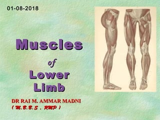
- 1. MusclesMuscles ofof LowerLower LimbLimb DR RAI M. AMMAR MADNIDR RAI M. AMMAR MADNI ( M.B.B.S , RMP )( M.B.B.S , RMP ) 01-08-2018
- 2. Larger and stronger than Upper limb muscles, Function in stability, locomotion, and maintenance of posture - In contrast, upper limb muscles are characterized by versatility of movement. Often cross two joints and can act equally on both. Divided into:- Muscles of Hip, Muscles of Thigh, Muscles of Leg & Muscles of Foot.
- 3. Strengthen and stabilize hip joint, Transmit weight from trunk, upper extremity, and head & neck, Help preserve erect posture, Move the body through space, Provide stable platform for trunk flexion Muscles that produce movement at Hip Joint
- 4. Anterior group Iliopsoas • iliacus • psoas major Psoas minor Tensor fasciae latae Posterior group Gluteus maximus Gluteus medius Gluteus minimus Piriformis Obturator internus Quadratus femoris Obturator externus
- 5. 10-5 Ilio-psoas Major hip flexor, Origin: Psoas major: transverse processes & lateral surface of lumbar vertebrae Iliacus: iliac fossa Insertion: lesser trochanter of femur,crosses hip joint anteriorly, Action: flexes thigh and vertebral column, laterally rotates thigh, keeps the upper body from falling backward when standing erect.
- 7. Muscles of the Gluteal Region that Move the Femur Most muscles that move the femur, originate on the pelvic girdle and insert on the femur. Major muscle groups that move the thigh include the gluteals, and adductor muscles. Most Gluteals laterally rotate the femur; Except → Gluteus minimus, that medially rotates the femur.
- 8. 10-8 Posterior Hip Muscles Gluteus maximus Origin: Gluteal surface of ilium and dorsal aspect of sacrum, Insertion: Gluteal tuberosity of femur and iliotibial tract (band of fascia lata(milky white) attached to the tibia). Action: Extends and laterally rotates thigh at hip joint; raises trunk when the lower limb is fixed Gluteus maximus Iliotibial band Superficial GlutealsSuperficial Gluteals
- 9. 10-9 Posterior Hip Muscles Gluteus medius Origin: Outer surface of ilium, Insertion: Greater trochanter of femur Action: Abducts thigh; Tilts pelvis when walking Gluteus mediusSuperficial GlutealsSuperficial Gluteals
- 10. Posterior Hip Muscles Gluteus minimus; Origin: Outer surface of ilium, Insertion: Greater trochanter of femur Action: * Abducts and medially rotates the femur, * Shifts body weight when foot is lifted;(When R leg is swinging, it holds up the L pelvis by pulling against the fixed thigh on the L side). Gluteus minimus Deep GlutealsDeep Gluteals
- 11. Posterior Hip Muscles Piriformis; Origin: anterior surface of sacrum Insertion: greater trochanter of femur Divides the greater sciatic foramen into suprapiriform foramen and infrapiriform foramen Action: Adducts and rotates thigh laterally at hip joint Piriformis Deep GlutealsDeep Gluteals
- 12. Posterior Hip Muscles Quadratus femoris; Origin: Lateral border of Ischial tuberosity, Insertion: Quadrate tubercle of Femur, Action: Rotates thigh laterally at hip joint. Quadratus femoris Deep GlutealsDeep Gluteals
- 13. Posterior Hip Muscles superior gemellus inferior gemellus obturator externus obturator internus ¤ All have common Insertion on greater trochanteric fossa ¤ All are lateral rotators of thigh. Deep GlutealsDeep Gluteals
- 14. Gluteal Muscles
- 15. gluteus maximus outer surface of ilium, sacrum, coccyx, sacrotuberous ligament iliotibial tract and gluteal tuberosity of femur extends & laterally rotates thigh; through iliotibial tract, it extends knee joint gluteus medius outer surface of ilium greater trochanter of femur abducts thigh. Tilts pelvis when walking gluteus minimus outer surface of ilium greater trochanter of femur abduct thigh; anterior fibers medially rotate thigh tensor fasciae latae iliac crest iliotibial tract assists gluteus major in locking the knee into full extension Quadratus femoris Lateral border of Ischial tuberosity Quadrate tubercle of Femur lateral rotator of thigh piriformis anterior surface of sacrum greater trochanteric fossa lateral rotator of thigh superior gemellus spine of ischium greater trochanteric fossa lateral rotator of thigh obturator internus inner surface of obturator membrane greater trochanteric fossa lateral rotator of thigh inferior gemellus ischial tuberosity greater trochanteric fossa lateral rotator of thigh obturator externus outer surface of obturator membrane greater trochanteric fossa of femur lateral rotator of thigh Muscles of the Gluteal Region
- 16. Muscle compartments of Thigh Deep fascia separate muscles that act on the femur, and tibia and fibula into medial, anterior, and posterior compartments. Medial (Adductor) compartment of the thigh adduct the femur at the hip joint. Anterior (Extensor) compartment of the thigh extend the leg (and flex the thigh). Posterior (Flexor) compartment of the thigh flex the leg (and extend the thigh).
- 17. Muscle Compartments of the Thigh Figure 11.6a
- 18. 10-18 Muscles of thigh MedialMedial (Adductor)(Adductor) groupgroup 5 muscles act as adductors Pectineus Adductor brevis Adductor longus Adductor magnus Gracilis
- 19. Muscles of thigh MedialMedial (Adductor)(Adductor) groupgroup Adductor magnus; is hip joint extensor Pectineus; is hip joint Flexor Gracilis; is flexor of knee Adductor magnus, Adductor longus & Adductor brevis assist in lateral rotation of femur
- 20. Medial Leg Muscles gastrocnemius soleus calcaneal (Achilles) tendon gracilis adductor longus sartoriussartorius adductor magnus
- 21. pectineus superior ramus of pubis upper end shaft of femur flexes and adducts thigh gracilis inferior ramus of pubis; ramus of ischium upper part of shaft of tibia on medial surface adducts thigh and flexes knee adductor longus body of pubis posterior surface of shaft of femur adducts thigh; assists in lateral rotation adductor brevis inferior ramus of pubis posterior surface of shaft of femur adducts thigh; assists in lateral rotation adductor magnus inferior ramus of pubis; ramus of ischium, ischial tuberosity posterior surface of shaft of femur near linea aspera; adductor tubercle of femur adducts thigh and assists in lateral rotation; hamstring part extends thigh MUSCLES OF MEDIAL (ADDUCTOR) COMPARTMENT OF THIGH
- 22. Muscles of thigh AnteriorAnterior (Extensor)(Extensor) compartmentcompartment Sartorius Quadricep Femoris • Rectus femoris • Vastus medialis • Vastus lateralis • Vastus intermedius
- 23. Origin: Anterior superior iliac spine Insertion: Upper medial surface of tibia Action: o Flexes hip and knee joints; o Rotates flexed knee medially. Sartorius Sartorius
- 24. Quadriceps femoris Powerful knee extensor Has four separate heads Origin: Rectus femoris: anterior inferior iliac spine Vastus medialis: medial lip of linea aspera Vastus lateralis: lateral lip of linea aspera Vastus intermedius: anterior surface of femur Insertion: Common insertion at the tibial tuberosity via patellar ligament, Action: Extends leg at knee joint; Rectus femoris also flexes Hip joint
- 25. Anterior Thigh Quadriceps femoris rectus femoris vastus lateralis vastus medials vastus intermedius (under rectus femoris)
- 26. sartorius pectineus (part of) quadriceps tendon patellar ligament Anterior Thigh
- 27. sartorius anterior superior iliac spine upper medial surface of tibia flexes, abducts, laterally rotates thigh; flexes & medially rotates leg iliacus iliac fossa with psoas into the lesser trochanter of femur flexes thigh on trunk; if thigh is fixed, it flexes the trunk onto the thigh as in sitting up psoas 12th thoracic body; transverse process, bodies & intervertebral discs of the 5 lumbar vertebrae lesser trochanter of femur along with iliacus flexes thigh on trunk; if thigh fixed, it flexes trunk onto thigh as in sitting up Anterior Muscles of Thigh
- 28. quadriceps femoris, Rectus femoris straight head from anterior inferior iliac spine; reflected head from ilium above acetabulum quadriceps tendon into patella; into tibial tuberosity by patellar tendon extension of leg quadriceps femoris, Vastus lateralis upper end and shaft of femur quadriceps tendon into patella; into tibial tuberosity by patellar tendon extension of leg quadriceps femoris, Vastus medialis upper end and shaft of femur quadriceps tendon into patella; into tibial tuberosity by patellar tendon extension of leg quadriceps femoris, Vastus intermedius shaft of femur quadriceps tendon into patella; into tibial tuberosity by patellar tendon extension of leg Anterior Muscles of Thigh
- 29. Muscles of thigh PosteriorPosterior (Flexor)(Flexor)compartmentcompartment Hamstring group— Thigh extensors and Knee flexors Biceps femoris Semitendinosus Semimembranosus
- 30. Posterior Thigh semimembranosus gluteus maximus iliotibial band Hamstring group biceps femoris semitendinosus
- 31. biceps femoris long head from ischial tuberosity; short head from shaft of femur head of fibula flexes and laterally rotates leg; long head extends thigh semitendinosus ischial tuberosity upper part medial surface of shaft of tibia flexes and medially rotates leg; extends thigh semimembranosus ischial tuberosity medial condyle of tibia; forms oblique popliteal ligament flexes and medially rotates leg; extends thigh adductor magnus (hamstring part) ischial tuberosity adductor tubercle of femur extends thigh Muscles of Posterior compartment of Thigh
- 32. Fascia lata of the leg surrounds muscles, Tightly binds muscles, Prevents swelling during exercise, Aids venous return, Divides leg into three compartments; anterior, lateral and posterior. Tendons are held in place by Extensor, fibular, and flexor retinacula Muscle movement at ankle and intertarsal joints. Muscles of the Leg
- 33. 10-33 Muscle compartments of Leg Leg (crural) muscles are separated into 3 compartments. Anterior compartment (Red) Lateral (fibular) compartment (Green) posterior (superficial = Pink) (deep = purple)
- 34. Muscles of the Leg Leg muscles, like those of the thigh, are divided by deep fascia into three compartments: anterior, lateral, and posterior. Anterior compartment: Contains digital extensors and foot dorsiflexors Lateral compartment: muscles plantar flex & evert the foot. Posterior compartment: Contains digital and plantar flexors • muscles are split between superficial (e.g., gastrocnemius) and deep (e.g., tibialis posterior) groups. • Superficial muscles share a common tendon of insertion, the calcaneal tendon.
- 35. Frolich, Human Anatomy, Lower LImb Muscle compartments of Leg
- 36. Muscles of Leg Anterior CompartmentAnterior Compartment Tibialis anterior - Dorsiflexes and Inverts foot Extensor hallucis longus - Extension of big toe and ankle Extensor digitorum longus - Extension of toes and ankle Peroneus (Fibularis) tertius - Dorsiflexes and Everts foot
- 37. tibialis anterior shaft of tibia and interosseous membrane medial cuneiform & base of first metatarsal extends the foot; inverts foot at subtalar and transverse tarsal joints; supports medial longitudinal arch extensor digitorum shaft of fibula and interosseous membrane extensor expansion of lateral four toes extends toes; dorsiflexes (extends) foot peroneus tertius shaft of fibula & interosseous membrane base of 5th metatarsal bone dorsiflexes (extends) foot; everts foot at subtalar and transverse tarsal joints extensor hallucis longus shaft of fibula & interosseous membrane base of distal phalanx of big toe extends big toe; dorsiflexes (extends) foot; inverts foot at subtalar and transverse tarsal joints Muscles of Anterior compartment of Leg
- 38. Muscles of Leg Lateral compartmentLateral compartment 22 muscles in this compartment Both plantar flex and evert the foot Provide lift and forward thrust Peroneus longus Peroneus brevis
- 39. Lateral Leg peroneus longus extensor digitorum tibialis anterior retinaculum
- 40. Muscles of the Lateral CompartmentMuscles of the Lateral Compartment
- 41. peroneus longus shaft of fibula base of 1st metatarsal & medial cuneiform plantar flexes foot; everts foot at subtalar & transverse tarsal joints; supports lateral longitudinal and transverse arches of foot peroneus brevis shaft of fibula base of 5th metatarsal bone plantar flexes foot; everts foot at subtalar & transverse tarsal joints; supports lateral longitudinal arch Muscles of Lateral compartment of Leg
- 42. Superficial group Triceps surae • Gastrocnemius • Soleus Plantaris Deep group Popliteus Flexor digitorum longus Flexor hallucis longus Tibialis posterior
- 43. 10-43 Posterior Compartment of Leg Superficial Group (Plantar Flexors)Superficial Group (Plantar Flexors) Gastrocnemius: Flexes knee and plantar-flexes ankle Soleus: Plantar-flexes ankle Plantaris: plantar flexes foot; flexes leg Gastrocnemius SoleusPlantaris
- 44. 10-44 Posterior Compartment of Leg Deep Group (Plantar Flexors)Deep Group (Plantar Flexors) Tibialis posterior, Flexor digitorum longus, Flexor hallucis longus and Popliteus - unlocks the knee joint for knee flexion.
- 45. Posterior Compartment of Leg - SuperficialSuperficial Triceps surae Origin: Gastrocnemius: medial and lateral condyles of femur Soleus: soleal line of tibia and upper third of fibula Insertion: calcaneum via tendo calcaneus Action: flexes knee joint and plantarflexes foot at ankle joint; steadies leg on foot during standing Gastrocnemius soleus
- 46. Posterior Compartment of Leg - DeepDeep Tibialis posterior Origin: posterior surface of tibia and fibula and interosseous membrane Insertion: tuberosity of navicular, all cuniforms Action: plantar-flexes and inverts foot Tibialis posterior
- 47. gastrocnemius medial and lateral condyles of femur by way of Achilles tendon to calcaneum plantar flexes foot; flexes leg plantaris lateral supracondylar ridge of femur calcaneum plantar flexes foot; flexes leg soleus shafts of tibia and fibula by way of achilles tendon into calcaneum with gastrocnemius & plantaris is powerful plantar flexor of foot; provides main propulsive force in walking & running popliteus lateral condyle of femur shaft of tibia flexes leg; unlocks full extension of knee by laterally rotating femur on tibia Muscles of Posterior compartment of Leg
- 48. flexor digitorum longus shaft of tibia distal phalanges of lateral four toes flexes distal phalanges of lateral four toes; plantar flexes foot; supports medial and lateral longitudinal arches of foot flexor hallucis longus shaft of fibula base of distal phalanx of big toe flexes distal phalanx of big toe; plantar flexes foot; supports medial longitudinal arch tibialis posterior shafts of tibia and fibula & interosseous membrane tuberosity of navicular bone plantar flexes foot; inverts foot at subtalar and transverse tarsal joints; supports medial longitudinal arch of foot Muscles of Posterior compartment of Leg
- 49. Intrinsic Muscles of the Foot These muscles are termed intrinsic because they originate & insert within the foot. These muscles are limited designed for Toe movements and support arches of the foot, Split into dorsal and plantar groups. There is a single dorsal foot muscle, the extensor digitorum brevis, which extends the toes 2–5 at the MTP joints. The plantar muscles are arranged in 4 layers, from superficial to deep.
- 50. Dorsal muscle ofDorsal muscle of foot:foot: Extensor digitorum brevis Intrinsic Muscles of the Foot Extensor digitorum brevis
- 51. Planter musclesPlanter muscles First layer • Flexor digitorum brevis • Abductor hallucis • Abductor digiti minimi Intrinsic Muscles of the Foot
- 52. Figure 11.25b Planter musclesPlanter muscles Second layer Flexor accessorius (Quadratus plantæ) Lumbricals Tendon of Flexor digitorum longus Tendon of Flexor hallucis longus Intrinsic Muscles of the Foot
- 53. Flexor hallucis brevis Adductor hallucis Flexor digiti minimi brevis Intrinsic Muscles of the Foot PlanterPlanter musclesmuscles Third layer
- 54. Planter musclesPlanter muscles Fourth layer Intrinsic Muscles of the Foot Dorsal Interossei (dab) meaning dorsal abduct Plantar Interossei (pad) meaning plantar adduct
- 55. Intrinsic Muscles of the Foot Muscle Origin Insertion Action Layer Extensor Digitorum Brevis Anterior part of calcaneus Base of proximal phalanx 1, extensor expansions of toes 2-4 Extend the MPJ Only one on dorsum of foot Flexor Digitorum Brevis Calcaneal tuber Middle phalanx of toes 2-4 Flex toes 1 Abductor Hallucis Tuber calcanei and flexor retinaculum Proximal phalanx of hallux Abducts hallux 1 Abductor digiti minimi Tuber calcanei Lateral side base of little toe proximal phalanx Abducts little toe 1 Quadratus Plantae calcaneus Tendon on FDL Straightens the pull of FDL 2
- 56. Intrinsic Muscles of the Foot Muscle Origin Insertion Action Layer Lumbricales Tendons of FDL Extensor expansion of proximal phalanx of toes 2-5 on medial side Flex at MPJ and extend at IPJ 2 Flexor Hallucis Brevis Lateral cuneiform and cuboid bones Base of proximal phalanx of hallux. Each tendon has asesamoid bone Flex hallux at MPJ 3 Adductor Hallucis Bases of metatarsals 2- 4, Base of proximal phalanx of great toe Weak adductor of great toe 3 Flexor Digiti Minimi Brevis Base of 5th metatarsal Base of 5th toe proximal phalanx Flexes little toe 3 Plantar and Dorsal Interossei Same as interossei of hand Same as interossei of hand Same as interossei of hand 4
- 57. Lower Limb MovementsLower Limb Movements Flexion: Bending on posterior side, except hip Extension: Bending on anterior , except hip Hip Flexion/extension Abduction/adduction Lateral/medial rotation Knee Flexion/extension Ankle Dorsiflexion/plantarflexion Inversion/eversion Toes Flexion/extension
- 58. 58 Common Disorders of MuscularCommon Disorders of Muscular SystemSystem Paralysis - loss of voluntary movement due to neurological damage such as stroke or trauma Multiple sclerosis – weakness of muscles due to loss of covering on nerves that control them. Atrophy – muscle mass decreases in size
- 59. 59 Common Disorders of MuscularCommon Disorders of Muscular SystemSystem(continued)(continued) Contracture - permanent shortening of muscle; joints become ankylosed (frozen) Muscle strain – damage caused by trauma Myalgia - muscle pain Torn muscle - tear caused by trauma
- 60. GET IN TOUCH AT: www.facebook.com/drraiammar www.twitter.com/drraiammar www.instagram.com/drraiammar www.linkedin.com/in/drraiammar www.themedicall.com/blog/auther/drraiammar/ For Any Book or Notes Visit Our Website: www.allmedicaldata.wordpress.com www.drraiammar.blogspot.com https://www.youtube.com/channel/UCu-oR9V3OdFNTJW5yqXWXxA YouTube Channel : DR RAI M. AMMAR MADNIDR RAI M. AMMAR MADNI ( M.B.B.S , RMP )( M.B.B.S , RMP )