
Recommended
Recommended
More Related Content
What's hot
What's hot (18)
Similar to The neuroprotective role of Panax notoginseng saponins in APP/PS1 transgenic mice through the modulation of cerebrovascular
Similar to The neuroprotective role of Panax notoginseng saponins in APP/PS1 transgenic mice through the modulation of cerebrovascular (20)
More from LucyPi1
More from LucyPi1 (20)
Recently uploaded
Recently uploaded (20)
The neuroprotective role of Panax notoginseng saponins in APP/PS1 transgenic mice through the modulation of cerebrovascular
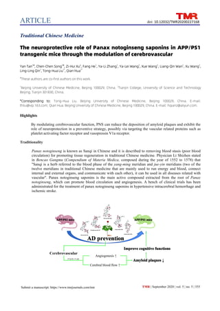
- 1. ARTICLE TMR | September 2020 | vol. 5 | no. 5 | 355 doi: 10.12032/TMR20200227168 Submit a manuscript: https://www.tmrjournals.com/tmr Traditional Chinese Medicine The neuroprotective role of Panax notoginseng saponins in APP/PS1 transgenic mice through the modulation of cerebrovascular Yan Tan1# , Chen-Chen Song1# , Zi-Hui Xu2 , Fang He1 , Ya-Li Zhang1 , Ya-Lei Wang1 , Xue Wang1 , Liang-Qin Wan1 , Xu Wang1 , Ling-Ling Qin1 , Tong-Hua Liu1* , Qian Hua1* # These authors are co-first authors on this work. 1 Beijing University of Chinese Medicine, Beijing 100029, China. 2 Tianjin College, University of Science and Technology Beijing, Tianjin 301830, China. *Corresponding to: Tong-Hua Liu. Beijing University of Chinese Medicine, Beijing 100029, China. E-mail: thliu@vip.163.com; Qian Hua. Beijing University of Chinese Medicine, Beijing 100029, China. E-mail: hqianz@aliyun.com. Highlights By modulating cerebrovascular function, PNS can reduce the deposition of amyloid plaques and exhibit the role of neuroprotection in a preventive strategy, possibly via targeting the vascular related proteins such as platelet activating factor receptor and vasopressin V1a receptor. Traditionality Panax notoginseng is known as Sanqi in Chinese and it is described to removing blood stasis (poor blood circulation) for promoting tissue regeneration in traditional Chinese medicine. Physician Li Shizhen stated in Bencao Gangmu (Compendium of Materia Medica, composed during the year of 1552 to 1578) that "Sanqi is a herb referred to the blood phase of the yang-ming meridian and jue-yin meridians (two of the twelve meridians in traditional Chinese medicine that are mainly used to run energy and blood, connect internal and external organs, and communicate with each other), it can be used in all diseases related with vascular". Panax notoginseng saponins is the main active compound extracted from the root of Panax notoginseng, which can promote blood circulation and angiogenesis. A bench of clinical trials has been administrated for the treatment of panax notoginseng saponins in hypertensive intracerebral hemorrhage and ischemic stroke.
- 2. ARTICLE TMR | September 2020 | vol. 5 | no. 5 | 356 doi: 10.12032/TMR20200227168 Submit a manuscript: https://www.tmrjournals.com/tmr Abstract Background: Panax notoginseng saponins (PNS) is extracted from Sanqi (Panax notoginseng), which is a valuable herb and has been widely used in traditional Chinese medicine for the treatment of cerebrovascular diseases and pain. PNS has been proved to promote blood circulation and angiogenesis by inhibiting platelet aggregation. In our previous study, PNS accompanied with geniposide can prevent Alzheimer’s disease (AD). However, the efficacy of PNS and its potential mechanism in AD remain unclear. Methods: Amyloid precursor protein/presenilin-1 (APP/PS1) transgenic (Tg) mice were used as AD-like animal models. Wild-type mice and APP/PS1 transgenic were administrated with saline solution while mice in PNS treatment group were administrated with PNS at a dosage of 17 mg/kg/day for three months. Morris water maze (MWM) was applied to evaluate the spatial learning and memory and step-down test was used to evaluate the cognitive function. 1% Thioflavin-S staining was used to calculate the average number amyloid plaques in cortex and hippocampus. CD31 staining was detected to observe the density of cerebrovascular in hippocampus areas and CD105 staining was further detected to evaluate angiogenesis. Laser Doppler PeriFlux 5000 was further measured the change of cerebrovascular blood flow. ChemDraw was used to draw the molecular structures of five main ingredients of PNS. AlzPlatform were used to estimate the potential targets of PNS. Results: By a bench of behavioral tests, PNS showed a better tendency in proving cognitive functions. In addition, the amyloid plaques in both cortex and hippocampus were significantly reduced after PNS intervention (P < 0.05 and P < 0.001 respectively). Furthermore, the density of cerebrovascular in the hippocampus areas was increased under PNS administration (P < 0.001), which accompanied with angiogenesis in dentate gyrus areas and cerebrovascular blood flow promotion (P < 0.05). By AlzPlatform docking serve, we screened five major ingredients of PNS—R1, Rd, Rb1, Re and Rg1. These screening data suggested that vascular related proteins could be the one of potential targets of PNS, such as platelet activating factor receptor and vasopressin V1a receptor. Conclusion: By modulating cerebrovascular function, PNS can reduce the deposition of amyloid plaques and exhibit the role of neuroprotection in a preventive strategy. Keywords: Alzheimer’s disease, Angiogenesis, Behavioral test, Cerebrovascular blood flow, Panax notoginseng saponins Acknowledgments: This study was supported by grants of National Natural Science Foundation of China Project (Project No. 81904049), Regional Collaborative Innovation Center of Tibetan Medicine (Project No. 2017XTCX012, 2018XTCX014) and Young Elite Scientists Sponsorship Program by CAST (Project No. CACM-2018-QNRCC2-C06). Abbreviations: PNS, Panax notoginseng saponins; AD, Alzheimer’s disease; APP/PS1, amyloid precursor protein/presenilin-1; Tg, transgenic; MWM, morris water maze; Aβ, amyloid-β; TCM, Traditional Chinese Medicine; GP, geniposide; WT, wild-type; PU, perfusion unit; HTDocking, high-throughput docking; DG, dentate gyrus; PAFR, platelet activating factor receptor; V1AR, vasopressin V1a receptor. Competing interests: There are no conflicts of interest. Citation: Yan Tan, Chen-Chen Song, Zi-Hui Xu, et al. The neuroprotective role of Panax notoginseng saponins in APP/PS1 transgenic mice through the modulation of cerebrovascular. Traditional Medicine Research 2020, 5 (5): 355–367. Executive editor: Nuo-Xi Pi. Submitted: 24 December 2019, Accepted: 24 February 2020, Online: 23 March 2020.
- 3. ARTICLE TMR | September 2020 | vol. 5 | no. 5 | 357 Submit a manuscript: https://www.tmrjournals.com/tmr doi: 10.12032/TMR20200227168 Background Alzheimer disease (AD) is an age-related neurodegenerative disorder, the progress of which usually starts slowly and worsens over time [1]. At the early stage of AD, patients usually have no evident clinical syndromes with normal cognition. But according to epidemic statistics, a series of pathophysiological changes observed at preclinical phase of AD, such as the gradual accumulation of amyloid-β (Aβ), the changes of cerebral hemodynamics and hippocampal atrophy [2, 3]. Since the failure of the present clinical trials target on later stages of AD, the treatment window should be re-considered to shift to an early stage. Currently, Food and Drug Administration (FDA) proposed that “in the clinical development of drugs for the treatment of the stages of sporadic AD that occur before the onset of overt dementia (collectively referred to as early AD …)…” in Early Alzheimer’s Disease: Developing Drugs for Treatment published in February 2018. This guideline provides us two important issues: preventive strategies and early diagnosis. In China, Traditional Chinese medicine (TCM) has a long history in preventing and treating cognitive decline [4–6]. Panax notoginseng is known as Sanqi in Chinese, the root of which has plenty of medicinal properties. In TCM, it is described that “removing blood stasis (poor blood circulation) for promoting tissue regeneration”, and physician Li Shizhen stated in Bencao Gangmu (Compendium of Materia Medica, composed during the year of 1552 to 1578) that "Sanqi is a herb referred to the blood phase of the yang-ming meridian and jue-yin meridians (two of the twelve meridians in traditional Chinese medicine that are mainly used to run energy and blood, connect internal and external organs, and communicate with each other), it can be used in all diseases related with vascular"; he also called Sanqi was “not to be exchanged even for gold”, showing its preciousness and importance in medication. Panax notoginseng saponins (PNS) is the main active compound extracted from the root of Panax notoginseng, which can promote blood circulation and angiogenesis [7, 8]. PNS has the effect of inhibiting platelet aggregation, increasing the heart and cerebral blood flow and plays a protective role in cardiovascular and cerebrovascular diseases [7, 9]. A bench of clinical trials has been administrated for the treatment of PNS in hypertensive intracerebral hemorrhage and ischemic stroke (Table 1). In addition, in our previous study, we found PNS accompanied with geniposide (GP) prevented cognitive decline in different AD animal models [10, 11]; in vitro, PNS protected endothelial cells from oxidative stress, suggesting that the neuroprotection role of PNS may work on vascular. Here, in order to evaluate the preventive role of PNS in AD, we made use of both virtual screening methods and amyloid precursor protein/presenilin-1 (APP/PS1) transgenic (Tg) mice to explore its potential targets and efficacy in neuroprotection. Materials and methods Animal APP/PS1 Tg mice were used as AD-like animal models. The double Tg mice expressing a human amyloid precursor protein (HuAPP695swe) and a mutant human presenilin 1 (PS1-dE9), both associated with early-onset AD. The typical pathological products, such as Aβ40 and Aβ42 were gradually accumulated around 4-month age; the deposition of amyloid plaques can be detected around 6-month age. Accordingly, the cognitive impairments can be observed at 7-month age [12, 13]. This model is useful in studying neurological disorders of the brain, specifically AD and amyloid plaque formation. In this study, 4-month age of APP/PS1 Tg mice and their littermates of wild-type (WT) mice were purchased from Model Animal Research Center of Nanjing University (Cat. No. N000175). These Tg mice were randomly divided into two groups—APP/PS1 group and PNS treatment group (n = 12). Littermate WT mice were considered as control, WT group (n = 14). The animals were maintained at a constant temperature on a 12-h light-dark cycle with accessing to food and water freely. All procedures concerning care, treatment and dissection are in accordance with the Animal Ethics Committee of Beijing University of Chinese Medicine (No. BUCM-2016103101-1008). Administration Based on previous studies [11], the dosage of PNS was converted according to body weight index between human being and mouse, orally administrating with a dosage of 17 mg/kg/day dissolved in saline solution. WT and APP/PS1 groups were administrated with same volumes of saline solution. A preventive strategy has been applied that the administration started at four-month age, and lasting for 3-month. PNS was purchased from National Institute for the Control of Pharmaceutical and Biological Products (Cat. No. 84527, UV95%, China). Morris water maze To evaluate hippocampal dependent spatial learning and memory, mice were tested in a standard morris water maze (MWM) task. Briefly, mice were trained 4 trials per day in 6 consecutive days. During the training section, mice were placed on the hidden platform for 10 sec at the end of each trial. 24 hours after the training section, a testing trial was carried out with the platform removed. Mice started the test from the opposite quadrant, and escape latency, percentage of dwelling time in the target quadrant were measured.
- 4. ARTICLE TMR | September 2020 | vol. 5 | no. 5 | 358 doi: 10.12032/TMR20200227168 Submit a manuscript: https://www.tmrjournals.com/tmr Step-down test To evaluate the fear memory, mice were tested in the step-down test. A inhibitory avoidance apparatus was used. Briefly, during training test, mice were placed into the apparatus for a 5-minute habituation. Then, an electric current of 36V was delivered to the copper grid, and lasted for another 5 min. The escape latency to step up to the platform with all paws was measured. 48 hours after the training test, a retention test was carried out without electronic shock. The escape latency of step down to the floor with all paws was measured. Brain tissue preparation Mice were deeply anesthetized with a cocktail of 1.25% avertin (0.6 mL/20 g) via intraperitoneal injection, and transcardially perfused with ice-cold phosphate-buffered saline, followed by ice-cold 4% paraformaldehyde (PFA) in PBS. Brains were carefully eviscerated and placed in 4% PFA at 4°C for overnight, following by 15% sucrose and 30% sucrose at 4℃ for overnight. Brains were embedded with OCT compound (Tissue Tek, Japan) in cryostat, and coronal cut at 10 μm thickness and mounted on positively charged microscope slides. The slides were kept at -80°C until used. Amyloid plaques deposition Cryostat slices prepared above were collected and stained by 1% Thioflavin-S (Sigma, Cat. No. T1867, USA). Briefly, three consecutive sections at three positions (15 slices interval) were picked, and calculated the average number amyloid plaques (in cortex and hippocampus, respectively) of each position, as average (Ave) 1, Ave 2 and Ave 3. Then, a sum total from Ave 1 to Ave 3 was calculated as one piece of data for one hemisphere. There were three animals in each group. All images were obtained by inverted fluorescence microscope (Olympus, CKX41, Japan). 10× or 20× objective lens have been used for each field. The strategy for data collection and calculation per animal as Figure 1. Figure1 A brief strategy for data collection and calculation per animal. Upper panel: sagittal plates were collected for amyloid plaque deposition (left-up corner). On the right hemisphere, we targeted on the parietal-temporal lobes and the hippocampus beneath (red box). Every 15 slices interval, 3 positions were collected. Each position was picked serial three slices. Lower panel: average number of each position was calculated, and Ave 1 for position 1, Ave 2 for position 2, and Ave 3 for position 3. Then, a sum total from Ave 1 to Ave 3 was calculated as one piece of data as one hemisphere. A, anterior; P, posterior; Ave, average.
- 5. ARTICLE TMR | September 2020 | vol. 5 | no. 5 | 359 Submit a manuscript: https://www.tmrjournals.com/tmr doi: 10.12032/TMR20200227168 Table 1 Clinical trials list of PNS mainly in cerebrovascular diseases Clinical trials. government identifier Condition or disease Intervention/treatment Phase Study completion date NCT02999048 Hematoma absorption and neurological function recovery Drug: PNS Phase 4 May 2016 NCT01636154 Ischemic stroke Drug: Ixeris of sonchifolia Hance Drug: PNS Drug: Ixeris of sonchifolia Hance combined with PNS Not applicable August 2016 NCT02544087 Ischemic stroke Drug: Ixeris of sonchifolia Hance Drug: PNS Drug: Ixeris of sonchifolia Hance combined with PNS Not applicable April 2018 PNS, Panax notoginseng saponins. Immunofluroscence staining Cryostat slides were prepared and incubated with CD31 or CD105 primary antibody (Abcam, Cat. No. ab24590 and ab107595, host in rabbit, USA) at a dilution of 1:100 at 4℃ overnight, and followed by Alexa Fluor488 labeled anti-rabbit secondary antibody (Thermo Fisher, Cat. No. A11008, USA) at room temperature for 1 h. The sections were mounted with DAPI (Solarbio, Cat. No. 28718-90-3, China). All images were obtained by inverted fluorescence microscope (Olympus, CKX41, Japan). 20× or 40× objective lens have been used for each field. Cerebral blood flow Mice were deeply anesthetized with isoflurane (1.0 L /min). Brains were fixed by stereotaxic apparatus, and local dissected to expose the skull of brains. The detector of Laser Doppler flowmetry (Perimed AB, PeriFlux System5000, German) was placed in the midline of the brain, then right-handed adjust to 2 mm and pointed on the right branch of superior saggital sinus on the right hemisphere. A continuous measurement for 120 seconds was collected to analysis cerebrovascular perfusion, and analyzed as perfusion unit (PU). The fluctuation of cerebral blood flow was further calculated. Ten points in every 12-second interval were picked. Each point contained 3-second long, and the difference values of maximum and minimum PU (∆PU) in these 3-second were calculated to evaluate the fluctuation of blood flow. Target prediction ChemDraw was used to draw the molecular structures of five main ingredients of PNS—notoginsenoside R1, ginsenoside Rb1, ginsenoside Rg1, ginsenoside Rd and ginsenoside Re. Both Corey-Padi-Koltun molecular models and 2D molecular structures were provided at a situation of their minimal energy statements. For virtual screening, AlzPlatform has been used to estimate the potential targets of PNS in the treatment of AD. AlzPlatform is an AD domain-specific database for target identification [14]. It is a free web-based computing tool to explore interactions between compounds and proteins. In the program of high-throughput docking (HTDocking) (http://www.cbligand.org/AD/docking_search. php), it automates docking procedure to search for targets between compound and protein. Briefly, water molecules and ligands were removed, hydrogen atoms were added. By using AutoDock utility scripts, the active sites of each protein were defined. AutoDock provides predicted binding affinity values (ΔG values) from different docking poses for each compound in a binding pocket of a protein. The overall score of a queried compound from each protein structure is used to assess and rank the potential protein partners or targets. Statistical analysis All data are reported as mean ± SEM. Paired or unpaired Student’s t-test (for two group means) or one or two-way analysis of variance (ANOVA) with post-hoc Tukey test, Scheffe’s test, or Bonferroni test, as appropriate, were conducted using the SPSS 17.0. The significance level for the two-sided analyses was set at P ≤ 0.05. Prism and Photoshop Illustrator were used for photographing. Results PNS improves cognitive functions of 7-month age of APP/PS1 Tg mice in both MWM and step-down test MWM was applied to evaluate the spatial learning and
- 6. ARTICLE TMR | September 2020 | vol. 5 | no. 5 | 360 doi: 10.12032/TMR20200227168 Submit a manuscript: https://www.tmrjournals.com/tmr memory. In the training trials, all the groups showed a progress in spatial learning at the first three days, but the deficits occurred at day 4 in APP/PS1 Tg mice. Compared with Tg mice, both WT and PNS groups significantly shortened the latency to platform (P < 0.001, P < 0.05, Figure 2a). In the testing trial, the time percentage in target quadrant has been measured. WT group showed a significant longer occupancy compared with Tg mice (P < 0.05), while there is a longer tendency in PNS groups but no significant change (Figure 2b, P = 0.068). Step-down test also has been used to evaluate the cognitive functions. 48h after the training trial, only WT mice maintained a significant longer latency than Tg mice (P < 0.05, Figure 2c). But, when measuring the percentage of animals in the safe zone up to 5 min, there is only 64.3% of Tg mice maintained the shock memory; both WT and PNS groups showed better performance—more than 85% of them maintained the shock memory (Figure 2d). Taken together, at 7-month age, the cognition is impaired in Tg mice; PNS can prevent the deficit in both spatial learning and fear memory. Figure 2 PNS improves cognitive functions of 7-month age of APP/PS1 Tg mice. a. During the 6-day training of MWM, all groups showed a reduced latency to platform at the first three days, but PNS and WT groups maintained the progress in the following days of training. Compared with APP/PS1 Tg in the training test, PNS and WT showed significant reduced latency to platform. b. In the probe test of MWM, the percentage in target quadrant was significantly higher in WT group compared with Tg mice. Although there was no significant change in PNS group, it enjoyed a better tendency in spatial memory (P = 0.068). c. In the step-down test after training, WT mice showed significant longer latency compared with Tg mice. d. 48h after step-down test training, the number of animals remained fear memory up to 5 min was measured. Like WT mice, PNS could maintain the number of animals to 86.7%, while Tg group only enjoyed a percentage of 64.3. Data are represented as mean ± SEM. *P < 0.05, ***P < 0.001. PNS, Panax notoginseng saponins; APP/PS1, amyloid precursor protein/presenilin-1; Tg, transgenic; MWM, morris water maze; WT: wild-type mice.
- 7. ARTICLE TMR | September 2020 | vol. 5 | no. 5 | 361 Submit a manuscript: https://www.tmrjournals.com/tmr doi: 10.12032/TMR20200227168 PNS reduces the number of amyloid plaques in both cortex and hippocampus In previous studies, we have proved that the combination of PNS and GP can significantly reduce the number of amyloid plaques in brains of several AD animal models [11, 15, 16]. In order to illustrate if PNS contributes to this efficacy, amyloid plaques deposits has been detected. By 1% Thioflavin-S staining, we chose three-consisting slices of every 10–15 slice interval, to estimate the deposition of amyloid plaques in both cortex and hippocampus areas. In WT mice, there was no amyloid plaque observed, while there were amount of plaque deposition in both cortex and hippocampus areas. Compared with Tg mice, PNS significantly reduced the number of plaques deposition (P < 0.05, P < 0.01, Figure 3). This result indicates that PNS can contribute the clearance of amyloid plaques in AD brains. Figure 3 PNS reduces the number of amyloid plaques in both cortex and hippocampus. By Thioflavin-S staining, there was no deposition of amyloid plaques detected in WT mice. Compared with APP/PS1 Tg mice, the number of amyloid deposits was significantly reduced under the treatment of PNS in both cortex and hippocampus areas. Data are represented as mean ± SEM. *P < 0.05, **P < 0.01, ***P < 0.001. Scale bar: 100µm. PNS, Panax notoginseng saponins; WT, wild-type mice; APP/PS1, amyloid precursor protein/presenilin-1; Tg, transgenic.
- 8. ARTICLE TMR | September 2020 | vol. 5 | no. 5 | 362 doi: 10.12032/TMR20200227168 Submit a manuscript: https://www.tmrjournals.com/tmr PNS promotes angiogenesis and cerebral blood flow in APP/PS1 Tg mice Since PNS is used in cerebrovascular diseases, we next want to detect if PNS can work on cerebrovascular function in APP/PS1 Tg mice. CD31 refers to a typical marker of the vascular. The structure of cerebral vascular in the hippocampus was detected. Compared with WT mice, the density of vessels was reduced in Tg mice; while PNS reversed the phenomena (Figure 4a). In addition, since CD105 is widely considered as a crucial role in angiogenesis, we next want to detect if the increased density of vessels is contributed by angiogenesis. By CD105 immunofluorescence staining, the number of CD105 positive cells was measured. In the dentate gyrus (DG) area of hippocampus, compared with Tg mice, the number of CD105 positive cells was significantly increased under the treatment of PNS (P < 0.001), which is identical to WT groups (Figure 4b). Figure 4 PNS increases the density of cerebrovascular and the number of CD105 positive cells in the hippocampus areas. a. CD31 staining was detected to observe the density of cerebrovascular in hippocampus areas. From the overview, the density of vessels was evidently reduced in APP/PS1 mice compared with WT mice, while PNS reversed the phenomena. Scale bar: 500 µm. b. CD105 staining was further detected to evaluate angiogenesis. In APP/PS1 Tg mice, few CD105 positive cells was detected (arrow indicator). Compared with Tg group, the number of CD105 positive cells was significantly increased in both WT and PNS mice.Scale bar: 50µm. c. The number of CD105 positive cells was counted. Data are represented as mean ± SEM. ***P < 0.001. PNS, Panax notoginseng saponins; APP/PS1, amyloid precursor protein/presenilin-1; WT, wild-type mice; Tg, transgenic.
- 9. ARTICLE TMR | September 2020 | vol. 5 | no. 5 | 363 Submit a manuscript: https://www.tmrjournals.com/tmr doi: 10.12032/TMR20200227168 By Laser Doppler PeriFlux 5000, we further measured the change of cerebrovascular blood flow in the right branch of superior saggital sinus, sketched according to Dorr’s work [17]. Within 120-second of whole view recording, the blood fluctuation was much smoother in WT mice, which indicated a normal vasoconstriction; however, Tg mice showed a severe variation. In addition, the blood fluctuation was calculated within 3 seconds of every 12 seconds interval. Again, compared with Tg mice, PNS significantly smoothed the vasoconstriction (Figure 5a). Compared with Tg mice, cerebrovascular blood perfusion was significantly higher in both WT and PNS groups (P < 0.001, P < 0.05, Figure 5b, 5c). Taken together, under a three-month treatment of PNS, the angiogenesis and cerebral blood flow are promoted at 7-month age of APP/PS1 mice. By virtual screening servers, vascular related proteins are found to be the potential targets of PNS Since PNS can work on cerebrovascular function, we next want to further explore if vascular related protein is its potential target. Firstly, by ChemDraw software, the structure of R1, Rd, Rb1, Re and Rg1, the five major ingredients of PNS has been drawn. At their minimal energy statements, both CPK molecular models and 2D molecular structures are provided (Figure 6a). Secondly, potential targets was predicted by a free serve for AD drugs. AlzPlatform (www.cbligand.org/AD/) provides a user-friendly interface with a HTDocking program on AD chemicals and targets. By docking analysis, , the five ingredients were input into the interface separately. Overall score refers to the degree of feasibility. We found that thyroid stimulating hormone receptor (TSHR), muscarinic acetylcholine receptor M1-5, platelet activating factor receptor (PAFR) and vasopressin V1a receptor (V1AR) were always listed on the top 10 of target identification (Figure 6b), suggesting that vascular related proteins could be the potential targets of PNS. Figure 5 PNS promotes cerebrovascular functions in 7-month age of APP/PS1 Tg mice. a. A graph was showed the perfusion and fluctuation of right branch of superior saggital sinus. Compared with APP/PS1 Tg and PNS groups, the fluctuation was more smoother in WT mice within 120 sec recording. b. To measure the cerebral blood flow, the signal of PU was qualified. Compared with Tg mice, the signal was significantly increased in both WT and PNS group. c. Since the changes of vasoconstriction was observed in recording. The fluctuation was measured within 3sec every 12sec interval. Again, both WT and PNS significantly smoothed the vasoconstriction compared with Tg mice. Data are represented as mean ± SEM. (*P < 0.05, ***P < 0.001). PNS, Panax notoginseng saponins; APP/PS1, amyloid precursor protein/presenilin-1; Tg, transgenic; WT, wild-type mice; PU, perfusion unit; ∆PU, the difference values of maximum and minimum PU.
- 10. ARTICLE TMR | September 2020 | vol. 5 | no. 5 | 364 doi: 10.12032/TMR20200227168 Submit a manuscript: https://www.tmrjournals.com/tmr Figure 6 The structures of five major ingredients of PNS and the potential targets of PNS. A. There are five main ingredients of PNS—R1, Rd, Rb1, Re and Rg1, by ChemDraw, the CPK molecular models and 2D molecular structures were provided as their minimal energy statement. B. By THDocking, vascular-related proteins were found to be the potential targets of PNS. PAFR and VIAR were in the top ten identifications. Overall score indicated the degree of feasibility based on THDocking database. PNS, Panax notoginseng saponins; TSHR, thyroid stimulating hormone receptor; PAFR, platelet activating factor receptor; VIAR, vasopressin V1a receptor. Discussions Currently, cerebrovascular diseases are getting increased attention. In clinic, insufficient cerebral perfusion is closely related with cognition impairment [18]. According to long-term clinical observation, Torre addressed that neurodegeneration could be caused by cerebrovascular diseases, and he pointed that insufficient cerebral perfusion could be considered as a diagnostic indicator of AD in clinic [19]. Further, Zlokovic has proposed “neurovascular hypothesis” — the dysfunction of neurovascular units could lead to neurological dysfunctions, which would result in neuron damage or even cognition impairment [20]. These suggest that cerebrovascular dysfunction is occurred on the early stage of AD and closely related with AD processing; it can be a new drug target of AD. Since there is no effect drugs anti-AD, when we come back to TCM, which has a long history prescribed in oriental countries, in China, a widely used herb, PNS has been administrated in the diseases of vascular dysfunctions, such as hemorrhage, stroke, pain [21, 22]. In our previous studies [11], accompanied with GP, the combination has been administrated for acute strokes and improved the cognition of stroke patients with memory deficits. In addition, we found the combination could improve impaired cognition in several AD animal models [11, 15, 16]. In vitro, the combination could protect vascular endothelial cells from oxidative stress [23]. These results indicate that PNS and its combination have a neuroprotective role in AD. Therefore, in order to further explore the efficacy and its potential targets of PNS, especially in preventive strategy, we mainly focused on the changes of vascular functions in both animal model and computer simulation technology. In vivo, we focused on cerebrovascular function of PNS in AD-like animal model. In behavioral tests, PNS could improve cognitive functions of spatial learning and fear memory; in addition, the number of amyloid plaques deposition was significantly reduced in both cortex and hippocampus in PNS group. Furthermore, under the three-month long administration, angiogenesis especially in hippocampus DG areas was observed. Adult neurogenesis contributes to hippocampal plasticity [24]. Accumulating evidence suggests that adult-born neurons in this area play distinct physiological roles in memory encoding and mood regulation [25]. Angiogenesis may control function of neuronal stem cells via VEGF factors [26]. VEGF shows a role in
- 11. ARTICLE TMR | September 2020 | vol. 5 | no. 5 | 365 Submit a manuscript: https://www.tmrjournals.com/tmr doi: 10.12032/TMR20200227168 learning and memory improvement by VEGF-induced modulations of neuronal activity [27]. What’s more, cerebral blood perfusion was significantly higher compared with APP/PS1 mice, and the fluctuation of blood flow was significantly smoother. These data suggest that PNS recovers the cerebrovascular function—angiogenesis and cerebrovascular blood flow in APP/PS1 Tg mice. There are five main ingredients of PNS—R1, Rd, Rb1, Re and Rg1. By HTDocking tools [14], we further found that its main gradients might work on two vascular-related proteins, PAFR and V1AR. In addition, according to PHAROS server (www.pharos.nih.gov/idg/targets), these two proteins might work on APP, which is the precursor protein of Aβ. In addition, V1AR is one of major receptors for vasopressin, highly present throughout brain, such as cortex, hippocampus, hypothalamus and adrenal gland [28]. Besides working on APP, V1AR also had a potential effect on one of subunits of γ-secretase—Aph1, screening by PHAROS server. Further, 20-year ago, it was found that there was a significant correlation between PAF binding and degree of cognitive impairment in AD patients [29]; in depressed coronary artery disease patients, platelet-activating factors were associated with cognitive deficits [30]; in addition, V1AR was indispensable for normal social recognition and cognitive empathy [31, 32]. By other virtual screening serves, such as ChamMapper and PharmMapper, VEGF receptors are also the top-level of potential targets of PNS, as well as other neuron-related proteins, such as dopamine receptors, estrogen receptor. However, computer simulation can provide an assistant analysis in target identification, but a validation required [33]. Conclusions PNS has been used for the treatment of intracerebral hemorrhage and ischemic stroke, and improves cognition deficits of stroke patients as well as in a bench of AD animal models. In this study, we find that PNS can partially contribute to the improvement of cognitive functions in 7-month age of APP/PS1 mice. In addition, PNS can promote angiogenesis in DG areas of hippocampus and cerebrovascular blood flow. These results indicate that the contribution of PNS in neuroprotection is via the modulation of cerebrovascular functions (Figure 7). Figure 7 The hypothesis of neuroprotective role of PNS in cerebrovascular targets. Based on experimental studies and virtual screening, the neuroprotective role of PNS may work on cerebrovascular to promote vascular function, the latter of which could lead to the reduction of amyloid plaques and eventually result in AD prevention. PNS, Panax notoginseng saponins; AD, Alzheimer’s disease.
- 12. ARTICLE TMR | September 2020 | vol. 5 | no. 5 | 366 doi: 10.12032/TMR20200227168 Submit a manuscript: https://www.tmrjournals.com/tmr References 1. Apostolova LG. Alzheimer Disease. Continuum 2016, 22: 419–434. 2. Ewers M, Sperling RA, Klunk WE, et al. Neuroimaging markers for the prediction and early diagnosis of Alzheimer's disease dementia. Trends Neurosci 2011, 34: 430–442. 3. Solomon A, Mangialasche F, Richard E, et al. Advances in the prevention of Alzheimer's disease and dementia. J Intern Med 2014, 275: 229–250. 4. Zeng Q, Li L, Jin Y, et al. A network pharmacology approach to reveal the underlying mechanisms of Paeonia lactiflora Pall. on the treatment of Alzheimer’s disease. Evid-Based Compl Alt 2019. 5. Qin W, Chen S, Yang S, et al. The effect of traditional Chinese medicine on neural stem cell proliferation and differentiation. Aging Dis 2017, 8: 792–811. 6. Peng A, Zhuang X, Gao Y, et al. Bazhu decoction, a traditional Chinese medical formula, ameliorates cognitive deficits in the 5xFAD mouse model of Alzheimer's disease. Front Pharmacol 2019, 10: 1391. 7. Dong Y, Duan L, Chen H, et al. Network pharmacology-based prediction and verification of the targets and mechanism for Panax notoginseng saponins against coronary heart disease. Evid-Based Compl Alt 2019. 8. Wang D, Jie Q, Liu B, et al. Saponin extract from Panax notoginseng promotesangiogenesis through AMPK‑ and eNOS‑ dependent pathways in HUVECs. Mol Med Rep 2017, 16: 5211–5218. 9. Liu Y, Weng W, Gao R, et al. New insights for cellular and molecular mechanisms of aging and aging-related diseases: herbal medicine as potential therapeutic approach. Oxid Med Cell Longev 2019. 10. Klenerova V, Hynie S, Li XJ, et al. Antianxiety effect of Tong Luo Jiu Nao, traditional Chinese medicinal preparation, in rats. J Appl Biomed 2015, 13: 33–38. 11. Yang K, Tan Y, Wang F, et al. The improvement of spatial memory deficits in APP/V717I transgenic mice by chronic anti-stroke herb treatment. Exp Biol Med 2014, 239: 1007–1017. 12. Jankowsky JL, Fadale DJ, Anderson J, et al. Mutant presenilins specifically elevate the levels of the 42 residue β-amyloid peptide in vivo: evidence for augmentation of a 42-specific γ secretase. Hum Mol Genet 2004, 13: 159–170. 13. Guo HB, Cheng YF, Wu JG, et al. Donepezil improves learning and memory deficits in APP/PS1 mice by inhibition of microglial activation. Neurosci 2015, 290: 530–542. 14. Liu H, Wang L, Lv M, et al. AlzPlatform: an Alzheimer’s disease domain-specific chemogenomics knowledgebase for polypharmacology and target identification research. J Chem Inf Model 2014, 54: 1050–1060. 15. Liu Y, Hua Q, Lei H, et al. Effect of Tong Luo Jiu Nao on Aβ-degrading enzymes in AD rat brains. J Ethnopharmacol 2011, 137: 1035–1046. 16. He P, Li P, Hua Q, et al. Chronic administration of anti-stroke herbal medicine TongLuoJiuNao reduces amyloidogenic processing of amyloid precursor protein in a mouse model of Alzheimer’s disease. Plos One 2013, 8: e58181. 17. Dorr A, Sled JG, Kabani N. Three-dimensional cerebral vasculature of the CBA mouse brain: a magnetic resonance imaging and micro computed tomography study. NeuroImage 2007, 35: 1409–1423. 18. Kisler K, Nelson A R, Montagne A, et al. Cerebral blood flow regulation and neurovascular dysfunction in Alzheimer disease. Nat Rev Neurosci 2017, 18: 419–434. 19. de la Torre J C. Are major dementias triggered by poor blood flow to the brain? Theoretical considerations. J Alzheimers Dis 2017, 57: 353–371. 20. Zlokovic BV. Neurovascular pathways to neurodegeneration in Alzheimer's disease and other disorders. Nat Rev Neurosci 2011, 12: 723–738. 21. Yang X, Xiong X, Wang H, et al. Protective effects of Panax notoginseng saponins on cardiovascular diseases: a comprehensive overview of experimental studies. Evid-Based Compl Alt 2014. 22. Xu D, Huang P, Yu Z, et al. Efficacy and safety of Panax notoginseng saponin therapy for acute intracerebral hemorrhage, meta-analysis, and mini review of potential mechanisms of action. Front Neurol 2015, 5: 274. 23. Li XJ, Hou JC, Sun P, et al. Neuroprotective effects of Tong Luo Jiu Nao in neurons exposed to oxygen and glucose deprivation. J Ethnopharmacol 2012, 141: 927–933. 24. Toda T, Gage FH. Adult neurogenesis contributes to hippocampal plasticity. Cell Tissue Res 2018, 373: 693–709. 25. Toda T, Parylak SL, Linker SB, et al. The role of adult hippocampal neurogenesis in brain health and disease. Mol Psychiatry 2019, 24: 67–87. 26. Licht T, Keshet E. The vascular niche in adult neurogenesis. Mech Development 2015, 138: 56–62. 27. Licht T, Goshen I, Avital A, et al. Reversible modulations of neuronal plasticity by VEGF. Proc Natl Acad Sci 2011, 108: 5081–5086. 28. Meyer-Lindenberg A, Kolachana B, Gold B, et al.
- 13. ARTICLE TMR | September 2020 | vol. 5 | no. 5 | 367 Submit a manuscript: https://www.tmrjournals.com/tmr doi: 10.12032/TMR20200227168 Genetic variants in AVPR1A linked to autism predict amygdala activation and personality traits in healthy humans. Mol Psychiatry 2009, 14: 968–975. 29. Hershkowitz M, Adunsky A. Binding of platelet-activating factor to platelets of Alzheimer's disease and multiinfarct dementia patients. Neurobiol Aging 1996, 17: 865–868. 30. Mazereeuw G, Herrmann N, Xu H, et al. Platelet-activating factors are associated with cognitive deficits in depressed coronary artery disease patients: a hypothesis-generating study. J Neuroinflammation 2014, 11: 119. 31. Bielsky IF, Hu SB, Ren X, et al. The V1a vasopressin receptor is necessary and sufficient for normal social recognition: a gene replacement study. Neuron 2005, 47: 503–513. 32. Uzefovsky F, Shalev I, Israel S, et al. Oxytocin receptor and vasopressin receptor 1a genes are respectively associated with emotional and cognitive empathy. Horm Behavior 2015, 67: 60–65. 33. Chen XP, Du GH. Target validation: a door to drug discovery. Drug Discov Ther 2007, 1: 23–29.