SlideShare a Scribd company logo
1 of 14
Download to read offline
P E D I A T R I C C A R D I O L OG Y
Neuregulin stimulation of cardiomyocyte regeneration
in mice and human myocardium reveals a
therapeutic window
Brian D. Polizzotti,1,2
* Balakrishnan Ganapathy,1,3
* Stuart Walsh,1,2
*†
Sangita Choudhury,1,2
*
Niyatie Ammanamanchi,1,3
David G. Bennett,4‡
Cristobal G. dos Remedios,5
Bernhard J. Haubner,6
Josef M. Penninger,6
Bernhard Kühn1,2,3,7,8,9§
Therapies developed for adult patients with heart failure have been shown to be ineffective in pediatric clinical
trials, leading to the recognition that new pediatric-specific therapies for heart failure must be developed. Ad-
ministration of the recombinant growth factor neuregulin-1 (rNRG1) stimulates regeneration of heart muscle
cells (cardiomyocytes) in adult mice. Because proliferation-competent cardiomyocytes are more abundant in
growing mammals, we hypothesized that administration of rNRG1 during the neonatal period might be more
effective than in adulthood. If so, neonatal rNRG1 delivery could be a new therapeutic strategy for treating
heart failure in pediatric patients. To evaluate the effectiveness of rNRG1 administration in cardiac regenera-
tion, newborn mice were subjected to cryoinjury, which induced myocardial dysfunction and scar formation
and decreased cardiomyocyte cell cycle activity. Early administration of rNRG1 to mice from birth to 34 days of
age improved myocardial function and reduced the prevalence of transmural scars. In contrast, administration
of rNRG1 from 4 to 34 days of age only transiently improved myocardial function. The mechanisms of early
administration involved cardiomyocyte protection (38%) and proliferation (62%). We also assessed the ability
of rNRG1 to stimulate cardiomyocyte proliferation in intact cultured myocardium from pediatric patients.
rNRG1 induced cardiomyocyte proliferation in myocardium from infants with heart disease who were less than
6 months of age. Our results identify an effective time period within which to execute rNRG1 clinical trials in
pediatric patients for the stimulation of cardiomyocyte regeneration.
INTRODUCTION
Congenital heart disease (CHD) is the leading cause of birth defect–
related morbidity and mortality (1, 2). Although corrective surgery
enables young patients to survive most forms of CHD, they are at risk
of developing chronic heart failure (3). The currently available thera-
pies for heart failure—such as b-blockers and angiotensin-converting
enzyme inhibitors—were developed for adult patients, but subsequently
were shown to be ineffective in controlled pediatric trials (4–6). There is
anincreasing awarenessthatthe underlying mechanismsofheart failure
are different in pediatric patients than in adults, and thus new therapeu-
tic paradigms must be developed (4).
The goal of cardiac regeneration is to provide new functionally
integrated heart muscle cells (cardiomyocytes). Three principal strate-
gies are being pursued: transplantation of stem cells, transdifferentiation
of fibroblasts into cardiomyocytes, and stimulation of endogenous car-
diomyocyte proliferation (7, 8). Although human adults show little or
no endogenous cardiomyocyte proliferation, this does not seem to be
true foryounger people under 20 yearsof age (9, 10). In fact, cardiomyo-
cyte proliferation contributes to physiological heart growth in young
humans (10). Cardiomyocyte proliferation during postnatal heart
growthwas also shown in mouse and rat pups, but proliferation declines
significantly during the first week of life (11, 12). The rate of endogenous
cardiomyocyte proliferation is higher in neonatal mice (1 to 4 days after
birth) than in preadolescent mice (14 to 18 days after birth) and adult
mice (60 days after birth) (11, 13, 14). Neonatal mice were reported to be
capable of scarless regeneration (15–17), in contrast to preadolescent
mice, which can only partially regenerate after myocardial injury (14).
Although endogenous cardiomyocyte proliferation can be detected in
humans without heart disease up to 20 years of age (10), the presence
of heart disease likely influences the rate of cardiomyocyte proliferation.
Neuregulin-1 (NRG1), a member of the epidermal growth factor
family, is required for cardiac development (18, 19) and for limiting in-
jury after myocardial ischemia (20). Administration of recombinant
NRG1 (rNRG1) preparations is beneficial in a variety of small and large
animal models of acquired heart disease (21–24). rNRG1 is currently
being pursued as an investigational new drug for the treatment of heart
failure (25, 26) and has been shown to be effective in adult patients with
a left ventricular ejection fraction (LVEF) <40% (27, 28). Thus, rNRG1
is available and suitable for administration in humans.
Different mechanisms of action have been proposed to explain the
beneficial effects of rNRG1 administration in heart failure (29, 30). Be-
cause rNRG1 stimulates cardiomyocyte proliferation (22, 24, 29, 30), it
has the potential to benefit pediatric patients. Here, we combine heart
regeneration experiments in neonatal mice with cell biological
experiments in intact human myocardium to characterize the period
of cardiomyocyte proliferation, the ability to stimulate proliferation
1
Department of Cardiology, Boston Children’s Hospital, Boston, MA 02115, USA. 2
Department
of Pediatrics, Harvard Medical School, Boston, MA 02115, USA. 3
Department of Pediatrics,
University of Pittsburgh, and Richard King Mellon Institute for Pediatric Research and Division
of Pediatric Cardiology, Children’s Hospital of Pittsburgh of UPMC, Pittsburgh, PA 15224, USA.
4
Preclinical MRI Core, Beth Israel Deaconess Medical Center, Boston, MA 02115, USA.
5
Department of Anatomy, Bosch Institute, The University of Sydney, Sydney, New South Wales
2006, Australia. 6
Institute of Molecular Biotechnology of the Austrian Academy of Sciences,
1030 Vienna, Austria. 7
Harvard Stem Cell Institute, Cambridge, MA 02138, USA. 8
Department of
Stem Cell and Regenerative Biology, Harvard University, Cambridge, MA 02138, USA.
9
McGowan Institute for Regenerative Medicine, Pittsburgh, PA 15219, USA.
*These authors contributed equally to this work.
†Present address: Bayer Healthcare, 42096 Wuppertal, Germany.
‡Present address: PAREXEL Informatics, Billerica, MA 01821, USA.
§Corresponding author. E-mail: bernhard.kuhn2@chp.edu
R E S E A R C H A R T I C L E
www.ScienceTranslationalMedicine.org 1 April 2015 Vol 7 Issue 281 281ra45 1
onApril1,2015stm.sciencemag.orgDownloadedfrom
with rNRG1, and the relationship between the timing of rNRG1 admin-
istration and myocardial repair.
RESULTS
Cryoinjury in neonatal mice induces myocardial cell death
We used cryoinjury to create a neonatal mouse model of myocardial
dysfunction and scarring and emulated key elements of previously pub-
lished amputation and cryoinjury methods used in zebrafish (31–34)
and in neonatal mice (35, 36). We fashioned a metal cryoprobe, cooled
it in liquidnitrogen for~20min, andappliedit to the surface ofthe heart
for 2 s. Using this technique, we performed more than 500 surgeries on
the first day of life (P1) and observed a 24-hour mortality of 20% and a
mortality of 25% between 24 hours and 7 days post-injury (dpi). Mor-
tality was largely associated with maternal cannibalism.
Having implemented a feasible and survivable injury technique, we
examined the extent and degree of tissue injury achieved by cryoinjury.
Bright-field microscopy showed superficial hemorrhages at 1 dpi (Fig. 1A),
and vital staining with triphenyltetrazolium chloride (TTC) demarcated
the injured region on the surface of the heart (Fig. 1B). The extent of
myocardial death was visualized by terminal deoxynucleotidyl transferase–
mediated deoxyuridine triphosphate nick end labeling (TUNEL) staining
on histological sections (Fig. 1C). The TUNEL-positive myocardial vol-
ume at 1 dpi was 0.58 ± 0.02 mm3
(n = 6), corresponding to ~18% of the
myocardium (Fig. 1D). Staining with a sarcomeric marker revealed
disorganized sarcomeres and an overall decreased abundance of sarco-
meric proteins in the cryoinjured area (fig. S2).
Cryoinjury induces myocardial dysfunction and scarring
We performed echocardiography to examine the degree and time
course of myocardial dysfunction after cryoinjury on P1. The left ven-
tricular ejection fraction (LVEF) decreased in the control hearts be-
tween 4 and 14 dpi (Fig. 1E), consistent with the previously reported
decrease in contractility within this age range (37). Cryoinjury reduced
the EF significantly at all time points examined. As an example, at 30 dpi,
the EF in the cryoinjury group was 45.7± 3% (n = 19) comparedto 57.9±
3.5% (n = 19; P < 0.05, ANOVA) in sham mice. Thus, cryoinjury induced
significant and sustained myocardial dysfunction.
We also examined the extent and time course of myocardial injury
and observed fibrin deposition at 1 dpi and significant fibrosis at 7 dpi,
which matured into transmural scars (that is, the radial myocardial wall
dimension is composed of scar) at 30 dpi (Fig. 1F and fig. S3). We
followed a group of mice for 7 months after cryoinjury, and they all
showed transmural scars (n = 6; Fig. 1G and fig. S4). Morphometric
analysis revealed that the relative scar volume after neonatal cryoinjury
was comparable to values obtained after ligation of the left anterior de-
scending artery (LAD ligation) in adult mice, which means that the
amount of scar produced after neonatal cryoinjury is equal to that
produced after experimental myocardial infarction in adult mice (Fig.
1H) (22). In summary, cryoinjury in neonatal mouse hearts on P1 in-
duced persistent myocardial dysfunction and scar formation.
Cryoinjury reduces cardiomyocyte cycling
Our data demonstrated a lack of significant regeneration at the struc-
tural (scar, Fig. 1F) and functional levels (echocardiography, Fig. 1E)
and raise the question of whether there is evidence of cardiac regenera-
tion at the cellular level. Because cardiomyocyte cell cycle activity was
shown to increase with myocardial regeneration in zebrafish (38) and
neonatal mice (15, 16), we evaluated this mechanism by visualizing car-
diomyocytes in karyokinesis using an anti–phospho-histone H3 (H3P)
antibody. In sham-operated and cryoinjured hearts, the overall number
of H3P-positive cardiomyocytes decreased significantly between 1 and
7 dpi (Fig. 1, I and J), consistent with prior studies showing that cardio-
myocyte cell cycle activity decreases in the first week of life (11, 12).
However, the numbers of H3P-positive cardiomyocytes in the injury
and border zones were significantly lower in cryoinjured hearts at 1,
4, and 7 dpi compared with the corresponding region in sham-operated
hearts (Fig. 1J). These results indicate that cryoinjury in neonatal mice
inhibits endogenous cardiomyocyte cell cycle activity and, thus, that neo-
natal hearts do not regenerate to the same degree after cryoinjury as re-
ported for hearts after myocardial resection or LAD ligation (15, 17).
Administration of rNRG1 improves myocardial function
Because administration of rNRG1 stimulates cardiomyocyte cycling and
proliferation in vitro and myocardial repair in vivo (22, 24, 29, 30), we inves-
tigated the effect of rNRG1 on cardiomyocyte cycling in our cryoinjury
mouse model. Toward this goal, we performed two independent pre-
clinical studies in which only the beginning of rNRG1 therapy was var-
ied (schematic shown in Fig. 2A). In both experiments, cryoinjury was
induced 1 day after birth in all animals, with control animals receiving
daily injections of bovine serum albumin (BSA) and test animals receiv-
ing daily injections of rNRG1 (100 ng/g). In the first study (referred to as
“early administration” from here on), therapy began at birth and
continued every day for 34 days. These mice showed a significant im-
provement in their EF (measured by echocardiography) beginning at 5 dpi
(Fig. 2B). The absolute improvement of the EF was increased by 14 dpi
and persisted for 30 days (64 dpi) after cessation of rNRG1 administration.
Cardiac magnetic resonance imaging (cMRI), performed by a blinded core
laboratoryat64dpi,showedanincreaseoftheEFfrom44.3±2.7(n=6)in
BSA-treated mice to 58.3 ± 1 (n = 5; P < 0.0001, Student’s t test) in rNRG1-
treated mice, in agreement with the echocardiography results (Fig. 2C, fig.
S5, and movies S1 and S2). The relative heart weights were significantly
lower in rNRG1-treated mice at 64 dpi (Fig. 2D), indicating that rNRG1
administration induced beneficial changes in the myocardium that per-
sisted for 30 days after cessation of therapy.
In the second study (referred to as “late administration” from here
on), we began therapy at 5 days after birth (4 dpi) and kept the remain-
der of the experimental design the same. Echocardiography showed a
significantincreaseintheEF,firstobservedat14dpi(Fig.2E).However,at
64 dpi (that is, 30 days after cessation of rNRG1 injections), echocardio-
graphy and cMRI showed that the EF was no longer different between
BSA- and rNRG1-treated mice (Fig. 2, E and F, fig. S6, and movies S3
and S4). There also were no significant changes in heart weight after late
administration (Fig. 2G). In summary, early administration of rNRG1
yielded sustained improvements in cardiac function, leading to the conclu-
sion that the timing of rNRG1 therapy is important for long-term results.
Early administration of rNRG1 prevents
transmural scar formation
We examined histological sections for scar formation at 10, 34, and
64 dpi (Fig. 2H and figs. S7 and S8). Quantification of the scar size at
10 dpi showed a statistically significant decrease in rNRG1-treated mice
after early administration but not after late administration (Fig. 2, I
and J). The relative scar size at 64 dpi (that is, when mice reached
adult age) was ~2.5% after early and late administration (Fig. 2, I
R E S E A R C H A R T I C L E
www.ScienceTranslationalMedicine.org 1 April 2015 Vol 7 Issue 281 281ra45 2
onApril1,2015stm.sciencemag.orgDownloadedfrom
and J), which is comparable to our measurements in adult mice after
LAD ligation (22). At 34 and 64 dpi after early administration of
rNRG1, only one heart showed a transmural scar (n = 10; Fig. 2K).
In contrast, 76.5% of mice starting rNRG1 late (n = 12) or receiving
BSA (n = 22) showed transmural scars at 34 and 64 dpi (P = 0.0003,
Fisher’s exact test on raw data; Fig. 2K). In conclusion, early rNRG1
2
4
6
0
H3P-positive
cardiomyocytes
aroundinjuryzone(mm–2)
Sham
4 71
Cryo****
**
**
Days after injury
2 4 14 30
0
20
40
60
80
100
Ejectionfraction(%)
Days after injury
****
** **
Sham
Cryo
****
0
2
4
6
8
10
Days after injury
7 30
Sham
Cryo
Scarsize
(%oftotalmyocardium)
****
*
TTC
A
F
CryoinjurySham
CryoinjurySham
7 dpi 30 dpi
C
B
DSham Cryoinjury
0.5
1.0
Volumeofinjuryzone
(TUNEL,mm3
)
Sham
C
ryo
*
0
TUNEL, Hoechst
E
I JH
G
7 months
H3P, -Actinin, Hoechst
YZ
XZ
Fig. 1. Cryoinjury induces cell death, myocardial dysfunction, and de-
creased cardiomyocyte cell cycle activity in neonatal mice. Mice
underwent sham surgery or cryoinjury on P1. (A) Hematoma at the injury site.
(B) Vital staining with TTC shows the injury zone indicated by yellow arrow-
head.(CandD)MyocardialcelldeathvisualizedbyTUNELstaining(red)andDNA
staining with Hoechst (blue) (C) and quantification (D). (E) Cryoinjury induces a
sustained decrease in EF. (F) Acid fuchsin–orange G (AFOG)–stained sec-
tions show scar (blue) formation (within 7 dpi and present 30 days later).
(G) Cryoinjury-induced scars, visualized on two sections of the same heart
(500 mm apart) by Masson trichrome staining, persist to 7 months after injury.
(H) Quantification of scar size. (I) Two cardiomyocytes in M phase visualized
with antibodies against H3P (green/yellow), a-actinin (red), and Hoecsht
(blue). The position of orthogonal reconstructions (along the XZ and YZ axes)
of the cardiomyocyte in the center are indicated by yellow arrowheads. (J)
Quantification of M-phase cardiomyocytes in the region around the injury
zone shows significant and sustained reduction after cryoinjury. Scar region
is indicated by black arrowheads (F and G). Scale bars, 1 mm (A, B, F, and G) and
20 mm (C and I). Statistical analysis by Student’s t test (D) and analysis of variance
(ANOVA) followed by Bonferroni’s multiple comparison test (E, H, and J).
*P < 0.05, **P < 0.01, ***P < 0.001, ****P < 0.0001. Cryo, cryoinjury.
R E S E A R C H A R T I C L E
www.ScienceTranslationalMedicine.org 1 April 2015 Vol 7 Issue 281 281ra45 3
onApril1,2015stm.sciencemag.orgDownloadedfrom
0
5
10
Scarsize
(%oftotalmyocardium)
Days after injury
****
10 34 64
BSA
rNRG1
5 14 34 45 64
0
20
40
60
80
100
Days after injury
± rNRG1
****************
*
BSA
rNRG1
Ejectionfraction(%)
10 dpi 34 dpi 64 dpiH
A
BSA
Early
rNRG1
Late
rNRG1
0
5
10
Scarsize
(%oftotalmyocardium)
Days after injury
10 34 64
BSA
rNRG1
0
50
100
ns
Injury
Remote
Leftventricular
freewallthickening(%)
D
G J
K
B C
L M
5 14 34 45 64
0
20
40
60
80
100
Days after injury
± rNRG1
******
***
BSA
rNRG1
Ejectionfraction(%)
0
20
40
60
80
100
Heartswithtransmural
scar(%)
BSA
Early
Late
FE
0
25
50
75
100
rN
R
G
1
BSA
Ejectionfraction(%)
ns
0
25
50
75
100
rN
R
G
1
BSA
Ejectionfraction(%)
*
0
2
4
6
8
10
10 34 64
Heartweight/bodyweight
(mg/g) Days after injury
±rNRG1
*
BSA
rNRG1
I
2
4
6
8
10
10 34 64
Days after injury
±rNRG1
Heartweight/bodyweight
(mg/g)
BSA
rNRG1
N - ctinin, Connexin 43, Hoechst
TransmuralNontransmural
A
Fig. 2. Early administration of rNRG1 improves myocardial function
and structure. (A) Experimental design of mouse preclinical trials. (B to N)
Mice underwent cryoinjury on P1 and were treated with BSA or rNRG1 from
P0 (early administration; B to D) or from P5 (late administration; E to G). Pro-
longed improvement in myocardial function after early administration
shown by echocardiography (B) and cMRI at 64 dpi (C). Late administration
of rNRG1 resulted in transient improvement of myocardial function mea-
sured by echocardiography (E) and cMRI at 64 dpi (F). (D and G) Indexed
heart weights showed that early rNRG1 administration reduced cardiac hy-
pertrophy at 64 dpi. (H) Time series of AFOG-stained section shows that scar
(blue) is formed within 10 dpi and is still present at 64 dpi. Note transmural
scars after cryoinjury in BSA and late-administration rNRG1 treatment
groups. Quantification of scar size after AFOG staining shows transient and
significant scar reduction after early rNRG1 administration (I) when com-
pared to late administration (J). (K) Early administration reduces the percent-
age of transmural scars at 34 and 64 dpi. (L) Nontransmural injury site
thickens in systole (64 dpi, early administration). (M) Relative thickening of
nontransmural scars is similar to remote LV free wall myocardium. (N) Trans-
mural and nontransmural scars were identified by AFOG sections (left
panels). Black rectangles indicate photomicrographs shown in the middle
panels. Nontransmural scars have cardiomyocytes connected by gap junc-
tions visualized with connexin 43 staining (64 dpi, early administration; mid-
dle and right panels). Yellow squares indicate zoomed-in areas of scar region
(right panels). Scale bars, 1 mm (H), 500 mm (N, center panel), and 50 mm (N,
far right). SC, subcutaneous injection. Statistical significance was tested with
Student’s t test (C, F, and M), ANOVA followed by Bonferroni’s multiple com-
parison test (B, D, E, G, I, and J), and Fisher’s exact test (K). *P< 0.05, **P < 0.01,
***P < 0.001, ****P < 0.0001; ns, not significant.
R E S E A R C H A R T I C L E
www.ScienceTranslationalMedicine.org 1 April 2015 Vol 7 Issue 281 281ra45 4
onApril1,2015stm.sciencemag.orgDownloadedfrom
administration prevents transmural scar formation without affecting
the total volume of scar tissue.
Nontransmural scars contract and contain
cardiomyocytes with electromechanical connections
We examined the functionality of the myocardium in nontransmural
scars by cMRI (Fig. 2L). To this end, we measured the thickness of non-
transmural scars in the relaxed (diastole) and contracted (systole) states
and calculated relative myocardial thickening, a measurement of myo-
cardial function (Fig. 2M). The thickening of nontransmural injury sites
was similar to the corresponding remote LV free wall myocardium (Fig.
2M). In contrast, the thickness of transmural scars in the late rNRG1 and
BSA groups was so low that it could not be measured, even after enhance-
ment by gadolinium injection, which highlights scar tissue (fig. S9).
We then examined the extent to which cardiomyocytes in nontrans-
mural scars exhibited gap junctions, which are structures that provide
electromechanical connections between adjacent cells. We visualized
gap junctions with an antibody against connexin 43 (a key structural
protein of gap junctions) (Fig. 2N) and found that transmural scars
did not contain gap junctions (Fig. 2N, upper panels, and movie S5).
In contrast, nontransmural scars had intact myocardium and gap junc-
tions (Fig. 2N, lower panels, and movie S5). These results show that ear-
ly administration of rNRG1 induces the formation of functionally
active myocardium.
Early administration of rNRG1 preserves myocardium
Because early administration of rNRG1 began before cryoinjury, we
considered a cardioprotective effect. The size of the hematoma demar-
cating the contact site between the myocardium and the cryoprobe was
similar in control and rNRG1-treated mice, indicating that the extent of
initial injury was not affected by rNRG1 (Fig. 3, A and B). However, at
1 dpi, mice treated with BSA had 0.88 ± 0.04 mm3
(n = 6) TUNEL-
positive myocardium and rNRG1-treated mice had 0.51 ± 0.07 mm3
(n = 5) TUNEL-positive myocardium, representing a significant de-
crease (P = 0.0009, Student’s t test; Fig. 3, C and D). The difference (that
is, 0.37 mm3
at 1 dpi) represents the myocardium protected by rNRG1
and corresponds to 13.6% of the heart. Assuming that ~1 × 10⁶
cardio-
myocytes exist in the heart at birth (14), rNRG1 rescues ~136,000 cardi-
omyocytes at 1 dpi (Supplementary Results). This suggests that the first
two rNRG1 injections of the early administration protocol elicited a small
cardioprotective effect.
Administration of rNRG1 stimulates cardiomyocyte
cell cycling
Because rNRG1 increases cardiomyocyte cell cycling in vitro (29, 30)
and in vivo (22, 24), we examined the effect of rNRG1 on the cell divi-
sion cycle in the cryoinjured mouse neonates by quantifying the num-
ber of H3P-positive cardiomyocytes on histological sections (Fig. 3, E
to G). Early administration of rNRG1 induced a twofold increase in
H3P-positive cardiomyocytes compared with BSA-injected animals at
1 and 10 dpi (Fig. 3F). Late administration of rNRG1 showed a similar
effect at 10 dpi (Fig. 3G); however, the increase in cardiomyocyte
cycling started 5 days later relative to the animals that received rNRG1
early administration. We examined histological sections for evidence
of cardiomyocyte mitosis at 1 dpi and found a significant increase in
the number of Aurora B kinase–positive cells in rNRG1 early treated
animals (Fig. 3, H and I). These findings show that rNRG1 increases
cardiomyocyte mitosis.
Early administration of rNRG1 stimulates significant
cardiomyocyte regeneration in the first 10 days of life
We used stereology to quantify cardiomyocyte nuclei on a-actinin– and
Hoechst-stained sections. Early administration of rNRG1 induced an
18% increase in cardiomyocyte nuclei density compared to BSA-treated
mice at 64 dpi (Fig. 3J). We correlated these directly determined stere-
ology results with the predicted number of regenerated cardiomyocytes
using our cell cycle data (H3P) and a linear regression model. We then
calculated the difference in cardiomyocyte volume density (in mm−3
)
between BSA- and rNRG1-treated mice for six age bins between birth
and 34 days of age and plotted the rNRG1-induced increase for each bin
(Fig. 3K and Supplementary Results). Between 1 and 10 dpi, there was a
steep slope for the rNRG1-induced increase in the number of cardio-
myocytes (slope, 3661 cardiomyocytes/mm3
per day; Fig. 3K). In other
words, rNRG1 treatment stimulated the generation of ~36,610 addi-
tional cardiomyocytes/mm3
during the first 10 days of life. This accounts
for ~78% of the total number of new cardiomyocytes generated during
this period. After 10 days of life, the rate of rNRG1-stimulated cardio-
myocyte proliferation declined significantly to ~ 96 cardiomyocytes/mm3
per day, resulting in 2,300 new cardiomyocytes/mm3
generated between
days10and34.ThesecalculationsindicatethatrNRG1-stimulatedcardio-
myocyte proliferation in mice was most active in the first 10 days of life
(Fig. 3K). In summary, both the timing of rNRG1 administration and
the animal age [P0 (day of birth) to P5] are critical determinates of the
ability to regenerate myocardium.
ErbB4 expression in cardiomyocytes is required for
rNRG1-stimulated cardiomyocyte cell cycling
We showed previously that adult cardiomyocytes require the receptor
tyrosine kinase ErbB4 for rNRG1-stimulated cycling (22). However, it is
possible that, in neonatal hearts, rNRG1 also acts on stem or progenitor
cells via ErbB4. To explore this possibility, we used ErbB4 floxed mice to
inactivate the NRG1 receptor gene (ErbB4) in cardiomyocytes. We
administered tamoxifen in aMHC-MerCreMer; ErbB4F/WT
mice (con-
trol group) and in aMHC-MerCreMer; ErbB4F/F
mice (test group) from
P0 to P3, which led to a significant reduction in ErbB4 mRNA levels (Fig.
4A). We then quantified H3P-positive cardiomyocytes at P12 and found
thatrNRG1increasedthenumberofH3P-positivecardiomyocytesin con-
trol mice (n = 3) but not in test mice (n = 3), indicating that ErbB4 is
required for rNRG1-stimulated cardiomyocyte cell cycling (Fig. 4, B
and C). Collectively, these data indicate that rNRG1 acts directly on
cardiomyocytes, rather than stem cells, via ErbB4 to induce the cell
division cycle.
Cryoinjury and rNRG1 administration induce
gene regulation patterns that are consistent with
structural and functional changes
We next investigated changes in gene expression profiles induced by
cryoinjury and rNRG1 administration. To this end, we induced cryoin-
jury on P1 and performed RNA sequencing (RNAseq) after early ad-
ministration of BSA (n = 5) or rNRG1 (n = 5). Compared with sham
(n = 5), cryoinjury (n= 5) wascorrelatedwitha change in the expression
of 2,867 genes. We examined a heat map of 622 genes that were regu-
lated in a significantly different manner in the BSA- and rNRG1-treated
groups relative to sham-operated mice (P < 0.05, Cuffdiff; Fig. 5A and
table S1). Genes that were significantly up-regulated (P < 0.05) by
rNRG1 treatment included cyclic adenosine monophosphate–dependent
transcription factor 3 (ATF3), geminin (Gmnn), centromere protein A
R E S E A R C H A R T I C L E
www.ScienceTranslationalMedicine.org 1 April 2015 Vol 7 Issue 281 281ra45 5
onApril1,2015stm.sciencemag.orgDownloadedfrom
(Cenpa), growth differentiation factor 15 (GDF15), connective tissue
growth factor (Ctgf), and apelin (Apln). In addition, gap junction pro-
tein g2 (Gjc2) was up-regulated in rNRG1-treated hearts, consistent
with our finding that early administration of rNRG1 induced the
formation of functional myocardium. Genes that were significantly
down-regulated (P < 0.05) included collagen 23a1 (Col23a1), suppressor
BSA
rN
R
G
1
0.5
1.0
0
***
Volumeofinjuryzone
(TUNEL,mm3)
0
1
2
H3P-positive
cardiomyocytes
aroundinjuryzone(mm–2)
dpi 10
BSA
rNRG1
****
34
TUNEL,Hoechst
Early rNRG1BSA
H3P, -Actinin, Hoechst
Aurora B kinase,
-Actinin, Hoechst
C D
H I J K
Early rNRG1BSAA
0
2
4
6
AuroraB–positive
cardiomyocytes
aroundinjuryzone(mm–2
)
rN
R
G
1
BSA
*
0
rN
R
G
1
BSA
Cardiomyocytenuclei
density(103
nuclei/mm3
)
20
40
60
80 *
E G
1
2
0
H3P-positive
cardiomyocytes
aroundinjuryzone(mm–2)
dpi 10 341
***
****
rNRG1
BSA
FYZ
XZ
0 20 40
5
15
25
Days after injury
rNRG1-inducedincrease
incardiomyocytes
(103CM/mm3)
BSA rNRG1
0
5
10
15
20
25
Hematomasize
(%oftotal)
ns
B
Fig. 3. Early administration of rNRG1 reduces myocardial death and stim-
ulates cardiomyocyte proliferation. (A) Hematomas are present at the zone of
injury at 1 dpi. (B) Hematoma size quantification shows no change after early ad-
ministration of rNRG1. (C and D) Photomicrographs (C) and quantification (D) of
myocardial cell death visualized by TUNEL staining at 1 dpi after early administra-
tion.(E)CardiomyocytesinMphasewerevisualizedwithanantibodyagainstH3P.
(F and G) H3P-positive cardiomyocytes were quantified around the injury zone
after early (F) and late (G) administration of rNRG1. Treatment with rNRG1 in-
creases cardiomyocyte cell cycle activity, and early administration captures the
regenerative phase (F). (H and I) Cardiomyocytes in cytokinesis were visualized
with an antibody against Aurora B kinase (H) and quantified around the injury
zone after early administration at 1 dpi (I). (J) Cardiomyocyte nuclear density is
increased after early administration of rNRG1 (34 dpi). (K) Early administration
of rNRG1 increases the cardiomyocyte density by ~62,000 cardiomyocytes/
mm3
withinthe first 8 days,comparedtoBSAcontrols.CM,cardiomyocytes.Scale
bars, 1 mm (A) and 20 mm (C, E, and H). Statistical significance was tested with
Student’s t test (B, D, I, and J) and ANOVA followed by Bonferroni’s multiple com-
parison test (F and G). *P < 0.05, ***P < 0.001, ****P < 0.0001; ns, not significant.
R E S E A R C H A R T I C L E
www.ScienceTranslationalMedicine.org 1 April 2015 Vol 7 Issue 281 281ra45 6
onApril1,2015stm.sciencemag.orgDownloadedfrom
of cytokine signaling 1 (SOCS1), bone morphogenic protein 10 (BMP-
10), interleukin-6 (IL-6), and glycerophosphodiester phosphodiesterase
domain–containing protein 3 (GDPD3). Functional annotation cluster-
ing revealed that the genes whose expression changed with rNRG1 treat-
ment encoded cytokines, cell growth
anddivisionregulators,andtranscrip-
tionfactors(Fig.5B).Insummary,the
broad changes in gene regulation par-
alleled the observed functional and
structural changes.
Cardiomyocyte cell cycling is
decreased in pediatric patients
with heart disease
The results from our mouse studies
suggestthatrNRG1therapyismostef-
fective during an early-administration
therapeuticperiod.Todeterminewheth-
er a similar trend is present in hu-
mans, we examined myocardium
obtained from patients with heart
disease (for clinical characteristics,
see tables S2 and S3). Although car-
diomyocyte proliferation occurs in
humans without heart disease up to
20 years of age, the presence of heart
disease might influence cardiomyo-
cyte cell cycle activity. We isolated
cardiomyocytesfrompatientsamples,
stained them with antibodies against
pan-cadherin to determine cell quali-
ty (Fig. 6A), and then used flow cy-
tometry to quantify the population
of cardiomyocytes that were H3P-
positive;H3P-positivecardiomyocytes
weredetectedinpatientsyoungerthan
6 months but not in patients older
than 6 months (Fig. 6, B and C). Fur-
thermore, cardiomyocytes from pa-
tients with heart disease exhibited
lower cell cycle activity compared to
nondiseased myocardium (Fig. 6C).
Together, these findings suggest that
pediatric patients with heart disease
exhibit lower levels and earlier cessa-
tion of cardiomyocyte cell cycle ac-
tivity relative to pediatric donor
hearts without disease.
Infants with heart disease have
the capacity for stimulated
cardiomyocyte cycling
We tested whether rNRG1 therapy
could stimulate the cycling of car-
diomyocytes from patients with heart
disease. To accomplish this, we mod-
ified an organotypic cell culture sys-
tem for primary human myocardium
(39). A parallel sheet-like arrangement of cardiomyocytes, sarcomeric
striations (Fig. 7A), and gap junctions (Fig. 7B) were present after 72 hours
of organotypic culture. Thus, the organotypic culture represents a three-
dimensional (3D) multicellular model of the human myocardium and
0.2
0.4
0.6
0.8
H3P-positive
cardiomyocytes(mm–2)
ErbB4: F/Wt F/F
*
*
BSA rNRG1
0
2
4
6
8
Relativefoldchange
ofErbB4mRNA
ErbB4: F/WtF/F
****
H3P, -Actinin, HoechstA B C
YZ
XZ
Fig. 4. rNRG1 acts through ErbB4 on cardiomyocytes in neonatal mouse hearts in vivo. (A to C) Experiments
were performed in aMHC-MerCreMer+/+
; ErbB4F/WT
(control) and aMHC-MerCreMer+/+
; ErbB4F/F
(test) mice. rNRG1 or
BSA was administered from P0 until P12. ErbB4 inactivation was induced with tamoxifen administration from days
P1 to P3, which caused a significant down-regulation of ErbB4 mRNA levels (A). Representative example of a car-
diomyocyte in M phase with orthogonal reconstructions along the XZ and YZ axes (shown by staining of heart
sections with an antibody against H3P, B). Quantification of these sections showed that rNRG1 increased cardio-
myocytecell cycleactivity inErbB4F/WT
but not in ErbB4F/F
mice (C).Statistical testby Student’st test (A) and ANOVA
followed by Bonferroni’s multiple comparison test (C). *P < 0.05, ****P < 0.0001. Scale bar, 50 mm.
ATF3
A B
SOCS1
BSA
rNRG1
Gjc2
GDF15
Gmnn
Col23a1
Apln
Ctgf
Cenpa
GDPD3
BMP-10
IL-6
Fig. 5. Cryoinjury and rNRG1 administration induce gene regulation patterns that are consistent with
structural and functional changes. Mice underwent cryoinjury on P1 and were treated with BSA or rNRG1
according to the early administration protocol. Expression profiling was performed at 10 dpi with five mice
per group and were normalized to sham (n = 5). (A) Heat map shows 622 genes whose expression was signifi-
cantly different (P < 0.05, Cuffdiff) in BSA- and rNRG1-treated mice relative to sham-operated mice. Selected genes
discussed in the text are indicated. The color chart indicates fold change of expression using a log2 scale. (B)
Functional annotation clustering of differentially expressed genes shows significant differences in the expression
of components of multiple biological pathways by rNRG1. GO, Gene Ontology, a major bioinformatics initiative to
unify the representation of gene and gene product attributes enabling functional interpretation of experimental
data through enrichment analysis. P values were generated by DAVID bioinformatics tools. Fisher’s exact test was
adopted to measure gene enrichment in annotation terms.
R E S E A R C H A R T I C L E
www.ScienceTranslationalMedicine.org 1 April 2015 Vol 7 Issue 281 281ra45 7
onApril1,2015stm.sciencemag.orgDownloadedfrom
is a suitable platform for studies of cardiomyocyte cell cycle stimula-
tion. The 3D culture enabled us to test molecular manipulations that
mimic therapeutic interventions and to determine the effect on cardi-
omyocyte proliferation in situ without disrupting the native myocar-
dial tissue architecture.
Myocardial samples were collected at the time of surgery and main-
tained for 72 hours with either rNRG1 at 100 ng/ml, the same concen-
trationthat stimulated the cycling of culturedcardiomyocytes(22,29, 30),
or fetal calf serum (FCS; 1 or 20%). Tissue samples were subsequently
sectioned and stained with antibodies to a-actinin and H3P (Fig. 7C),
and the number of mitotic cardiomyocytes was quantified manually
using fluorescence microscopy (Fig. 7D and table S4). Compared with
controls (1% FCS; n = 8 hearts studied, all <6 months old), rNRG1 in-
duced an increase in the M-phase cardiomyocyte population in the my-
ocardium by 13- to 31-fold from patients 2 to 5 months of age (n = 8
hearts studied; Fig. 7D and table S4). In samples from 14 patients ≥6
months of age [age range, 6 months to 66 years (Fig. 7D and table S4)], no
M-phase cardiomyocytes were detected. These results indicate that in-
fants with heart disease who are <6 months of age have cardiomyocytes
that can be induced with rNRG1 to reenter the cell cycle.
Infants with heart disease show the capacity for stimulated
cardiomyocyte proliferation
We used a nongenetic labeling technique with the fluorescent dye car-
boxyfluorescein succinimidyl ester (CFSE; fig. S10) to identify prolifer-
ating human cardiomyocytes. In the CFSE assay, fluorescein-protein
adducts are retained by cells and diluted by half with every cell division.
Using our organotypic culture system, we labeled the myocardium with
CFSE and identified cardiomyocytes that had divided with the use of
flow cytometry and a forward and side scatter gating strategy to enrich
for large cells (cardiomyocytes; Fig. 8A, left panel) and to exclude
doublets and cell aggregates (Fig. 8A, middle panel) (13); dead cells were
excluded with 7-aminoactinomycin D (7-AAD) dye (Fig. 8A, right
panel). CFSE populations were quantified in the FL1 channel and
revealed two distinct populations (CFSElo
and CFSEhi
). Figure 8B
shows representative data from a 3-month-old infant who exhibited a
CFSElo
population of 4.1%. The CFSElo
population represents cardio-
myocytes that have undergone several cell divisions, whereas the CFSEhi
population represents quiescent cardiomyocytes. A CFSEhi
population
was found in every heart (fig. S11). We isolated the CFSElo
population
for lineage reanalysis and stained it with an isotype antibody to exam-
ine the nonspecific binding of the cardiac troponin T (cTnT) antibody
(Fig. 8C, left panel); staining with an antibody against cTnT showed that
94.7% of this cell population was cardiomyocytes (Fig. 8C, right panel).
These cardiomyocytes had similar forward and side scatter charac-
teristics (Fig. 8D) compared to the original forward and side scatter
gate settings (Fig. 8A, left panel). These experiments demonstrate
that the CFSElo
cells in Fig. 8B are indeed cardiomyocytes.
To further characterize the CFSElo
population, we bulk-sorted the
cells, isolated RNA, and performed reverse transcription polymerase
chain reaction (RT-PCR) analysis to examine the expression of selected
marker genes. CFSElo
cardiomyocytes expressed the heart-specific mar-
kers cTnT and b-myosin heavy chain (b-MHC) but not the stem cell
marker c-Kit, suggesting that this population contained mature differ-
entiated cardiomyocytes and no dividing stem cells (40) (Fig. 8E). Fur-
thermore, cell cycle–related genes were expressed in the CFSElo
but not
in the CFSEhi
population (Fig. 8F). After this extensive validation of the
CFSE assay, we examined by flow cytometry the CFSElo
population in
infant patients with heart disease (n = 16). Quantification showed that
theCFSElo
populationwas5.8±1.7%(n=4)at2months,5±1.4%(n=3)
at 3 months, and 1.2 ± 0.9% (n = 4) at 6 months of age (Fig. 8G and fig.
S11). CFSElo
populations were not detectable in patients over 6 months
of age, indicating that patient age is an important determinant in the
proliferative response to rNRG1 (Fig. 8G).
Last, we examined the possibility that rNRG1 stimulation increased
the proportion of cardiomyocytes with higher DNA contents, which
may arise from DNA synthesis without subsequent nuclear division
(endocycling) or nuclear division without subsequent cell division (en-
domitosis). Addition of rNRG1didnotchange theproportionof mono-
nucleated cardiomyocytes (Fig. 8H) or ploidy in the mononucleated
portion (Fig. 8I). This supports the conclusion that rNRG1 stimulates
cardiomyocyte division in intact infant human myocardium.
A B
C
Pan-cadherin,
-actinin, Hoechst
0 6 12 10 20 30
0
1
2
H3P-positivecardiomyocytes(%)
Age (years)Age (months)
1
1
2 2 3 1
No heart disease
Heart disease
2 2 2 11 11 111
Fig. 6. Pediatric patients with heart disease show decreased cardio-
myocyte cell cycle activity. Cardiomyocytes from patients were iso-
lated, stained, and analyzed by flow cytometry. (A) Isolated human
cardiomyocytes were intact as evidenced by staining with antibodies
to pan-cadherin and a-actinin. Intact desmosomes are indicated by
white arrowheads. (B) Representative double-marker plot of a 3-month-
old patient showing flow cytometric analysis of cardiomyocyte cell cy-
cle activity using cardiomyocyte (a-actinin) and cell cycle (H3P) markers.
(C) Summary graph showing that patients with heart disease exhibited
decreased cell cycling compared to age-matched controls without heart
disease. Red dots connected with solid lines indicate results from pa-
tients with heart disease, and numbers of corresponding patients are
indicated in red. Each donor heart control without heart disease is in-
dicated with an open black symbol connected with dotted lines. Circles
represent right ventricular and triangles represent LV samples. Scale bar,
50 mm.
R E S E A R C H A R T I C L E
www.ScienceTranslationalMedicine.org 1 April 2015 Vol 7 Issue 281 281ra45 8
onApril1,2015stm.sciencemag.orgDownloadedfrom
DISCUSSION
The findings herein advance a strategy for inducing myocardial repair
in pediatric patients. Myocardial regeneration experiments in neonatal
mice were not feasible until the recent introduction of techniques for
inducing myocardial injury (15, 17, 41). LAD ligation (16, 17) and am-
putation injury (15, 42) in neonatal mice were reported to lead to scar-
less repair, although these results are controversial (43, 44). Here, we
verify that cryoinjury is a technically feasible method that produces scar
formation and dysfunction, which is in line with the scar formation and
delayed repair process observed in zebrafish (31–33) and neonatal mice
(35, 36) after cryoinjury. We also show that cryoinjury in neonatal mice
is a useful model for human infants with heart disease because it recap-
itulates the scar formation, dysfunc-
tion, and decrease in cardiomyocyte
cell cycle activity frequently seen in
young patients with heart disease.
The observed decrease in cardiomy-
ocyte proliferation after cryoinjury
contrasts with the reported increase
in cardiomyocyte cycling after LAD
ligation (16) and amputation (15),
possibly because of technical differ-
ences in the scoring of cardiomyocyte
cell cycle events (45). Our results sug-
gestthatapopulationofproliferation-
competent cardiomyocytes exists in
young mammals and is inhibited or
depleted by heart disease. Adminis-
tration of rNRG1 rescued this inhibi-
tion to near-normal levels (about
twofold increase) and stimulated the
generation of new heart muscle in
neonatal mice after cryoinjury and
in organotypic cultures of myocardi-
um from infants with heart disease
(2 to 5 months of age).
Ourmodelofthe mechanismsac-
tivated by early administration of
rNRG1 in mice involves myocardial
protection and regeneration. What
are their relative contributions to
the overall reparative process? Com-
parison of the extent of myocardial
deathbetweenBSA-andrNRG1-treated
miceintheearlyadministrationgroup
at 1 dpi showed a difference of ~14%,
corresponding to ~136,000 cardio-
myocytes. On the basis of the H3P
analyses, 0.04% of these 136,000 pro-
tected cardiomyocytes actively cycle,
resulting in ~25,000 new cardiomyo-
cytes over a 34-day period (Supple-
mentary Results). On the other hand,
direct quantification of cardiomyo-
cyte nuclear density at 34 dpi revealed
3.1 × 10⁴
and 3.8 ×10⁴
cardiomyocytes/
mm3
forBSA-andrNRG1-treatedani-
mals, respectively, corresponding to a difference of 7,000 cardiomyocytes/
mm3
. Subtracting the number of cardiomyocytes contributed by pro-
tection, this corresponds to an additional ~224,000 new cardiomyo-
cytes in rNRG1-treated animals in the early administration regimen.
From these data, we calculated that the new cardiomyocyte popu-
lation generated by stimulated proliferation of uninjured cardio-
myocytes accounts for 89% [(199,000/224,000) × 100] and the new
cardiomyocyte population generated by proliferation of protected car-
diomyocytes accounts for 11% [(25,000/224,000) × 100] of the total
new cardiomyocyte population. We calculated the relative contribu-
tions of rNRG1-induced cardiomyocyte protection as 38% {[136,000/
(334,000 + 25,000)] × 100}, with the remaining 62% resulting from
rNRG1-stimulated cardiomyocyte proliferation (Supplementary Results).
D
Preculture Post-72 h culture
-Actinin,DAPI
A
HoechstConnexin 43 -Actinin MergeB
C
H3P, -actinin, Hoechst
0.2
0.4
0.6
0
H3P-positivecardiomyocytes
(%)
Neuregulin-1 (100 ng/ml)
1 1 1 1 1 1 1 1424
3
3
2
8 1
Months
1%FCS(<5mo)
2 3 5 6 71517 3 4 1116 29 66
20%FCS(>5mo)
Years
Age
XZ
YZ
Fig. 7. rNRG1 stimulates cardiomyocyte cycling in myocardium from infants with heart disease (younger
than 6monthsofage).For organotypicculture,chunksofmyocardiumweremaintainedinthepresenceof1%FCS
or rNRG1 for 3 days, fixed, and analyzed by immunofluorescence microscopy. (A and B) Preculture is fresh myocar-
dium. Post–72 hour culture is after 72 hours of organotypic culture. Incubation of cells in organotypic culture for
3 days did not change microscopic architecture (A). Gap junctions and electromechanical connections were identi-
fied by connexin 43 staining and were present after 72 hours of organotypic culture (B). (C and D) rNRG1 stimulates
cardiomyocytes to enter M phase in a 2-month-old patient with Tetralogy of Fallot. The position of orthogonal
reconstructions of the cardiomyocyte in the center are indicated by yellow arrowheads (along the XZ and YZ axes)
(C). Quantitative analysis showed that rNRG1 increased M-phase cardiomyocytes in an age-dependent manner (D).
Numbers of patients per data point are indicated (D). Scale bars, 20 mm (A and C) and 50 mm (B).
R E S E A R C H A R T I C L E
www.ScienceTranslationalMedicine.org 1 April 2015 Vol 7 Issue 281 281ra45 9
onApril1,2015stm.sciencemag.orgDownloadedfrom
Inadditiontotheincreaseinthenumberofcardiomyocytes,rNRG1 might
activate other beneficial mechanisms, as suggested by the transient im-
provement of myocardial function after late administration. The broad
range of genes that displayed changes in expression levels after rNRG1
administration (Fig. 5) is consistent with the function of the NRG1
gene product in sustaining the cardiac gene regulatory network dur-
ing development (19). The rNRG1-
regulated genes might hold clues for
a deeper understanding of the bio-
logical mechanism(s) activated with
rNRG1 administration.
The timing of the rNRG1 thera-
peutic period identified in neonatal
mice after cryoinjury was different
from that in human infants. In mice,
this period ended within the first
5 days of life and is similar to the re-
ported transient period of scarless re-
pair after LAD ligation (16, 17). In
humans, this therapeutic window
ended at about 6 months of age (Fig.
7D), which is significantly shorter
than would have been predicted
from our results in humans without
heart disease (10). The timing of the
rNRG1 therapeutic window in hu-
mans leads to two conclusions: (i)
administration of rNRG1 in human
infants <6 months of age may in-
duce cardiomyocyte regeneration,
and (ii) because our mechanistic
model involves cardiomyocyte pro-
tection, rNRG1 could be adminis-
tered in infants with congenital heart
disease before surgical repair, which
is feasible because most of these sur-
geries are scheduled electively.
It is intriguing to note that human
cardiomyocyte cell cycle reentry was
not stimulated by rNRG1 in patients
older than 6 months (Fig. 7D, Fig.
8G). We demonstratedpreviouslythat
rNRG1stimulatescardiomyocytedivi-
sion in young adult mice (22). Setting
aside potential species differences, how
could this apparent discrepancy be re-
conciled? As shown in Fig. 6C, pre-
mature cessation of cardiomyocyte
cycling occurred in the presence of
heart disease, indicating that heart
disease in infants drives proliferation-
competent cardiomyocytes out of
the cell cycle and into a permanently
quiescent phenotype. In contrast, the
young adult mice characterized in
(22) were free fromheart disease until
8 weeks of life. In other words, these
mice were not exposed to signals that
drive cardiomyocytes prematurely into a permanently quiescent pheno-
type. It is likely that, as a result, cardiomyocytes with proliferative potential
remain present into adulthood and could be stimulated by rNRG1.
Because induction of cardiomyocyte proliferation with rNRG1 appears
to be a conserved mechanism in neonatal mice and in myocardium from
human infants, targeting the growth factor could represent a therapeutic
Age (months)
0
20
40
60
80
100
Cardiomyocytes(%)
Ctrl
2 3 5 2 3 5
1% FCS rNRG1
2N
4N
8N
IsotypeFITC
FL-4
cTNT-FITC
FL-4
SSC
FSCCFSE
%Max
B
4.1%
C D
3months
CFSEhighCFSElowE F G
I
A
H
0
20
40
60
80
100
Mononucleated
cardiomyocytes(%)
Preculture
Postculture
21 21 21
Age (months)
0
5
10
Proliferating
cardiomyocytes
(%oftotal) 4
4
3
1 1 1 1 1
2 3 6 15 16 2.5 5 10
Months Years
Fig. 8. rNRG1 stimulates cardiomyocyte proliferation in myocardium from infants with heart disease
(younger than 6 months). Organotypic cultures of human myocardium were metabolically labeled with CFSE
and then maintained in the presence of 1% FBS or rNRG1 for 3 days. Cardiomyocytes were analyzed and isolated
by fluorescence-activated cell sorting (FACS). (A) FACS strategy for enrichment by size (left panel), doublet dis-
crimination (middle panel), and viability (right panel). (B) Flow cytometric analysis of a 3-month-old infant reveals
a CFSElo
population of 4.1%. (C and D) After fixation, the CFSElo
population was stained with isotype control (C, left
panel) and antibodies against cTnT (C, right panel). Analysis by flow cytometry shows that 94.7% of the population
were cardiomyocytes (C, right panel) with forward and side scatter characteristics (D) similar to (A). (E and F) RT-
PCR showed that CFSElo
cardiomyocytes expressed markers of mature differentiated cardiomyocytes (E) and cell
cycle–associated genes (F). (G) The graph of proportion of CFSElo
populations shows that stimulation of cardio-
myocyte proliferation in patients with heart disease is age-dependent. Numbers of patients per data point are
indicated. (H and I) Laser scanning cytometry shows that administration of rNRG1 in organotypic culture did not
change the overall percentage of mononucleated cardiomyocytes (H) or the ploidy pattern of mononucleated
cardiomyocytes (I). Red boxes indicate the gating parameters used for data acquisition and analysis (A to D). FSC,
forward scatter; SSC, side scatter; FSC-H, forward scatter height; SSC-W, side scatter width; FL4, detector for
488-nm laser with a 695/40 bandpass filter; FITC, fluorescein isothiocyanate; Ctrl, control.
R E S E A R C H A R T I C L E
www.ScienceTranslationalMedicine.org 1 April 2015 Vol 7 Issue 281 281ra45 10
onApril1,2015stm.sciencemag.orgDownloadedfrom
strategy for pediatric patients with heart disease. rNRG1 therapy might
have risksassociatedwithitsproliferative effectsinotherorgans.However,
clinical studies that investigate the safety and efficacy of rNRG1 as a car-
diac therapeutic in adult patients with heart failure have thus far
provided no evidence of uncontrolled growth effects (27, 28). Further-
more, the systemic administration of other recombinant growth factors
(for example, insulin, granulocyte-macrophage colony-stimulating
factor, erythropoetin, thrombopoetin, and insulin-like growth factor 1)
that modify the activity of well-defined signaling pathways is a success-
ful therapeutic paradigm and has a good track record for safety in pe-
diatric patients.
MATERIALS AND METHODS
Study design
For the mouse experiments, the study design, including the number of
animals to be included and the type and time points of analyses, was
predefined by the investigators. Neonatal mouse cryoinjury was per-
formed by B.D.P., and future assignment to rNRG1 and BSA adminis-
tration was independently performed by B.G. cMRI was performed and
analyzed independently by D.G.B. Investigators were blinded with the
use of coded samples. Study objects (neonatal mice and human heart
samples) were not excluded on the basis of results being outliers. The
number of biological and technical replicates is provided in table S9.
Cryoinjury
Mouse experiments were approved by Boston Children’s Hospital and
Institute of Molecular Biotechnology. ICR mice were used. Pups born
after 5:00 p.m. were considered to be P0 the following day and subjected
to cryoablation the next day (P1). Pups received a subcutaneous in-
jection of 0.1% bupivacaine, were placed inside a sleeve, and were put
in an ice-water bath until they were nonresponsive to the paw reflex.
Ventrolateral thoracotomy was performed between the fourth and fifth
ribs, the pericardium wasremoved, and the heart wasexposed. A 1.5-mm
diameter vanadium probe was equilibrated in liquid nitrogen for 20 min
and applied to the LV epicardium for 2 s. Sham injury consisted of
opening the chest and removing the pericardium. The chest was closed
with 6-0 Prolene sutures, and the skin was sealed with Webglue (Webster
Veterinary) or 8-0 Prolene sutures. Pups recovered under a heating lamp,
were placed on a warming blanket until they became responsive, were
rubbed with bedding, and were returned to their mothers.
Visualization of injury by TTC
Freshly excised mouseheartswere washedin ice-cold50 mM potassium
chloride in phosphate-buffered saline (PBS), immediately placed in 1%
TTC(w/v;inphosphate buffer, pH7.4)at 37°Cfor20min, andthenfixed
in 10% phosphate-buffered formaldehyde overnight. Photomicrographs
were taken using a Nikon SMZ1000 stereomicroscope at ×0.8, ×2, or ×3
magnification and an Olympus DP70 charge-coupled device camera.
Characterization of scar structure and function
We defined a transmural scar as the presence of transmural blue colla-
gen on any of 6 to 12 AFOG-stained sections per heart. We measured
the radial thickness of nontransmural scars and remote LV myocardi-
um in diastole and systole on cMRI images using ImageJ and calculated
relative systolic thickening as percent systolic change = (length in systole −
length in diastole)/length in systole.
Cardiomyocyte-specific deletion of ErbB4
Experiments were performed in aMHC-MerCreMer+/+
; ErbB4F/WT
(control) and in aMHC-MerCreMer+/+
; ErbB4F/F
(test) mice. ErbB4 in-
activation was induced with tamoxifen on days P1 to P3 (30 mg/g sub-
cutaneously, dissolved in peanut oil). rNRG1 or BSA was administered
from P0 for 12 days (P12).
Quantification of myocardial function in vivo by
echocardiography and cMRI
We performed transthoracic echocardiography under anesthesia with a
Vevo 2100 device (VisualSonics). cMRI was performed under isoflur-
ane anesthesia with a 9.4-T small-animal MRI scanner (Bruker BioSpin
MRI). Details of imaging are provided in Supplementary materials and
methods.
RNAseq sample preparation and data analysis
Total RNA was extracted using the RNeasy Micro kit (Qiagen).
Complementary DNA (cDNA) libraries were prepared using the TruSeq
Stranded mRNA kit (Illumina). Sequencing was performed on NextSeq
500 instruments (single-strand, single-end indexed, 75 base pairs per
read) at a depth of 35 × 106
per sample. Single reads were mapped to
the mouse genome (m10) using STAR in a strand-specific manner.
Cufflinks was used to determine the FPKM (fragments per kilobase
of exon per million fragments mapped) levels for each gene from
the STAR alignment and was used as input for Cuffdiff. All treatment
groups were normalized to sham. Differential gene expression was cal-
culated using Cuffdiff. Read counts were normalized between all sam-
ples. Genes with significant change of expression were defined by an
adjusted P value <0.05 (Maverix Biomics). Functional clustering was
performed with DAVID online software. Supplementary Materials
and Methods provide additional information.
Human myocardial samples
Discarded and de-identified human myocardial samples were collected
from patients undergoing heart surgery [Institutional Review Board
(IRB) protocol #Z06-10-0489]. Clinical information was retrieved
through an honest broker (IRB-P00000126). Normal human heart
samples, including one fetal heart, were provided by the Sydney Heart
Bank at The University of Sydney (Project No: 2012/2814). When
known, patients with a 22q11 microdeletion were excluded. For detailed
information, please refer to tables S2 to S5.
Human cardiomyocyte dissociation
Myocardial tissue was washed and resuspended in cold isolation buffer
[130 mM NaCl, 5 mM potassium chloride, 1.2 mM monopotassium
phosphate, 6 mM Hepes, 5 mM sodium bicarbonate, 1 mM magnesium
chloride, and 5 mM glucose (pH 7.4)]. Isolation buffer was supplemen-
ted with 0.36 mM calcium chloride for enzyme activity. Cardiac tissue
was incubated for 15 to 20 min in isolation buffer supplemented with
collagenase IV (Sigma). After each incubation step, the supernatants
were transferred into a tube and centrifuged at 600 rpm for 4 min. Cell
pellets were resuspended in ice-cold isolation buffer. Several rounds of
digestion were performed until the tissue was fully digested.
Laser scanning cytometry
Myocardial cell preparations were prepared as described above, and
laser scanning cytometric analysis for nucleation and ploidy was per-
formed as previously described (10).
R E S E A R C H A R T I C L E
www.ScienceTranslationalMedicine.org 1 April 2015 Vol 7 Issue 281 281ra45 11
onApril1,2015stm.sciencemag.orgDownloadedfrom
CFSE assay
CFSE is a fluorescein-derived intracellular fluorescent label that is di-
vided between daughter cells upon cell division. Human myocardial
samples were washed twice in warm PBS, incubated in PBS/2% FBS
containing 0.5 mM CFSE (Molecular Probes, C1157) at 37°C for 30 min,
and washed with 10× volume of ice-cold PBS/2% FBS. Samples were
washed once with warm Dulbecco’s modified Eagle’s medium (DMEM)
and incubated in supplemented DMEM for 72 hours. Cardiomyocytes
were then isolated as described in “Human cardiomyocyte dissociation”
section and analyzed on a FACSAria II (BD Bioscience) in the FL-
1 channel. Viable populations were distinguished by 7-AAD. For lineage
reanalysis of the CFSElo
population, cells were gated according to CFSE-
loaded but non–rNRG1-stimulated controls, and 10,000 cells were bulk-
sorted into PBS/5% FCS from two patient samples. Cells were fixed in
4% paraformaldehyde (PFA) for 15 min, washed, stained with either
primary antibodies to cTnT (Neomarkers) or an isotype control (immu-
noglobulin G1), and conjugated to FITC fluorophore. Cells were analyzed
on a FACSAria II.
Flow cytometry and FACS
Dissociated cardiomyocytes were prepared as previously described (10),
washed, and pelleted for antibody staining. For cell cycle analysis, sar-
comeric a-actinin (Sigma) antibody was conjugated to a FITC sec-
ondary antibody and H3P (Upstate) antibody was conjugated to a
Pacific Blue secondary antibody (both from Invitrogen, Molecular
Probes monoclonal antibody conjugation kits). Cells were fixed in 4%
PFA and washed twice with PBS/5% FCS. Cells were stained in PBS/5%
FCS and antibody cocktail for 1 hour at 4°C, washed twice, and resus-
pended in PBS/5% FCS. Samples were acquired on a FACSAria Cell
Sorter (BD Biosciences), and data were analyzed with FlowJo software.
Quantitative RT-PCR
Cardiomyocytes were isolated and sorted with a FACSAria II (100 mm
nozzle) into TRIzol buffer and were frozen at −80°C. RNA extraction
was performed with an RNeasy Micro Kit (Qiagen) according to the
manufacturer’s instructions, including on-column DNase I digestion.
cDNA was synthesized from 200 ng of total RNA. Eluted RNA samples
were reverse-transcribed using SuperScript II and random hexamers
(Invitrogen). PCR was performed using iQ5 real-time PCR thermal cy-
cler or Bio-Rad CFX384 Touch thermal cycler and iQ SYBR Green
Supermix (Bio-Rad) or iTaq UniversalSYBR Green Supermix. The nor-
malized values of each biological replicate were averaged before the cal-
culation of fold change in expression levels. The primer sequences are
provided in tables S10 and S11.
Statistical analyses
Numerical results are presented as means ± SEM. Statistical testing was
performed with Student’s t test, Fisher’s exact test, and ANOVA
followed by Bonferroni post hoc testing. Statistical significance was
achieved with a two-sided P value ≤0.05. Statistical analyses were per-
formed with GraphPad Prism, version 6.
SUPPLEMENTARY MATERIALS
www.sciencetranslationalmedicine.org/cgi/content/full/7/281/281ra45/DC1
Materials and Methods
Results
Fig. S1. Contribution of cardiomyocyte proliferation to regeneration.
Fig. S2. Cryoinjured hearts showed loss of sarcomeric organization.
Fig. S3. Characterization of myocardial repair after cryoinjury.
Fig. S4. Transmural scar persists even after 7 months after cryoinjury.
Fig. S5. Visualization of scar and quantification of cardiac function by MRI from early admin-
istration group.
Fig. S6. Visualization of scar and quantification of cardiac function by MRI for late administration
group.
Fig. S7. Time course of myocardial repair after cryoinjury from early rNRG1 administration group.
Fig. S8. Time course of myocardial repair after cryoinjury from late rNRG1 administration group.
Fig. S9. Failure to visualize transmural scars with late gadolinium enhancement due to low
spatial resolution of cMRI in mice.
Fig. S10. Schematic representation illustrating the nongenetic labeling technique with CFSE.
Fig. S11. rNRG1-stimulated cardiomyocyte proliferation in infants is age-dependent (2-month-,
6-month-, 1.5- to 5-year-, and 10-year-old patients).
Table S1. List of all differentially expressed genes between the BSA and rNRG1 treatment
groups relative to sham mice (P < 0.05).
Table S2. Clinical information of patients with heart disease analyzed for Fig. 6C (H3P activity
over age).
Table S3. Clinical information for normal hearts analyzed for Fig. 6C (H3P activity over age).
Table S4. Clinical information of patients with heart disease analyzed for Fig. 7D (rNRG1
stimulation).
Table S5. Clinical information of patients with heart disease analyzed for Fig. 8G (CFSE assay).
Table S6. Comparison of tissue response after cryoinjury in mice and myocardial disease in human
infants (myocardial dysfunction, scar formation, and decreased cardiomyocyte cycling).
Table S7. Antibody manufacturers and dilutions.
Table S8. Image acquisition hardware and settings.
Table S9. Quantification of numeric data.
Table S10. Human primers for quantitative RT-PCR for calculation of fold change in expression
levels.
Table S11. Mouse primers for quantitative PCR for calculation of fold change in expression
levels.
Movie S1. BSA-treated mouse from early administration.
Movie S2. rNRG1-treated mouse from early administration.
Movie S3. BSA-treated mouse from late administration.
Movie S4. rNRG1-treated mouse from late administration.
Movie S5. 3D reconstructions show myocardial syncytium adjacent to the scar after early
administration (64 dpi).
Reference (46)
REFERENCES AND NOTES
1. P. W. Tennant, M. S. Pearce, M. Bythell, J. Rankin, 20-year survival of children born with
congenital anomalies: A population-based study. Lancet 375, 649–656 (2010).
2. J. I. Hoffman, S. Kaplan, The incidence of congenital heart disease. J. Am. Coll. Cardiol. 39,
1890–1900 (2002).
3. A. P. Bolger, A. J. Coats, M. A. Gatzoulis, Congenital heart disease: The original heart failure
syndrome. Eur. Heart J. 24, 970–976 (2003).
4. K. M. Burns, B. J. Byrne, B. D. Gelb, B. Kühn, L. A. Leinwand, S. Mital, G. D. Pearson, M. Rodefeld,
J. W. Rossano, B. L. Stauffer, M. D. Taylor, J. A. Towbin, A. N. Redington, New mechanistic and
therapeutic targets for pediatric heart failure: Report from a National Heart, Lung, and Blood
Institute working group. Circulation 130, 79–86 (2014).
5. R. E. Shaddy, M. M. Boucek, D. T. Hsu, R. J. Boucek, C. E. Canter, L. Mahony, R. D. Ross, E. Pahl,
E. D. Blume, D. A. Dodd, D. N. Rosenthal, J. Burr, B. LaSalle, R. Holubkov, M. A. Lukas, L. Y. Tani;
Pediatric Carvedilol Study Group, Carvedilol for children and adolescents with heart failure:
A randomized controlled trial. JAMA 298, 1171–1179 (2007).
6. D. T. Hsu, V. Zak, L. Mahony, L. A. Sleeper, A. M. Atz, J. C. Levine, P. C. Barker, C. Ravishankar,
B. W. McCrindle, R. V. Williams, K. Altmann, N. S. Ghanayem, R. Margossian, W. K. Chung,
W. L. Border, G. D. Pearson, M. P. Stylianou; Pediatric Heart Network Investigators, S. Mital,
Enalapril in infants with single ventricle: Results of a multicenter randomized trial. Circulation
122, 333–340 (2010).
7. J. C. Garbern, R. T. Lee, Cardiac stem cell therapy and the promise of heart regeneration.
Cell Stem Cell 12, 689–698 (2013).
8. Z. Lin, W. T. Pu, Strategies for cardiac regeneration and repair. Sci. Transl. Med. 6, 239rv1 (2014).
9. O. Bergmann, R. D. Bhardwaj, S. Bernard, S. Zdunek, F. Barnabé-Heider, S. Walsh, J. Zupicich,
K. Alkass, B. A. Buchholz, H. Druid, S. Jovinge, J. Frisén, Evidence for cardiomyocyte renewal
in humans. Science 324, 98–102 (2009).
10. M. Mollova, K. Bersell, S. Walsh, J. Savla, L. T. Das, S. Y. Park, L. E. Silberstein, C. G. Dos
Remedios, D. Graham, S. Colan, B. Kühn, Cardiomyocyte proliferation contributes to heart
growth in young humans. Proc. Natl. Acad. Sci. U.S.A. 110, 1446–1451 (2013).
R E S E A R C H A R T I C L E
www.ScienceTranslationalMedicine.org 1 April 2015 Vol 7 Issue 281 281ra45 12
onApril1,2015stm.sciencemag.orgDownloadedfrom
11. M. H. Soonpaa, K. K. Kim, L. Pajak, M. Franklin, L. J. Field, Cardiomyocyte DNA synthesis and
binucleation during murine development. Am. J. Physiol. 271, H2183–H2189 (1996).
12. F. Li, X. Wang, J. M. Capasso, A. M. Gerdes, Rapid transition of cardiac myocytes from hy-
perplasia to hypertrophy during postnatal development. J. Mol. Cell. Cardiol. 28, 1737–1746
(1996).
13. S. Walsh, A. Ponten, B. K. Fleischmann, S. Jovinge, Cardiomyocyte cell cycle control and
growth estimation in vivo―An analysis based on cardiomyocyte nuclei. Cardiovasc. Res.
86, 365–373 (2010).
14. N. Naqvi, M. Li, J. W. Calvert, T. Tejada, J. P. Lambert, J. Wu, S. H. Kesteven, S. R. Holman, T. Matsuda,
J. D. Lovelock, W. W. Howard, S. E. Iismaa, A. Y. Chan, B. H. Crawford, M. B. Wagner, D. I. Martin,
D. J. Lefer, R. M. Graham, A. Husain, A proliferative burst during preadolescence establishes the
final cardiomyocyte number. Cell 157, 795–807 (2014).
15. E. R. Porrello, A. I. Mahmoud, E. Simpson, J. A. Hill, J. A. Richardson, E. N. Olson, H. A. Sadek,
Transient regenerative potential of the neonatal mouse heart. Science 331, 1078–1080
(2011).
16. E. R. Porrello, A. I. Mahmoud, E. Simpson, B. A. Johnson, D. Grinsfelder, D. Canseco, P. P. Mammen,
B. A. Rothermel, E. N. Olson, H. A. Sadek, Regulation of neonatal and adult mammalian heart
regeneration by the miR-15 family. Proc. Natl. Acad. Sci. U.S.A. 110, 187–192 (2013).
17. B. J. Haubner, M. Adamowicz-Brice, S. Khadayate, V. Tiefenthaler, B. Metzler, T. Aitman,
J. M. Penninger, Complete cardiac regeneration in a mouse model of myocardial infarction.
Aging 4, 966–977 (2012).
18. D. Meyer, C. Birchmeier, Multiple essential functions of neuregulin in development. Nature
378, 386–390 (1995).
19. D. Lai, A. Forrai, X. Liu, O. Wolstein, J. Michalicek, I. Ahmed, A. N. Garratt, C. Birchmeier, M. Zhou,
L. Hartley, L. Robb, M. P. Feneley, D. Fatkin, R. P. Harvey, Neuregulin 1 sustains the gene regulatory
network in both trabecular and nontrabecular myocardium. Circ. Res. 107, 715–727 (2010).
20. N. Hedhli, Q. Huang, A. Kalinowski, M. Palmeri, X. Hu, R. R. Russell, K. S. Russell, Endothelium-
derived neuregulin protects the heart against ischemic injury. Circulation 123, 2254–2262
(2011).
21. X. Liu, X. Gu, Z. Li, X. Li, H. Li, J. Chang, P. Chen, J. Jin, B. Xi, D. Chen, D. Lai, R. M. Graham, M. Zhou,
Neuregulin-1/erbB-activation improves cardiac function and survival in models of ischemic,
dilated, and viral cardiomyopathy. J. Am. Coll. Cardiol. 48, 1438–1447 (2006).
22. K. Bersell, S. Arab, B. Haring, B. Kühn, Neuregulin1/ErbB4 signaling induces cardiomyocyte
proliferation and repair of heart injury. Cell 138, 257–270 (2009).
23. S. M. Jay, A. C. Murthy, J. F. Hawkins, J. R. Wortzel, M. L. Steinhauser, L. M. Alvarez, J. Gannon,
C. A. Macrae, L. G. Griffith, R. T. Lee, An engineered bivalent neuregulin protects against
doxorubicin-induced cardiotoxicity with reduced proneoplastic potential. Circulation 128,
152–161 (2013).
24. J. E. Cohen, B. P. Purcell, J. W. MacArthur Jr., A. Mu, Y. Shudo, J. B. Patel, C. M. Brusalis, A. Trubelja,
A. S. Fairman, B. B. Edwards, M. S. Davis, G. Hung, W. Hiesinger, P. Atluri, K. B. Margulies,
J. A. Burdick, Y. J. Woo, A bioengineered hydrogel system enables targeted and sustained in-
tramyocardial delivery of neuregulin, activating the cardiomyocyte cell cycle and enhancing
ventricular function in a murine model of ischemic cardiomyopathy. Circ. Heart Fail. 7,
619–626 (2014).
25. B. Wadugu, B. Kühn, The role of neuregulin/ErbB2/ErbB4 signaling in the heart with special
focus on effects on cardiomyocyte proliferation. Am. J. Physiol. Heart Circ. Physiol. 302,
H2139–H2147 (2012).
26. E. M. Parodi, B. Kuhn, Signalling between microvascular endothelium and cardiomyocytes
through neuregulin. Cardiovasc. Res. 102, 194–204 (2014).
27. R. Gao, J. Zhang, L. Cheng, X. Wu, W. Dong, X. Yang, T. Li, X. Liu, Y. Xu, X. Li, M. Zhou, A phase II,
randomized, double-blind, multicenter, based on standard therapy, placebo-controlled study
of the efficacy and safety of recombinant human neuregulin-1 in patients with chronic heart
failure. J. Am. Coll. Cardiol. 55, 1907–1914 (2010).
28. A. Jabbour, C. S. Hayward, A. M. Keogh, E. Kotlyar, J. A. McCrohon, J. F. England, R. Amor, X. Liu,
X. Y. Li, M. D. Zhou, R. M. Graham, P. S. Macdonald, Parenteral administration of recombinant
human neuregulin-1 to patients with stable chronic heart failure produces favourable acute
and chronic haemodynamic responses. Eur. J. Heart Fail. 13, 83–92 (2011).
29. Y. Y. Zhao, D. R. Sawyer, R. R. Baliga, D. J. Opel, X. Han, M. A. Marchionni, R. A. Kelly, Neuregulins
promote survival and growth of cardiac myocytes. Persistence of ErbB2 and ErbB4 expression
in neonatal and adult ventricular myocytes. J. Biol. Chem. 273, 10261–10269 (1998).
30. F. B. Engel, M. Schebesta, M. T. Duong, G. Lu, S. Ren, J. B. Madwed, H. Jiang, Y. Wang, M. T. Keating,
p38 MAP kinase inhibition enables proliferation of adult mammalian cardiomyocytes. Genes
Dev. 19, 1175–1187 (2005).
31. F. Chablais, J. Veit, G. Rainer, A. Jaźwińska, The zebrafish heart regenerates after cryoinjury-
induced myocardial infarction. BMC Dev. Biol. 11, 21 (2011).
32. K. Schnabel, C. C. Wu, T. Kurth, G. Weidinger, Regeneration of cryoinjury induced necrotic
heart lesions in zebrafish is associated with epicardial activation and cardiomyocyte pro-
liferation. PLOS One 6, e18503 (2011).
33. J. M. González-Rosa, V. Martin, M. Peralta, M. Torres, N. Mercader, Extensive scar formation
and regression during heart regeneration after cryoinjury in zebrafish. Development 138,
1663–1674 (2011).
34. J. M. González-Rosa, N. Mercader, Cryoinjury as a myocardial infarction model for the study
of cardiac regeneration in the zebrafish. Nat. Protoc. 7, 782–788 (2012).
35. S. A. Jesty, M. A. Steffey, F. K. Lee, M. Breitbach, M. Hesse, S. Reining, J. C. Lee, R. M. Doran,
A. Y. Nikitin, B. K. Fleischmann, M. I. Kotlikoff, c-kit+
precursors support postinfarction myogen-
esis in the neonatal, but not adult, heart. Proc. Natl. Acad. Sci. U.S.A. 109, 13380–13385 (2012).
36. A. Darehzereshki, N. Rubin, L. Gamba, J. Kim, J. Fraser, Y. Huang, J. Billings, R. Mohammadzadeh,
J. Wood, D. Warburton, V. Kaartinen, C. L. Lien, Differential regenerative capacity of neonatal
mouse hearts after cryoinjury. Dev. Biol. 399, 91–99 (2015).
37. A. K. Bose, J. W. Mathewson, B. E. Anderson, A. M. Andrews, A. Martin Gerdes, M. Benjamin
Perryman, P. D. Grossfeld, Initial experience with high frequency ultrasound for the
newborn C57BL mouse. Echocardiography 24, 412–419 (2007).
38. K. D. Poss, L. G. Wilson, M. T. Keating, Heart regeneration in zebrafish. Science 298, 2188–2190
(2002).
39. M. Brandenburger, J. Wenzel, R. Bogdan, D. Richardt, F. Nguemo, M. Reppel, J. Hescheler,
H. Terlau, A. Dendorfer, Organotypic slice culture from human adult ventricular myocardium.
Cardiovasc. Res. 93, 50–59 (2012).
40. M. M. Zaruba, M. Soonpaa, S. Reuter, L. J. Field, Cardiomyogenic potential of C-kit+
–expressing
cells derived from neonatal and adult mouse hearts. Circulation 121, 1992–2000 (2010).
41. A. I. Mahmoud, E. R. Porrello, W. Kimura, E. N. Olson, H. A. Sadek, Surgical models for car-
diac regeneration in neonatal mice. Nat. Protoc. 9, 305–311 (2014).
42. D. M. Bryant, C. C. O’Meara, N. N. Ho, J. Gannon, L. Cai, R. T. Lee, A systematic analysis of neonatal
mouse heart regeneration after apical resection. J. Mol. Cell. Cardiol. 79, 315–318 (2015).
43. D. C. Andersen, S. Ganesalingam, C. H. Jensen, S. P. Sheikh, Do neonatal mouse hearts
regenerate following heart apex resection? Stem Cell Reports 2, 406–413 (2014).
44. T. Konfino, N. Landa, T. Ben-Mordechai, J. Leor, The type of injury dictates the mode of
repair in neonatal and adult heart. J. Am. Heart Assoc. 4, e001320 (2015).
45. K. L. Ang, L. T. Shenje, S. Reuter, M. H. Soonpaa, M. Rubart, L. J. Field, M. Galiñanes, Limita-
tions of conventional approaches to identify myocyte nuclei in histologic sections of the
heart. Am. J. Physiol. Cell Physiol. 298, C1603–C1609 (2010).
46. J. Frahm, A. Haase, D. Matthaei, Rapid NMR imaging of dynamic processes using the FLASH
technique. Magn. Reson. Med. 3, 321 (1986).
Acknowledgments: We thank H. Sadek and M. Ahmad (University of Texas Southwestern, Dallas)
for the training in mouse surgery; M. Farley and D. Burstein [Beth Israel Deaconess Medical Center
(BIDMC), Boston, MA] for their help with cMRI; the members of the Kuhn laboratory for helpful dis-
cussions; the cardiac surgeons at Boston Children’s Hospital for providing de-identified human my-
ocardium (F. Pigula, F. Fynn-Thompson, S. Emani, J. Mayer, C. Baird, and P. Del Nido); E. McCusty
(Boston Children’s Hospital) for retrieval of clinical information; the staff at the laser scanning cytom-
eter core at the Department of Transfusion Medicine (Boston Children’s Hospital), especially
L.Silberstein and S.-Y.Park;N. Francis and D. Bennett (Boston Children’s Hospital) for FACSassistance;
and W. Horne and A. Ferguson (Rangos Genomics Facility at Children’s Hospital of Pittsburgh of
UPMC) for their assistance with RNAseq. Funding: This research was supported by the Department
of Cardiology and the Translational Research Program at Boston Children’s Hospital and NIH grants
R01HL106302 and K08HL085143 (to B.K.). B.D.P. and S.C. were supported by the Office of Faculty
Development (Boston Children’s Hospital), and B.D.P. by T32HL007572 from the NIH. S.W. received
anAHA postdoctoralfellowshipaward. We acknowledge supportfrom an NIHinstrumentationgrant
(RR028792) to BIDMC Boston, MA. B.G., N.A., and B.K. were supported by the Richard King Mellon
Institute for Pediatric Research (Children’s Hospital of Pittsburgh of UPMC). J.M.P. and B.J.H. were
funded by a Fondation Leducq Transatlantic Network of Excellence Award. Author contributions:
B.D.P., B.G., S.W., S.C., B.J.H., C.G.R., J.M.P., and B.K. developed the research strategy. B.D.P., B.G., S.C.,
N.A., D.G.B., and B.J.H. designed, performed, and analyzed the animal experiments. S.W., S.C., and
N.A. designed, performed, and analyzed the experiments with human heart samples. C.G.R.
provided nondiseased human heart tissue. B.D.P., B.G., S.W., and B.K. wrote and all authors edited
the manuscript. Competing interests: The authors declare that they have no competing interests.
Submitted 18 December 2014
Accepted 27 February 2015
Published 1 April 2015
10.1126/scitranslmed.aaa5171
Citation: B. D. Polizzotti, B. Ganapathy, S. Walsh, S. Choudhury, N. Ammanamanchi,
D. G. Bennett, C. G. dos Remedios, B. J. Haubner, J. M. Penninger, B. Kühn, Neuregulin
stimulation of cardiomyocyte regeneration in mice and human myocardium reveals a
therapeutic window. Sci. Transl. Med. 7, 281ra45 (2015).
R E S E A R C H A R T I C L E
www.ScienceTranslationalMedicine.org 1 April 2015 Vol 7 Issue 281 281ra45 13
onApril1,2015stm.sciencemag.orgDownloadedfrom
DOI: 10.1126/scitranslmed.aaa5171
, 281ra45 (2015);7Sci Transl Med
et al.Brian D. Polizzotti
myocardium reveals a therapeutic window
Neuregulin stimulation of cardiomyocyte regeneration in mice and human
Editor's Summary
period might be a new therapeutic strategy for pediatric heart disease. Now that would be brilliant.
not in tissue from older pediatric patients. These findings suggest that rNRG1 administration during the neonatal
proliferation in heart muscle isolated from human infants with heart disease who were less than 6 months of age, but
a persistent manner relative to treatment regimens that began at 4 days after birth. rNRG1 also drove cardiomyocyte
injury and found that early treatment starting at 1 day of age boosted cardiomyocyte cell division and heart function in
neonatal period. To test their hypothesis, the authors treated newborn mice with rNRG1 at various times after heart
. asked whether rNRG1 might put cardiomyocyte proliferation into overdrive if given to mice during theet alPolizzotti
(cardiomyocytes). Because young mice bear more proliferation-competent cardiomyocytes than do adult animals,
growth factor neuregulin-1 (rNRG1) stimulates heart regeneration by driving the proliferation of heart muscle cells
and the need for pediatric-specific treatment regimens became clear. In adult mice, the recombinant−−disappointing
hilarious. But when children with heart disease were given adult medicines in clinical trials, the results were
When children are given adult roles in TV commercials, the results range from adorable to brilliant to simply
Young at heart: Restoring cardiac function in children
http://stm.sciencemag.org/content/7/281/281ra45.full.html
can be found at:
and other services, including high-resolution figures,A complete electronic version of this article
http://stm.sciencemag.org/content/suppl/2015/03/30/7.281.281ra45.DC1.html
can be found in the online version of this article at:Supplementary Material
http://www.sciencemag.org/about/permissions.dtl
in whole or in part can be found at:article
permission to reproduce thisof this article or about obtainingreprintsInformation about obtaining
is a registered trademark of AAAS.Science Translational Medicinerights reserved. The title
NW, Washington, DC 20005. Copyright 2015 by the American Association for the Advancement of Science; all
last week in December, by the American Association for the Advancement of Science, 1200 New York Avenue
(print ISSN 1946-6234; online ISSN 1946-6242) is published weekly, except theScience Translational Medicine
onApril1,2015stm.sciencemag.orgDownloadedfrom

More Related Content

What's hot

Impact Of Breast Feeding On Admission For Pneumonia During Postneonatal Perio...
Impact Of Breast Feeding On Admission For Pneumonia During Postneonatal Perio...Impact Of Breast Feeding On Admission For Pneumonia During Postneonatal Perio...
Impact Of Breast Feeding On Admission For Pneumonia During Postneonatal Perio...Biblioteca Virtual
 
Expression of Isocitrate Dehydrogenase-1 (IDH-1) Mutant Protein in Gliomas_Cr...
Expression of Isocitrate Dehydrogenase-1 (IDH-1) Mutant Protein in Gliomas_Cr...Expression of Isocitrate Dehydrogenase-1 (IDH-1) Mutant Protein in Gliomas_Cr...
Expression of Isocitrate Dehydrogenase-1 (IDH-1) Mutant Protein in Gliomas_Cr...CrimsonPublishersTNN
 
Gene therapy
Gene therapyGene therapy
Gene therapyalejaxela
 
Traumatic Brain Injury
Traumatic Brain InjuryTraumatic Brain Injury
Traumatic Brain InjuryRoss Finesmith
 
Chaoui 2016 csp 22q11 giãn khoang vách trong suốt
Chaoui 2016 csp 22q11 giãn khoang vách trong suốtChaoui 2016 csp 22q11 giãn khoang vách trong suốt
Chaoui 2016 csp 22q11 giãn khoang vách trong suốtVõ Tá Sơn
 
Evaluation the efficacy of IVIgG in treatment of Hemolytic Disease of Newborn
Evaluation the efficacy of IVIgG in treatment of Hemolytic Disease of NewbornEvaluation the efficacy of IVIgG in treatment of Hemolytic Disease of Newborn
Evaluation the efficacy of IVIgG in treatment of Hemolytic Disease of Newborniosrphr_editor
 
Therapeutic plasma exchange in patients with neurologic diseases: Retrospecti...
Therapeutic plasma exchange in patients with neurologic diseases: Retrospecti...Therapeutic plasma exchange in patients with neurologic diseases: Retrospecti...
Therapeutic plasma exchange in patients with neurologic diseases: Retrospecti...Apollo Hospitals
 
정밀의료와 다차원 의료데이터(유전자, Ehr, 국가자료, 영상, 센서-웨어러블)
정밀의료와 다차원 의료데이터(유전자, Ehr, 국가자료, 영상, 센서-웨어러블)정밀의료와 다차원 의료데이터(유전자, Ehr, 국가자료, 영상, 센서-웨어러블)
정밀의료와 다차원 의료데이터(유전자, Ehr, 국가자료, 영상, 센서-웨어러블)Hyung Jin Choi
 
Persistent monocytosis and CAL in Kawasaki disease, Ho-Chang Kuo, MD (郭和昌醫師 川崎症)
Persistent monocytosis and CAL in Kawasaki disease, Ho-Chang Kuo, MD (郭和昌醫師 川崎症)Persistent monocytosis and CAL in Kawasaki disease, Ho-Chang Kuo, MD (郭和昌醫師 川崎症)
Persistent monocytosis and CAL in Kawasaki disease, Ho-Chang Kuo, MD (郭和昌醫師 川崎症)Ho-Chang Kuo (郭和昌 醫師)
 
Stem Cell Transplantation for Stroke
Stem Cell Transplantation for StrokeStem Cell Transplantation for Stroke
Stem Cell Transplantation for StrokeAnkita-rastogi
 
Chronotropic Incompetence
Chronotropic Incompetence Chronotropic Incompetence
Chronotropic Incompetence Frank Meissner
 

What's hot (13)

Impact Of Breast Feeding On Admission For Pneumonia During Postneonatal Perio...
Impact Of Breast Feeding On Admission For Pneumonia During Postneonatal Perio...Impact Of Breast Feeding On Admission For Pneumonia During Postneonatal Perio...
Impact Of Breast Feeding On Admission For Pneumonia During Postneonatal Perio...
 
Expression of Isocitrate Dehydrogenase-1 (IDH-1) Mutant Protein in Gliomas_Cr...
Expression of Isocitrate Dehydrogenase-1 (IDH-1) Mutant Protein in Gliomas_Cr...Expression of Isocitrate Dehydrogenase-1 (IDH-1) Mutant Protein in Gliomas_Cr...
Expression of Isocitrate Dehydrogenase-1 (IDH-1) Mutant Protein in Gliomas_Cr...
 
Mechanism of Rhein Inhibiting Acute Myocardial Infarction–Induced Endoplasmic...
Mechanism of Rhein Inhibiting Acute Myocardial Infarction–Induced Endoplasmic...Mechanism of Rhein Inhibiting Acute Myocardial Infarction–Induced Endoplasmic...
Mechanism of Rhein Inhibiting Acute Myocardial Infarction–Induced Endoplasmic...
 
Gene therapy
Gene therapyGene therapy
Gene therapy
 
Traumatic Brain Injury
Traumatic Brain InjuryTraumatic Brain Injury
Traumatic Brain Injury
 
Chaoui 2016 csp 22q11 giãn khoang vách trong suốt
Chaoui 2016 csp 22q11 giãn khoang vách trong suốtChaoui 2016 csp 22q11 giãn khoang vách trong suốt
Chaoui 2016 csp 22q11 giãn khoang vách trong suốt
 
Evaluation the efficacy of IVIgG in treatment of Hemolytic Disease of Newborn
Evaluation the efficacy of IVIgG in treatment of Hemolytic Disease of NewbornEvaluation the efficacy of IVIgG in treatment of Hemolytic Disease of Newborn
Evaluation the efficacy of IVIgG in treatment of Hemolytic Disease of Newborn
 
Therapeutic plasma exchange in patients with neurologic diseases: Retrospecti...
Therapeutic plasma exchange in patients with neurologic diseases: Retrospecti...Therapeutic plasma exchange in patients with neurologic diseases: Retrospecti...
Therapeutic plasma exchange in patients with neurologic diseases: Retrospecti...
 
정밀의료와 다차원 의료데이터(유전자, Ehr, 국가자료, 영상, 센서-웨어러블)
정밀의료와 다차원 의료데이터(유전자, Ehr, 국가자료, 영상, 센서-웨어러블)정밀의료와 다차원 의료데이터(유전자, Ehr, 국가자료, 영상, 센서-웨어러블)
정밀의료와 다차원 의료데이터(유전자, Ehr, 국가자료, 영상, 센서-웨어러블)
 
Persistent monocytosis and CAL in Kawasaki disease, Ho-Chang Kuo, MD (郭和昌醫師 川崎症)
Persistent monocytosis and CAL in Kawasaki disease, Ho-Chang Kuo, MD (郭和昌醫師 川崎症)Persistent monocytosis and CAL in Kawasaki disease, Ho-Chang Kuo, MD (郭和昌醫師 川崎症)
Persistent monocytosis and CAL in Kawasaki disease, Ho-Chang Kuo, MD (郭和昌醫師 川崎症)
 
Stem Cell Transplantation for Stroke
Stem Cell Transplantation for StrokeStem Cell Transplantation for Stroke
Stem Cell Transplantation for Stroke
 
Chronotropic Incompetence
Chronotropic Incompetence Chronotropic Incompetence
Chronotropic Incompetence
 
Immunotherapy for gbs
Immunotherapy for gbsImmunotherapy for gbs
Immunotherapy for gbs
 

Similar to Neuregulin stimulation reveals a window for pediatric heart regeneration

Ganapathy et al_rNRG1_safety_pone.0155456.PDF
Ganapathy et al_rNRG1_safety_pone.0155456.PDFGanapathy et al_rNRG1_safety_pone.0155456.PDF
Ganapathy et al_rNRG1_safety_pone.0155456.PDFBalakrishnan Ganapathy S
 
The Heart is a Target Organ in Offspring Rats Due to Maternal Hypertension
The Heart is a Target Organ in Offspring Rats Due to Maternal HypertensionThe Heart is a Target Organ in Offspring Rats Due to Maternal Hypertension
The Heart is a Target Organ in Offspring Rats Due to Maternal HypertensionSymbiosis Group
 
Prediction for rashkind procedure in fetuses with d tgvb
Prediction for rashkind procedure in fetuses with d tgvbPrediction for rashkind procedure in fetuses with d tgvb
Prediction for rashkind procedure in fetuses with d tgvbgisa_legal
 
Novo metodo prediz rashkind em tgvb
Novo metodo prediz rashkind em tgvbNovo metodo prediz rashkind em tgvb
Novo metodo prediz rashkind em tgvbgisa_legal
 
The neuroprotective role of Panax notoginseng saponins in APP/PS1 transgenic ...
The neuroprotective role of Panax notoginseng saponins in APP/PS1 transgenic ...The neuroprotective role of Panax notoginseng saponins in APP/PS1 transgenic ...
The neuroprotective role of Panax notoginseng saponins in APP/PS1 transgenic ...LucyPi1
 
The Complete Guide to Know That RNA Sequencing of Cardiac Cells May Elucidate...
The Complete Guide to Know That RNA Sequencing of Cardiac Cells May Elucidate...The Complete Guide to Know That RNA Sequencing of Cardiac Cells May Elucidate...
The Complete Guide to Know That RNA Sequencing of Cardiac Cells May Elucidate...Bennie George
 
Rna sequencing of cardiac cells may elucidate cardiac developmental mechanism...
Rna sequencing of cardiac cells may elucidate cardiac developmental mechanism...Rna sequencing of cardiac cells may elucidate cardiac developmental mechanism...
Rna sequencing of cardiac cells may elucidate cardiac developmental mechanism...Bennie George
 
Abstract world congress
Abstract world congressAbstract world congress
Abstract world congressSergio Pinski
 
Does Type of Dialysis Affect BNP in Fluid Overload Patients?
Does Type of Dialysis Affect BNP in Fluid Overload Patients?Does Type of Dialysis Affect BNP in Fluid Overload Patients?
Does Type of Dialysis Affect BNP in Fluid Overload Patients?Premier Publishers
 
Estudio clínico randomizado para prevenir fibrilación auricular post operator...
Estudio clínico randomizado para prevenir fibrilación auricular post operator...Estudio clínico randomizado para prevenir fibrilación auricular post operator...
Estudio clínico randomizado para prevenir fibrilación auricular post operator...Cirugias
 
Copeptin as a Novel Biomarker in the Diagnosis of Acute Myocardial Infarction...
Copeptin as a Novel Biomarker in the Diagnosis of Acute Myocardial Infarction...Copeptin as a Novel Biomarker in the Diagnosis of Acute Myocardial Infarction...
Copeptin as a Novel Biomarker in the Diagnosis of Acute Myocardial Infarction...Premier Publishers
 
Clinical pharmacology of DP-b99 in healthy volunteers
Clinical pharmacology of DP-b99 in healthy volunteersClinical pharmacology of DP-b99 in healthy volunteers
Clinical pharmacology of DP-b99 in healthy volunteersAdina Chen Bar
 

Similar to Neuregulin stimulation reveals a window for pediatric heart regeneration (20)

Ganapathy et al_rNRG1_safety_pone.0155456.PDF
Ganapathy et al_rNRG1_safety_pone.0155456.PDFGanapathy et al_rNRG1_safety_pone.0155456.PDF
Ganapathy et al_rNRG1_safety_pone.0155456.PDF
 
Cryoinjury_nature protocols
Cryoinjury_nature protocolsCryoinjury_nature protocols
Cryoinjury_nature protocols
 
The Heart is a Target Organ in Offspring Rats Due to Maternal Hypertension
The Heart is a Target Organ in Offspring Rats Due to Maternal HypertensionThe Heart is a Target Organ in Offspring Rats Due to Maternal Hypertension
The Heart is a Target Organ in Offspring Rats Due to Maternal Hypertension
 
Prediction for rashkind procedure in fetuses with d tgvb
Prediction for rashkind procedure in fetuses with d tgvbPrediction for rashkind procedure in fetuses with d tgvb
Prediction for rashkind procedure in fetuses with d tgvb
 
Novo metodo prediz rashkind em tgvb
Novo metodo prediz rashkind em tgvbNovo metodo prediz rashkind em tgvb
Novo metodo prediz rashkind em tgvb
 
The neuroprotective role of Panax notoginseng saponins in APP/PS1 transgenic ...
The neuroprotective role of Panax notoginseng saponins in APP/PS1 transgenic ...The neuroprotective role of Panax notoginseng saponins in APP/PS1 transgenic ...
The neuroprotective role of Panax notoginseng saponins in APP/PS1 transgenic ...
 
The Complete Guide to Know That RNA Sequencing of Cardiac Cells May Elucidate...
The Complete Guide to Know That RNA Sequencing of Cardiac Cells May Elucidate...The Complete Guide to Know That RNA Sequencing of Cardiac Cells May Elucidate...
The Complete Guide to Know That RNA Sequencing of Cardiac Cells May Elucidate...
 
Rna sequencing of cardiac cells may elucidate cardiac developmental mechanism...
Rna sequencing of cardiac cells may elucidate cardiac developmental mechanism...Rna sequencing of cardiac cells may elucidate cardiac developmental mechanism...
Rna sequencing of cardiac cells may elucidate cardiac developmental mechanism...
 
Abstract world congress
Abstract world congressAbstract world congress
Abstract world congress
 
Does Type of Dialysis Affect BNP in Fluid Overload Patients?
Does Type of Dialysis Affect BNP in Fluid Overload Patients?Does Type of Dialysis Affect BNP in Fluid Overload Patients?
Does Type of Dialysis Affect BNP in Fluid Overload Patients?
 
doc-20119558
doc-20119558doc-20119558
doc-20119558
 
Estudio clínico randomizado para prevenir fibrilación auricular post operator...
Estudio clínico randomizado para prevenir fibrilación auricular post operator...Estudio clínico randomizado para prevenir fibrilación auricular post operator...
Estudio clínico randomizado para prevenir fibrilación auricular post operator...
 
Wet cupping therapy restores sympathovagal
Wet cupping therapy restores sympathovagalWet cupping therapy restores sympathovagal
Wet cupping therapy restores sympathovagal
 
RRBP1 Expression in Paired Central and Peripheral Astrocytoma Tissue Microarray
RRBP1 Expression in Paired Central and Peripheral Astrocytoma Tissue MicroarrayRRBP1 Expression in Paired Central and Peripheral Astrocytoma Tissue Microarray
RRBP1 Expression in Paired Central and Peripheral Astrocytoma Tissue Microarray
 
Medicine care
Medicine careMedicine care
Medicine care
 
Copeptin as a Novel Biomarker in the Diagnosis of Acute Myocardial Infarction...
Copeptin as a Novel Biomarker in the Diagnosis of Acute Myocardial Infarction...Copeptin as a Novel Biomarker in the Diagnosis of Acute Myocardial Infarction...
Copeptin as a Novel Biomarker in the Diagnosis of Acute Myocardial Infarction...
 
Metocard cnic trial
Metocard cnic trialMetocard cnic trial
Metocard cnic trial
 
Clinical pharmacology of DP-b99 in healthy volunteers
Clinical pharmacology of DP-b99 in healthy volunteersClinical pharmacology of DP-b99 in healthy volunteers
Clinical pharmacology of DP-b99 in healthy volunteers
 
Nejmoa061235
Nejmoa061235Nejmoa061235
Nejmoa061235
 
CCTGA dobutamine
CCTGA dobutamineCCTGA dobutamine
CCTGA dobutamine
 

Neuregulin stimulation reveals a window for pediatric heart regeneration

  • 1. P E D I A T R I C C A R D I O L OG Y Neuregulin stimulation of cardiomyocyte regeneration in mice and human myocardium reveals a therapeutic window Brian D. Polizzotti,1,2 * Balakrishnan Ganapathy,1,3 * Stuart Walsh,1,2 *† Sangita Choudhury,1,2 * Niyatie Ammanamanchi,1,3 David G. Bennett,4‡ Cristobal G. dos Remedios,5 Bernhard J. Haubner,6 Josef M. Penninger,6 Bernhard Kühn1,2,3,7,8,9§ Therapies developed for adult patients with heart failure have been shown to be ineffective in pediatric clinical trials, leading to the recognition that new pediatric-specific therapies for heart failure must be developed. Ad- ministration of the recombinant growth factor neuregulin-1 (rNRG1) stimulates regeneration of heart muscle cells (cardiomyocytes) in adult mice. Because proliferation-competent cardiomyocytes are more abundant in growing mammals, we hypothesized that administration of rNRG1 during the neonatal period might be more effective than in adulthood. If so, neonatal rNRG1 delivery could be a new therapeutic strategy for treating heart failure in pediatric patients. To evaluate the effectiveness of rNRG1 administration in cardiac regenera- tion, newborn mice were subjected to cryoinjury, which induced myocardial dysfunction and scar formation and decreased cardiomyocyte cell cycle activity. Early administration of rNRG1 to mice from birth to 34 days of age improved myocardial function and reduced the prevalence of transmural scars. In contrast, administration of rNRG1 from 4 to 34 days of age only transiently improved myocardial function. The mechanisms of early administration involved cardiomyocyte protection (38%) and proliferation (62%). We also assessed the ability of rNRG1 to stimulate cardiomyocyte proliferation in intact cultured myocardium from pediatric patients. rNRG1 induced cardiomyocyte proliferation in myocardium from infants with heart disease who were less than 6 months of age. Our results identify an effective time period within which to execute rNRG1 clinical trials in pediatric patients for the stimulation of cardiomyocyte regeneration. INTRODUCTION Congenital heart disease (CHD) is the leading cause of birth defect– related morbidity and mortality (1, 2). Although corrective surgery enables young patients to survive most forms of CHD, they are at risk of developing chronic heart failure (3). The currently available thera- pies for heart failure—such as b-blockers and angiotensin-converting enzyme inhibitors—were developed for adult patients, but subsequently were shown to be ineffective in controlled pediatric trials (4–6). There is anincreasing awarenessthatthe underlying mechanismsofheart failure are different in pediatric patients than in adults, and thus new therapeu- tic paradigms must be developed (4). The goal of cardiac regeneration is to provide new functionally integrated heart muscle cells (cardiomyocytes). Three principal strate- gies are being pursued: transplantation of stem cells, transdifferentiation of fibroblasts into cardiomyocytes, and stimulation of endogenous car- diomyocyte proliferation (7, 8). Although human adults show little or no endogenous cardiomyocyte proliferation, this does not seem to be true foryounger people under 20 yearsof age (9, 10). In fact, cardiomyo- cyte proliferation contributes to physiological heart growth in young humans (10). Cardiomyocyte proliferation during postnatal heart growthwas also shown in mouse and rat pups, but proliferation declines significantly during the first week of life (11, 12). The rate of endogenous cardiomyocyte proliferation is higher in neonatal mice (1 to 4 days after birth) than in preadolescent mice (14 to 18 days after birth) and adult mice (60 days after birth) (11, 13, 14). Neonatal mice were reported to be capable of scarless regeneration (15–17), in contrast to preadolescent mice, which can only partially regenerate after myocardial injury (14). Although endogenous cardiomyocyte proliferation can be detected in humans without heart disease up to 20 years of age (10), the presence of heart disease likely influences the rate of cardiomyocyte proliferation. Neuregulin-1 (NRG1), a member of the epidermal growth factor family, is required for cardiac development (18, 19) and for limiting in- jury after myocardial ischemia (20). Administration of recombinant NRG1 (rNRG1) preparations is beneficial in a variety of small and large animal models of acquired heart disease (21–24). rNRG1 is currently being pursued as an investigational new drug for the treatment of heart failure (25, 26) and has been shown to be effective in adult patients with a left ventricular ejection fraction (LVEF) <40% (27, 28). Thus, rNRG1 is available and suitable for administration in humans. Different mechanisms of action have been proposed to explain the beneficial effects of rNRG1 administration in heart failure (29, 30). Be- cause rNRG1 stimulates cardiomyocyte proliferation (22, 24, 29, 30), it has the potential to benefit pediatric patients. Here, we combine heart regeneration experiments in neonatal mice with cell biological experiments in intact human myocardium to characterize the period of cardiomyocyte proliferation, the ability to stimulate proliferation 1 Department of Cardiology, Boston Children’s Hospital, Boston, MA 02115, USA. 2 Department of Pediatrics, Harvard Medical School, Boston, MA 02115, USA. 3 Department of Pediatrics, University of Pittsburgh, and Richard King Mellon Institute for Pediatric Research and Division of Pediatric Cardiology, Children’s Hospital of Pittsburgh of UPMC, Pittsburgh, PA 15224, USA. 4 Preclinical MRI Core, Beth Israel Deaconess Medical Center, Boston, MA 02115, USA. 5 Department of Anatomy, Bosch Institute, The University of Sydney, Sydney, New South Wales 2006, Australia. 6 Institute of Molecular Biotechnology of the Austrian Academy of Sciences, 1030 Vienna, Austria. 7 Harvard Stem Cell Institute, Cambridge, MA 02138, USA. 8 Department of Stem Cell and Regenerative Biology, Harvard University, Cambridge, MA 02138, USA. 9 McGowan Institute for Regenerative Medicine, Pittsburgh, PA 15219, USA. *These authors contributed equally to this work. †Present address: Bayer Healthcare, 42096 Wuppertal, Germany. ‡Present address: PAREXEL Informatics, Billerica, MA 01821, USA. §Corresponding author. E-mail: bernhard.kuhn2@chp.edu R E S E A R C H A R T I C L E www.ScienceTranslationalMedicine.org 1 April 2015 Vol 7 Issue 281 281ra45 1 onApril1,2015stm.sciencemag.orgDownloadedfrom
  • 2. with rNRG1, and the relationship between the timing of rNRG1 admin- istration and myocardial repair. RESULTS Cryoinjury in neonatal mice induces myocardial cell death We used cryoinjury to create a neonatal mouse model of myocardial dysfunction and scarring and emulated key elements of previously pub- lished amputation and cryoinjury methods used in zebrafish (31–34) and in neonatal mice (35, 36). We fashioned a metal cryoprobe, cooled it in liquidnitrogen for~20min, andappliedit to the surface ofthe heart for 2 s. Using this technique, we performed more than 500 surgeries on the first day of life (P1) and observed a 24-hour mortality of 20% and a mortality of 25% between 24 hours and 7 days post-injury (dpi). Mor- tality was largely associated with maternal cannibalism. Having implemented a feasible and survivable injury technique, we examined the extent and degree of tissue injury achieved by cryoinjury. Bright-field microscopy showed superficial hemorrhages at 1 dpi (Fig. 1A), and vital staining with triphenyltetrazolium chloride (TTC) demarcated the injured region on the surface of the heart (Fig. 1B). The extent of myocardial death was visualized by terminal deoxynucleotidyl transferase– mediated deoxyuridine triphosphate nick end labeling (TUNEL) staining on histological sections (Fig. 1C). The TUNEL-positive myocardial vol- ume at 1 dpi was 0.58 ± 0.02 mm3 (n = 6), corresponding to ~18% of the myocardium (Fig. 1D). Staining with a sarcomeric marker revealed disorganized sarcomeres and an overall decreased abundance of sarco- meric proteins in the cryoinjured area (fig. S2). Cryoinjury induces myocardial dysfunction and scarring We performed echocardiography to examine the degree and time course of myocardial dysfunction after cryoinjury on P1. The left ven- tricular ejection fraction (LVEF) decreased in the control hearts be- tween 4 and 14 dpi (Fig. 1E), consistent with the previously reported decrease in contractility within this age range (37). Cryoinjury reduced the EF significantly at all time points examined. As an example, at 30 dpi, the EF in the cryoinjury group was 45.7± 3% (n = 19) comparedto 57.9± 3.5% (n = 19; P < 0.05, ANOVA) in sham mice. Thus, cryoinjury induced significant and sustained myocardial dysfunction. We also examined the extent and time course of myocardial injury and observed fibrin deposition at 1 dpi and significant fibrosis at 7 dpi, which matured into transmural scars (that is, the radial myocardial wall dimension is composed of scar) at 30 dpi (Fig. 1F and fig. S3). We followed a group of mice for 7 months after cryoinjury, and they all showed transmural scars (n = 6; Fig. 1G and fig. S4). Morphometric analysis revealed that the relative scar volume after neonatal cryoinjury was comparable to values obtained after ligation of the left anterior de- scending artery (LAD ligation) in adult mice, which means that the amount of scar produced after neonatal cryoinjury is equal to that produced after experimental myocardial infarction in adult mice (Fig. 1H) (22). In summary, cryoinjury in neonatal mouse hearts on P1 in- duced persistent myocardial dysfunction and scar formation. Cryoinjury reduces cardiomyocyte cycling Our data demonstrated a lack of significant regeneration at the struc- tural (scar, Fig. 1F) and functional levels (echocardiography, Fig. 1E) and raise the question of whether there is evidence of cardiac regenera- tion at the cellular level. Because cardiomyocyte cell cycle activity was shown to increase with myocardial regeneration in zebrafish (38) and neonatal mice (15, 16), we evaluated this mechanism by visualizing car- diomyocytes in karyokinesis using an anti–phospho-histone H3 (H3P) antibody. In sham-operated and cryoinjured hearts, the overall number of H3P-positive cardiomyocytes decreased significantly between 1 and 7 dpi (Fig. 1, I and J), consistent with prior studies showing that cardio- myocyte cell cycle activity decreases in the first week of life (11, 12). However, the numbers of H3P-positive cardiomyocytes in the injury and border zones were significantly lower in cryoinjured hearts at 1, 4, and 7 dpi compared with the corresponding region in sham-operated hearts (Fig. 1J). These results indicate that cryoinjury in neonatal mice inhibits endogenous cardiomyocyte cell cycle activity and, thus, that neo- natal hearts do not regenerate to the same degree after cryoinjury as re- ported for hearts after myocardial resection or LAD ligation (15, 17). Administration of rNRG1 improves myocardial function Because administration of rNRG1 stimulates cardiomyocyte cycling and proliferation in vitro and myocardial repair in vivo (22, 24, 29, 30), we inves- tigated the effect of rNRG1 on cardiomyocyte cycling in our cryoinjury mouse model. Toward this goal, we performed two independent pre- clinical studies in which only the beginning of rNRG1 therapy was var- ied (schematic shown in Fig. 2A). In both experiments, cryoinjury was induced 1 day after birth in all animals, with control animals receiving daily injections of bovine serum albumin (BSA) and test animals receiv- ing daily injections of rNRG1 (100 ng/g). In the first study (referred to as “early administration” from here on), therapy began at birth and continued every day for 34 days. These mice showed a significant im- provement in their EF (measured by echocardiography) beginning at 5 dpi (Fig. 2B). The absolute improvement of the EF was increased by 14 dpi and persisted for 30 days (64 dpi) after cessation of rNRG1 administration. Cardiac magnetic resonance imaging (cMRI), performed by a blinded core laboratoryat64dpi,showedanincreaseoftheEFfrom44.3±2.7(n=6)in BSA-treated mice to 58.3 ± 1 (n = 5; P < 0.0001, Student’s t test) in rNRG1- treated mice, in agreement with the echocardiography results (Fig. 2C, fig. S5, and movies S1 and S2). The relative heart weights were significantly lower in rNRG1-treated mice at 64 dpi (Fig. 2D), indicating that rNRG1 administration induced beneficial changes in the myocardium that per- sisted for 30 days after cessation of therapy. In the second study (referred to as “late administration” from here on), we began therapy at 5 days after birth (4 dpi) and kept the remain- der of the experimental design the same. Echocardiography showed a significantincreaseintheEF,firstobservedat14dpi(Fig.2E).However,at 64 dpi (that is, 30 days after cessation of rNRG1 injections), echocardio- graphy and cMRI showed that the EF was no longer different between BSA- and rNRG1-treated mice (Fig. 2, E and F, fig. S6, and movies S3 and S4). There also were no significant changes in heart weight after late administration (Fig. 2G). In summary, early administration of rNRG1 yielded sustained improvements in cardiac function, leading to the conclu- sion that the timing of rNRG1 therapy is important for long-term results. Early administration of rNRG1 prevents transmural scar formation We examined histological sections for scar formation at 10, 34, and 64 dpi (Fig. 2H and figs. S7 and S8). Quantification of the scar size at 10 dpi showed a statistically significant decrease in rNRG1-treated mice after early administration but not after late administration (Fig. 2, I and J). The relative scar size at 64 dpi (that is, when mice reached adult age) was ~2.5% after early and late administration (Fig. 2, I R E S E A R C H A R T I C L E www.ScienceTranslationalMedicine.org 1 April 2015 Vol 7 Issue 281 281ra45 2 onApril1,2015stm.sciencemag.orgDownloadedfrom
  • 3. and J), which is comparable to our measurements in adult mice after LAD ligation (22). At 34 and 64 dpi after early administration of rNRG1, only one heart showed a transmural scar (n = 10; Fig. 2K). In contrast, 76.5% of mice starting rNRG1 late (n = 12) or receiving BSA (n = 22) showed transmural scars at 34 and 64 dpi (P = 0.0003, Fisher’s exact test on raw data; Fig. 2K). In conclusion, early rNRG1 2 4 6 0 H3P-positive cardiomyocytes aroundinjuryzone(mm–2) Sham 4 71 Cryo**** ** ** Days after injury 2 4 14 30 0 20 40 60 80 100 Ejectionfraction(%) Days after injury **** ** ** Sham Cryo **** 0 2 4 6 8 10 Days after injury 7 30 Sham Cryo Scarsize (%oftotalmyocardium) **** * TTC A F CryoinjurySham CryoinjurySham 7 dpi 30 dpi C B DSham Cryoinjury 0.5 1.0 Volumeofinjuryzone (TUNEL,mm3 ) Sham C ryo * 0 TUNEL, Hoechst E I JH G 7 months H3P, -Actinin, Hoechst YZ XZ Fig. 1. Cryoinjury induces cell death, myocardial dysfunction, and de- creased cardiomyocyte cell cycle activity in neonatal mice. Mice underwent sham surgery or cryoinjury on P1. (A) Hematoma at the injury site. (B) Vital staining with TTC shows the injury zone indicated by yellow arrow- head.(CandD)MyocardialcelldeathvisualizedbyTUNELstaining(red)andDNA staining with Hoechst (blue) (C) and quantification (D). (E) Cryoinjury induces a sustained decrease in EF. (F) Acid fuchsin–orange G (AFOG)–stained sec- tions show scar (blue) formation (within 7 dpi and present 30 days later). (G) Cryoinjury-induced scars, visualized on two sections of the same heart (500 mm apart) by Masson trichrome staining, persist to 7 months after injury. (H) Quantification of scar size. (I) Two cardiomyocytes in M phase visualized with antibodies against H3P (green/yellow), a-actinin (red), and Hoecsht (blue). The position of orthogonal reconstructions (along the XZ and YZ axes) of the cardiomyocyte in the center are indicated by yellow arrowheads. (J) Quantification of M-phase cardiomyocytes in the region around the injury zone shows significant and sustained reduction after cryoinjury. Scar region is indicated by black arrowheads (F and G). Scale bars, 1 mm (A, B, F, and G) and 20 mm (C and I). Statistical analysis by Student’s t test (D) and analysis of variance (ANOVA) followed by Bonferroni’s multiple comparison test (E, H, and J). *P < 0.05, **P < 0.01, ***P < 0.001, ****P < 0.0001. Cryo, cryoinjury. R E S E A R C H A R T I C L E www.ScienceTranslationalMedicine.org 1 April 2015 Vol 7 Issue 281 281ra45 3 onApril1,2015stm.sciencemag.orgDownloadedfrom
  • 4. 0 5 10 Scarsize (%oftotalmyocardium) Days after injury **** 10 34 64 BSA rNRG1 5 14 34 45 64 0 20 40 60 80 100 Days after injury ± rNRG1 **************** * BSA rNRG1 Ejectionfraction(%) 10 dpi 34 dpi 64 dpiH A BSA Early rNRG1 Late rNRG1 0 5 10 Scarsize (%oftotalmyocardium) Days after injury 10 34 64 BSA rNRG1 0 50 100 ns Injury Remote Leftventricular freewallthickening(%) D G J K B C L M 5 14 34 45 64 0 20 40 60 80 100 Days after injury ± rNRG1 ****** *** BSA rNRG1 Ejectionfraction(%) 0 20 40 60 80 100 Heartswithtransmural scar(%) BSA Early Late FE 0 25 50 75 100 rN R G 1 BSA Ejectionfraction(%) ns 0 25 50 75 100 rN R G 1 BSA Ejectionfraction(%) * 0 2 4 6 8 10 10 34 64 Heartweight/bodyweight (mg/g) Days after injury ±rNRG1 * BSA rNRG1 I 2 4 6 8 10 10 34 64 Days after injury ±rNRG1 Heartweight/bodyweight (mg/g) BSA rNRG1 N - ctinin, Connexin 43, Hoechst TransmuralNontransmural A Fig. 2. Early administration of rNRG1 improves myocardial function and structure. (A) Experimental design of mouse preclinical trials. (B to N) Mice underwent cryoinjury on P1 and were treated with BSA or rNRG1 from P0 (early administration; B to D) or from P5 (late administration; E to G). Pro- longed improvement in myocardial function after early administration shown by echocardiography (B) and cMRI at 64 dpi (C). Late administration of rNRG1 resulted in transient improvement of myocardial function mea- sured by echocardiography (E) and cMRI at 64 dpi (F). (D and G) Indexed heart weights showed that early rNRG1 administration reduced cardiac hy- pertrophy at 64 dpi. (H) Time series of AFOG-stained section shows that scar (blue) is formed within 10 dpi and is still present at 64 dpi. Note transmural scars after cryoinjury in BSA and late-administration rNRG1 treatment groups. Quantification of scar size after AFOG staining shows transient and significant scar reduction after early rNRG1 administration (I) when com- pared to late administration (J). (K) Early administration reduces the percent- age of transmural scars at 34 and 64 dpi. (L) Nontransmural injury site thickens in systole (64 dpi, early administration). (M) Relative thickening of nontransmural scars is similar to remote LV free wall myocardium. (N) Trans- mural and nontransmural scars were identified by AFOG sections (left panels). Black rectangles indicate photomicrographs shown in the middle panels. Nontransmural scars have cardiomyocytes connected by gap junc- tions visualized with connexin 43 staining (64 dpi, early administration; mid- dle and right panels). Yellow squares indicate zoomed-in areas of scar region (right panels). Scale bars, 1 mm (H), 500 mm (N, center panel), and 50 mm (N, far right). SC, subcutaneous injection. Statistical significance was tested with Student’s t test (C, F, and M), ANOVA followed by Bonferroni’s multiple com- parison test (B, D, E, G, I, and J), and Fisher’s exact test (K). *P< 0.05, **P < 0.01, ***P < 0.001, ****P < 0.0001; ns, not significant. R E S E A R C H A R T I C L E www.ScienceTranslationalMedicine.org 1 April 2015 Vol 7 Issue 281 281ra45 4 onApril1,2015stm.sciencemag.orgDownloadedfrom
  • 5. administration prevents transmural scar formation without affecting the total volume of scar tissue. Nontransmural scars contract and contain cardiomyocytes with electromechanical connections We examined the functionality of the myocardium in nontransmural scars by cMRI (Fig. 2L). To this end, we measured the thickness of non- transmural scars in the relaxed (diastole) and contracted (systole) states and calculated relative myocardial thickening, a measurement of myo- cardial function (Fig. 2M). The thickening of nontransmural injury sites was similar to the corresponding remote LV free wall myocardium (Fig. 2M). In contrast, the thickness of transmural scars in the late rNRG1 and BSA groups was so low that it could not be measured, even after enhance- ment by gadolinium injection, which highlights scar tissue (fig. S9). We then examined the extent to which cardiomyocytes in nontrans- mural scars exhibited gap junctions, which are structures that provide electromechanical connections between adjacent cells. We visualized gap junctions with an antibody against connexin 43 (a key structural protein of gap junctions) (Fig. 2N) and found that transmural scars did not contain gap junctions (Fig. 2N, upper panels, and movie S5). In contrast, nontransmural scars had intact myocardium and gap junc- tions (Fig. 2N, lower panels, and movie S5). These results show that ear- ly administration of rNRG1 induces the formation of functionally active myocardium. Early administration of rNRG1 preserves myocardium Because early administration of rNRG1 began before cryoinjury, we considered a cardioprotective effect. The size of the hematoma demar- cating the contact site between the myocardium and the cryoprobe was similar in control and rNRG1-treated mice, indicating that the extent of initial injury was not affected by rNRG1 (Fig. 3, A and B). However, at 1 dpi, mice treated with BSA had 0.88 ± 0.04 mm3 (n = 6) TUNEL- positive myocardium and rNRG1-treated mice had 0.51 ± 0.07 mm3 (n = 5) TUNEL-positive myocardium, representing a significant de- crease (P = 0.0009, Student’s t test; Fig. 3, C and D). The difference (that is, 0.37 mm3 at 1 dpi) represents the myocardium protected by rNRG1 and corresponds to 13.6% of the heart. Assuming that ~1 × 10⁶ cardio- myocytes exist in the heart at birth (14), rNRG1 rescues ~136,000 cardi- omyocytes at 1 dpi (Supplementary Results). This suggests that the first two rNRG1 injections of the early administration protocol elicited a small cardioprotective effect. Administration of rNRG1 stimulates cardiomyocyte cell cycling Because rNRG1 increases cardiomyocyte cell cycling in vitro (29, 30) and in vivo (22, 24), we examined the effect of rNRG1 on the cell divi- sion cycle in the cryoinjured mouse neonates by quantifying the num- ber of H3P-positive cardiomyocytes on histological sections (Fig. 3, E to G). Early administration of rNRG1 induced a twofold increase in H3P-positive cardiomyocytes compared with BSA-injected animals at 1 and 10 dpi (Fig. 3F). Late administration of rNRG1 showed a similar effect at 10 dpi (Fig. 3G); however, the increase in cardiomyocyte cycling started 5 days later relative to the animals that received rNRG1 early administration. We examined histological sections for evidence of cardiomyocyte mitosis at 1 dpi and found a significant increase in the number of Aurora B kinase–positive cells in rNRG1 early treated animals (Fig. 3, H and I). These findings show that rNRG1 increases cardiomyocyte mitosis. Early administration of rNRG1 stimulates significant cardiomyocyte regeneration in the first 10 days of life We used stereology to quantify cardiomyocyte nuclei on a-actinin– and Hoechst-stained sections. Early administration of rNRG1 induced an 18% increase in cardiomyocyte nuclei density compared to BSA-treated mice at 64 dpi (Fig. 3J). We correlated these directly determined stere- ology results with the predicted number of regenerated cardiomyocytes using our cell cycle data (H3P) and a linear regression model. We then calculated the difference in cardiomyocyte volume density (in mm−3 ) between BSA- and rNRG1-treated mice for six age bins between birth and 34 days of age and plotted the rNRG1-induced increase for each bin (Fig. 3K and Supplementary Results). Between 1 and 10 dpi, there was a steep slope for the rNRG1-induced increase in the number of cardio- myocytes (slope, 3661 cardiomyocytes/mm3 per day; Fig. 3K). In other words, rNRG1 treatment stimulated the generation of ~36,610 addi- tional cardiomyocytes/mm3 during the first 10 days of life. This accounts for ~78% of the total number of new cardiomyocytes generated during this period. After 10 days of life, the rate of rNRG1-stimulated cardio- myocyte proliferation declined significantly to ~ 96 cardiomyocytes/mm3 per day, resulting in 2,300 new cardiomyocytes/mm3 generated between days10and34.ThesecalculationsindicatethatrNRG1-stimulatedcardio- myocyte proliferation in mice was most active in the first 10 days of life (Fig. 3K). In summary, both the timing of rNRG1 administration and the animal age [P0 (day of birth) to P5] are critical determinates of the ability to regenerate myocardium. ErbB4 expression in cardiomyocytes is required for rNRG1-stimulated cardiomyocyte cell cycling We showed previously that adult cardiomyocytes require the receptor tyrosine kinase ErbB4 for rNRG1-stimulated cycling (22). However, it is possible that, in neonatal hearts, rNRG1 also acts on stem or progenitor cells via ErbB4. To explore this possibility, we used ErbB4 floxed mice to inactivate the NRG1 receptor gene (ErbB4) in cardiomyocytes. We administered tamoxifen in aMHC-MerCreMer; ErbB4F/WT mice (con- trol group) and in aMHC-MerCreMer; ErbB4F/F mice (test group) from P0 to P3, which led to a significant reduction in ErbB4 mRNA levels (Fig. 4A). We then quantified H3P-positive cardiomyocytes at P12 and found thatrNRG1increasedthenumberofH3P-positivecardiomyocytesin con- trol mice (n = 3) but not in test mice (n = 3), indicating that ErbB4 is required for rNRG1-stimulated cardiomyocyte cell cycling (Fig. 4, B and C). Collectively, these data indicate that rNRG1 acts directly on cardiomyocytes, rather than stem cells, via ErbB4 to induce the cell division cycle. Cryoinjury and rNRG1 administration induce gene regulation patterns that are consistent with structural and functional changes We next investigated changes in gene expression profiles induced by cryoinjury and rNRG1 administration. To this end, we induced cryoin- jury on P1 and performed RNA sequencing (RNAseq) after early ad- ministration of BSA (n = 5) or rNRG1 (n = 5). Compared with sham (n = 5), cryoinjury (n= 5) wascorrelatedwitha change in the expression of 2,867 genes. We examined a heat map of 622 genes that were regu- lated in a significantly different manner in the BSA- and rNRG1-treated groups relative to sham-operated mice (P < 0.05, Cuffdiff; Fig. 5A and table S1). Genes that were significantly up-regulated (P < 0.05) by rNRG1 treatment included cyclic adenosine monophosphate–dependent transcription factor 3 (ATF3), geminin (Gmnn), centromere protein A R E S E A R C H A R T I C L E www.ScienceTranslationalMedicine.org 1 April 2015 Vol 7 Issue 281 281ra45 5 onApril1,2015stm.sciencemag.orgDownloadedfrom
  • 6. (Cenpa), growth differentiation factor 15 (GDF15), connective tissue growth factor (Ctgf), and apelin (Apln). In addition, gap junction pro- tein g2 (Gjc2) was up-regulated in rNRG1-treated hearts, consistent with our finding that early administration of rNRG1 induced the formation of functional myocardium. Genes that were significantly down-regulated (P < 0.05) included collagen 23a1 (Col23a1), suppressor BSA rN R G 1 0.5 1.0 0 *** Volumeofinjuryzone (TUNEL,mm3) 0 1 2 H3P-positive cardiomyocytes aroundinjuryzone(mm–2) dpi 10 BSA rNRG1 **** 34 TUNEL,Hoechst Early rNRG1BSA H3P, -Actinin, Hoechst Aurora B kinase, -Actinin, Hoechst C D H I J K Early rNRG1BSAA 0 2 4 6 AuroraB–positive cardiomyocytes aroundinjuryzone(mm–2 ) rN R G 1 BSA * 0 rN R G 1 BSA Cardiomyocytenuclei density(103 nuclei/mm3 ) 20 40 60 80 * E G 1 2 0 H3P-positive cardiomyocytes aroundinjuryzone(mm–2) dpi 10 341 *** **** rNRG1 BSA FYZ XZ 0 20 40 5 15 25 Days after injury rNRG1-inducedincrease incardiomyocytes (103CM/mm3) BSA rNRG1 0 5 10 15 20 25 Hematomasize (%oftotal) ns B Fig. 3. Early administration of rNRG1 reduces myocardial death and stim- ulates cardiomyocyte proliferation. (A) Hematomas are present at the zone of injury at 1 dpi. (B) Hematoma size quantification shows no change after early ad- ministration of rNRG1. (C and D) Photomicrographs (C) and quantification (D) of myocardial cell death visualized by TUNEL staining at 1 dpi after early administra- tion.(E)CardiomyocytesinMphasewerevisualizedwithanantibodyagainstH3P. (F and G) H3P-positive cardiomyocytes were quantified around the injury zone after early (F) and late (G) administration of rNRG1. Treatment with rNRG1 in- creases cardiomyocyte cell cycle activity, and early administration captures the regenerative phase (F). (H and I) Cardiomyocytes in cytokinesis were visualized with an antibody against Aurora B kinase (H) and quantified around the injury zone after early administration at 1 dpi (I). (J) Cardiomyocyte nuclear density is increased after early administration of rNRG1 (34 dpi). (K) Early administration of rNRG1 increases the cardiomyocyte density by ~62,000 cardiomyocytes/ mm3 withinthe first 8 days,comparedtoBSAcontrols.CM,cardiomyocytes.Scale bars, 1 mm (A) and 20 mm (C, E, and H). Statistical significance was tested with Student’s t test (B, D, I, and J) and ANOVA followed by Bonferroni’s multiple com- parison test (F and G). *P < 0.05, ***P < 0.001, ****P < 0.0001; ns, not significant. R E S E A R C H A R T I C L E www.ScienceTranslationalMedicine.org 1 April 2015 Vol 7 Issue 281 281ra45 6 onApril1,2015stm.sciencemag.orgDownloadedfrom
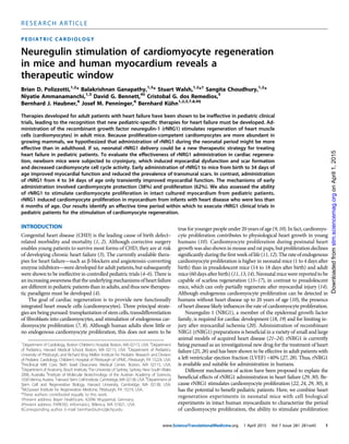
  • 7. of cytokine signaling 1 (SOCS1), bone morphogenic protein 10 (BMP- 10), interleukin-6 (IL-6), and glycerophosphodiester phosphodiesterase domain–containing protein 3 (GDPD3). Functional annotation cluster- ing revealed that the genes whose expression changed with rNRG1 treat- ment encoded cytokines, cell growth anddivisionregulators,andtranscrip- tionfactors(Fig.5B).Insummary,the broad changes in gene regulation par- alleled the observed functional and structural changes. Cardiomyocyte cell cycling is decreased in pediatric patients with heart disease The results from our mouse studies suggestthatrNRG1therapyismostef- fective during an early-administration therapeuticperiod.Todeterminewheth- er a similar trend is present in hu- mans, we examined myocardium obtained from patients with heart disease (for clinical characteristics, see tables S2 and S3). Although car- diomyocyte proliferation occurs in humans without heart disease up to 20 years of age, the presence of heart disease might influence cardiomyo- cyte cell cycle activity. We isolated cardiomyocytesfrompatientsamples, stained them with antibodies against pan-cadherin to determine cell quali- ty (Fig. 6A), and then used flow cy- tometry to quantify the population of cardiomyocytes that were H3P- positive;H3P-positivecardiomyocytes weredetectedinpatientsyoungerthan 6 months but not in patients older than 6 months (Fig. 6, B and C). Fur- thermore, cardiomyocytes from pa- tients with heart disease exhibited lower cell cycle activity compared to nondiseased myocardium (Fig. 6C). Together, these findings suggest that pediatric patients with heart disease exhibit lower levels and earlier cessa- tion of cardiomyocyte cell cycle ac- tivity relative to pediatric donor hearts without disease. Infants with heart disease have the capacity for stimulated cardiomyocyte cycling We tested whether rNRG1 therapy could stimulate the cycling of car- diomyocytes from patients with heart disease. To accomplish this, we mod- ified an organotypic cell culture sys- tem for primary human myocardium (39). A parallel sheet-like arrangement of cardiomyocytes, sarcomeric striations (Fig. 7A), and gap junctions (Fig. 7B) were present after 72 hours of organotypic culture. Thus, the organotypic culture represents a three- dimensional (3D) multicellular model of the human myocardium and 0.2 0.4 0.6 0.8 H3P-positive cardiomyocytes(mm–2) ErbB4: F/Wt F/F * * BSA rNRG1 0 2 4 6 8 Relativefoldchange ofErbB4mRNA ErbB4: F/WtF/F **** H3P, -Actinin, HoechstA B C YZ XZ Fig. 4. rNRG1 acts through ErbB4 on cardiomyocytes in neonatal mouse hearts in vivo. (A to C) Experiments were performed in aMHC-MerCreMer+/+ ; ErbB4F/WT (control) and aMHC-MerCreMer+/+ ; ErbB4F/F (test) mice. rNRG1 or BSA was administered from P0 until P12. ErbB4 inactivation was induced with tamoxifen administration from days P1 to P3, which caused a significant down-regulation of ErbB4 mRNA levels (A). Representative example of a car- diomyocyte in M phase with orthogonal reconstructions along the XZ and YZ axes (shown by staining of heart sections with an antibody against H3P, B). Quantification of these sections showed that rNRG1 increased cardio- myocytecell cycleactivity inErbB4F/WT but not in ErbB4F/F mice (C).Statistical testby Student’st test (A) and ANOVA followed by Bonferroni’s multiple comparison test (C). *P < 0.05, ****P < 0.0001. Scale bar, 50 mm. ATF3 A B SOCS1 BSA rNRG1 Gjc2 GDF15 Gmnn Col23a1 Apln Ctgf Cenpa GDPD3 BMP-10 IL-6 Fig. 5. Cryoinjury and rNRG1 administration induce gene regulation patterns that are consistent with structural and functional changes. Mice underwent cryoinjury on P1 and were treated with BSA or rNRG1 according to the early administration protocol. Expression profiling was performed at 10 dpi with five mice per group and were normalized to sham (n = 5). (A) Heat map shows 622 genes whose expression was signifi- cantly different (P < 0.05, Cuffdiff) in BSA- and rNRG1-treated mice relative to sham-operated mice. Selected genes discussed in the text are indicated. The color chart indicates fold change of expression using a log2 scale. (B) Functional annotation clustering of differentially expressed genes shows significant differences in the expression of components of multiple biological pathways by rNRG1. GO, Gene Ontology, a major bioinformatics initiative to unify the representation of gene and gene product attributes enabling functional interpretation of experimental data through enrichment analysis. P values were generated by DAVID bioinformatics tools. Fisher’s exact test was adopted to measure gene enrichment in annotation terms. R E S E A R C H A R T I C L E www.ScienceTranslationalMedicine.org 1 April 2015 Vol 7 Issue 281 281ra45 7 onApril1,2015stm.sciencemag.orgDownloadedfrom
  • 8. is a suitable platform for studies of cardiomyocyte cell cycle stimula- tion. The 3D culture enabled us to test molecular manipulations that mimic therapeutic interventions and to determine the effect on cardi- omyocyte proliferation in situ without disrupting the native myocar- dial tissue architecture. Myocardial samples were collected at the time of surgery and main- tained for 72 hours with either rNRG1 at 100 ng/ml, the same concen- trationthat stimulated the cycling of culturedcardiomyocytes(22,29, 30), or fetal calf serum (FCS; 1 or 20%). Tissue samples were subsequently sectioned and stained with antibodies to a-actinin and H3P (Fig. 7C), and the number of mitotic cardiomyocytes was quantified manually using fluorescence microscopy (Fig. 7D and table S4). Compared with controls (1% FCS; n = 8 hearts studied, all <6 months old), rNRG1 in- duced an increase in the M-phase cardiomyocyte population in the my- ocardium by 13- to 31-fold from patients 2 to 5 months of age (n = 8 hearts studied; Fig. 7D and table S4). In samples from 14 patients ≥6 months of age [age range, 6 months to 66 years (Fig. 7D and table S4)], no M-phase cardiomyocytes were detected. These results indicate that in- fants with heart disease who are <6 months of age have cardiomyocytes that can be induced with rNRG1 to reenter the cell cycle. Infants with heart disease show the capacity for stimulated cardiomyocyte proliferation We used a nongenetic labeling technique with the fluorescent dye car- boxyfluorescein succinimidyl ester (CFSE; fig. S10) to identify prolifer- ating human cardiomyocytes. In the CFSE assay, fluorescein-protein adducts are retained by cells and diluted by half with every cell division. Using our organotypic culture system, we labeled the myocardium with CFSE and identified cardiomyocytes that had divided with the use of flow cytometry and a forward and side scatter gating strategy to enrich for large cells (cardiomyocytes; Fig. 8A, left panel) and to exclude doublets and cell aggregates (Fig. 8A, middle panel) (13); dead cells were excluded with 7-aminoactinomycin D (7-AAD) dye (Fig. 8A, right panel). CFSE populations were quantified in the FL1 channel and revealed two distinct populations (CFSElo and CFSEhi ). Figure 8B shows representative data from a 3-month-old infant who exhibited a CFSElo population of 4.1%. The CFSElo population represents cardio- myocytes that have undergone several cell divisions, whereas the CFSEhi population represents quiescent cardiomyocytes. A CFSEhi population was found in every heart (fig. S11). We isolated the CFSElo population for lineage reanalysis and stained it with an isotype antibody to exam- ine the nonspecific binding of the cardiac troponin T (cTnT) antibody (Fig. 8C, left panel); staining with an antibody against cTnT showed that 94.7% of this cell population was cardiomyocytes (Fig. 8C, right panel). These cardiomyocytes had similar forward and side scatter charac- teristics (Fig. 8D) compared to the original forward and side scatter gate settings (Fig. 8A, left panel). These experiments demonstrate that the CFSElo cells in Fig. 8B are indeed cardiomyocytes. To further characterize the CFSElo population, we bulk-sorted the cells, isolated RNA, and performed reverse transcription polymerase chain reaction (RT-PCR) analysis to examine the expression of selected marker genes. CFSElo cardiomyocytes expressed the heart-specific mar- kers cTnT and b-myosin heavy chain (b-MHC) but not the stem cell marker c-Kit, suggesting that this population contained mature differ- entiated cardiomyocytes and no dividing stem cells (40) (Fig. 8E). Fur- thermore, cell cycle–related genes were expressed in the CFSElo but not in the CFSEhi population (Fig. 8F). After this extensive validation of the CFSE assay, we examined by flow cytometry the CFSElo population in infant patients with heart disease (n = 16). Quantification showed that theCFSElo populationwas5.8±1.7%(n=4)at2months,5±1.4%(n=3) at 3 months, and 1.2 ± 0.9% (n = 4) at 6 months of age (Fig. 8G and fig. S11). CFSElo populations were not detectable in patients over 6 months of age, indicating that patient age is an important determinant in the proliferative response to rNRG1 (Fig. 8G). Last, we examined the possibility that rNRG1 stimulation increased the proportion of cardiomyocytes with higher DNA contents, which may arise from DNA synthesis without subsequent nuclear division (endocycling) or nuclear division without subsequent cell division (en- domitosis). Addition of rNRG1didnotchange theproportionof mono- nucleated cardiomyocytes (Fig. 8H) or ploidy in the mononucleated portion (Fig. 8I). This supports the conclusion that rNRG1 stimulates cardiomyocyte division in intact infant human myocardium. A B C Pan-cadherin, -actinin, Hoechst 0 6 12 10 20 30 0 1 2 H3P-positivecardiomyocytes(%) Age (years)Age (months) 1 1 2 2 3 1 No heart disease Heart disease 2 2 2 11 11 111 Fig. 6. Pediatric patients with heart disease show decreased cardio- myocyte cell cycle activity. Cardiomyocytes from patients were iso- lated, stained, and analyzed by flow cytometry. (A) Isolated human cardiomyocytes were intact as evidenced by staining with antibodies to pan-cadherin and a-actinin. Intact desmosomes are indicated by white arrowheads. (B) Representative double-marker plot of a 3-month- old patient showing flow cytometric analysis of cardiomyocyte cell cy- cle activity using cardiomyocyte (a-actinin) and cell cycle (H3P) markers. (C) Summary graph showing that patients with heart disease exhibited decreased cell cycling compared to age-matched controls without heart disease. Red dots connected with solid lines indicate results from pa- tients with heart disease, and numbers of corresponding patients are indicated in red. Each donor heart control without heart disease is in- dicated with an open black symbol connected with dotted lines. Circles represent right ventricular and triangles represent LV samples. Scale bar, 50 mm. R E S E A R C H A R T I C L E www.ScienceTranslationalMedicine.org 1 April 2015 Vol 7 Issue 281 281ra45 8 onApril1,2015stm.sciencemag.orgDownloadedfrom
  • 9. DISCUSSION The findings herein advance a strategy for inducing myocardial repair in pediatric patients. Myocardial regeneration experiments in neonatal mice were not feasible until the recent introduction of techniques for inducing myocardial injury (15, 17, 41). LAD ligation (16, 17) and am- putation injury (15, 42) in neonatal mice were reported to lead to scar- less repair, although these results are controversial (43, 44). Here, we verify that cryoinjury is a technically feasible method that produces scar formation and dysfunction, which is in line with the scar formation and delayed repair process observed in zebrafish (31–33) and neonatal mice (35, 36) after cryoinjury. We also show that cryoinjury in neonatal mice is a useful model for human infants with heart disease because it recap- itulates the scar formation, dysfunc- tion, and decrease in cardiomyocyte cell cycle activity frequently seen in young patients with heart disease. The observed decrease in cardiomy- ocyte proliferation after cryoinjury contrasts with the reported increase in cardiomyocyte cycling after LAD ligation (16) and amputation (15), possibly because of technical differ- ences in the scoring of cardiomyocyte cell cycle events (45). Our results sug- gestthatapopulationofproliferation- competent cardiomyocytes exists in young mammals and is inhibited or depleted by heart disease. Adminis- tration of rNRG1 rescued this inhibi- tion to near-normal levels (about twofold increase) and stimulated the generation of new heart muscle in neonatal mice after cryoinjury and in organotypic cultures of myocardi- um from infants with heart disease (2 to 5 months of age). Ourmodelofthe mechanismsac- tivated by early administration of rNRG1 in mice involves myocardial protection and regeneration. What are their relative contributions to the overall reparative process? Com- parison of the extent of myocardial deathbetweenBSA-andrNRG1-treated miceintheearlyadministrationgroup at 1 dpi showed a difference of ~14%, corresponding to ~136,000 cardio- myocytes. On the basis of the H3P analyses, 0.04% of these 136,000 pro- tected cardiomyocytes actively cycle, resulting in ~25,000 new cardiomyo- cytes over a 34-day period (Supple- mentary Results). On the other hand, direct quantification of cardiomyo- cyte nuclear density at 34 dpi revealed 3.1 × 10⁴ and 3.8 ×10⁴ cardiomyocytes/ mm3 forBSA-andrNRG1-treatedani- mals, respectively, corresponding to a difference of 7,000 cardiomyocytes/ mm3 . Subtracting the number of cardiomyocytes contributed by pro- tection, this corresponds to an additional ~224,000 new cardiomyo- cytes in rNRG1-treated animals in the early administration regimen. From these data, we calculated that the new cardiomyocyte popu- lation generated by stimulated proliferation of uninjured cardio- myocytes accounts for 89% [(199,000/224,000) × 100] and the new cardiomyocyte population generated by proliferation of protected car- diomyocytes accounts for 11% [(25,000/224,000) × 100] of the total new cardiomyocyte population. We calculated the relative contribu- tions of rNRG1-induced cardiomyocyte protection as 38% {[136,000/ (334,000 + 25,000)] × 100}, with the remaining 62% resulting from rNRG1-stimulated cardiomyocyte proliferation (Supplementary Results). D Preculture Post-72 h culture -Actinin,DAPI A HoechstConnexin 43 -Actinin MergeB C H3P, -actinin, Hoechst 0.2 0.4 0.6 0 H3P-positivecardiomyocytes (%) Neuregulin-1 (100 ng/ml) 1 1 1 1 1 1 1 1424 3 3 2 8 1 Months 1%FCS(<5mo) 2 3 5 6 71517 3 4 1116 29 66 20%FCS(>5mo) Years Age XZ YZ Fig. 7. rNRG1 stimulates cardiomyocyte cycling in myocardium from infants with heart disease (younger than 6monthsofage).For organotypicculture,chunksofmyocardiumweremaintainedinthepresenceof1%FCS or rNRG1 for 3 days, fixed, and analyzed by immunofluorescence microscopy. (A and B) Preculture is fresh myocar- dium. Post–72 hour culture is after 72 hours of organotypic culture. Incubation of cells in organotypic culture for 3 days did not change microscopic architecture (A). Gap junctions and electromechanical connections were identi- fied by connexin 43 staining and were present after 72 hours of organotypic culture (B). (C and D) rNRG1 stimulates cardiomyocytes to enter M phase in a 2-month-old patient with Tetralogy of Fallot. The position of orthogonal reconstructions of the cardiomyocyte in the center are indicated by yellow arrowheads (along the XZ and YZ axes) (C). Quantitative analysis showed that rNRG1 increased M-phase cardiomyocytes in an age-dependent manner (D). Numbers of patients per data point are indicated (D). Scale bars, 20 mm (A and C) and 50 mm (B). R E S E A R C H A R T I C L E www.ScienceTranslationalMedicine.org 1 April 2015 Vol 7 Issue 281 281ra45 9 onApril1,2015stm.sciencemag.orgDownloadedfrom
  • 10. Inadditiontotheincreaseinthenumberofcardiomyocytes,rNRG1 might activate other beneficial mechanisms, as suggested by the transient im- provement of myocardial function after late administration. The broad range of genes that displayed changes in expression levels after rNRG1 administration (Fig. 5) is consistent with the function of the NRG1 gene product in sustaining the cardiac gene regulatory network dur- ing development (19). The rNRG1- regulated genes might hold clues for a deeper understanding of the bio- logical mechanism(s) activated with rNRG1 administration. The timing of the rNRG1 thera- peutic period identified in neonatal mice after cryoinjury was different from that in human infants. In mice, this period ended within the first 5 days of life and is similar to the re- ported transient period of scarless re- pair after LAD ligation (16, 17). In humans, this therapeutic window ended at about 6 months of age (Fig. 7D), which is significantly shorter than would have been predicted from our results in humans without heart disease (10). The timing of the rNRG1 therapeutic window in hu- mans leads to two conclusions: (i) administration of rNRG1 in human infants <6 months of age may in- duce cardiomyocyte regeneration, and (ii) because our mechanistic model involves cardiomyocyte pro- tection, rNRG1 could be adminis- tered in infants with congenital heart disease before surgical repair, which is feasible because most of these sur- geries are scheduled electively. It is intriguing to note that human cardiomyocyte cell cycle reentry was not stimulated by rNRG1 in patients older than 6 months (Fig. 7D, Fig. 8G). We demonstratedpreviouslythat rNRG1stimulatescardiomyocytedivi- sion in young adult mice (22). Setting aside potential species differences, how could this apparent discrepancy be re- conciled? As shown in Fig. 6C, pre- mature cessation of cardiomyocyte cycling occurred in the presence of heart disease, indicating that heart disease in infants drives proliferation- competent cardiomyocytes out of the cell cycle and into a permanently quiescent phenotype. In contrast, the young adult mice characterized in (22) were free fromheart disease until 8 weeks of life. In other words, these mice were not exposed to signals that drive cardiomyocytes prematurely into a permanently quiescent pheno- type. It is likely that, as a result, cardiomyocytes with proliferative potential remain present into adulthood and could be stimulated by rNRG1. Because induction of cardiomyocyte proliferation with rNRG1 appears to be a conserved mechanism in neonatal mice and in myocardium from human infants, targeting the growth factor could represent a therapeutic Age (months) 0 20 40 60 80 100 Cardiomyocytes(%) Ctrl 2 3 5 2 3 5 1% FCS rNRG1 2N 4N 8N IsotypeFITC FL-4 cTNT-FITC FL-4 SSC FSCCFSE %Max B 4.1% C D 3months CFSEhighCFSElowE F G I A H 0 20 40 60 80 100 Mononucleated cardiomyocytes(%) Preculture Postculture 21 21 21 Age (months) 0 5 10 Proliferating cardiomyocytes (%oftotal) 4 4 3 1 1 1 1 1 2 3 6 15 16 2.5 5 10 Months Years Fig. 8. rNRG1 stimulates cardiomyocyte proliferation in myocardium from infants with heart disease (younger than 6 months). Organotypic cultures of human myocardium were metabolically labeled with CFSE and then maintained in the presence of 1% FBS or rNRG1 for 3 days. Cardiomyocytes were analyzed and isolated by fluorescence-activated cell sorting (FACS). (A) FACS strategy for enrichment by size (left panel), doublet dis- crimination (middle panel), and viability (right panel). (B) Flow cytometric analysis of a 3-month-old infant reveals a CFSElo population of 4.1%. (C and D) After fixation, the CFSElo population was stained with isotype control (C, left panel) and antibodies against cTnT (C, right panel). Analysis by flow cytometry shows that 94.7% of the population were cardiomyocytes (C, right panel) with forward and side scatter characteristics (D) similar to (A). (E and F) RT- PCR showed that CFSElo cardiomyocytes expressed markers of mature differentiated cardiomyocytes (E) and cell cycle–associated genes (F). (G) The graph of proportion of CFSElo populations shows that stimulation of cardio- myocyte proliferation in patients with heart disease is age-dependent. Numbers of patients per data point are indicated. (H and I) Laser scanning cytometry shows that administration of rNRG1 in organotypic culture did not change the overall percentage of mononucleated cardiomyocytes (H) or the ploidy pattern of mononucleated cardiomyocytes (I). Red boxes indicate the gating parameters used for data acquisition and analysis (A to D). FSC, forward scatter; SSC, side scatter; FSC-H, forward scatter height; SSC-W, side scatter width; FL4, detector for 488-nm laser with a 695/40 bandpass filter; FITC, fluorescein isothiocyanate; Ctrl, control. R E S E A R C H A R T I C L E www.ScienceTranslationalMedicine.org 1 April 2015 Vol 7 Issue 281 281ra45 10 onApril1,2015stm.sciencemag.orgDownloadedfrom
  • 11. strategy for pediatric patients with heart disease. rNRG1 therapy might have risksassociatedwithitsproliferative effectsinotherorgans.However, clinical studies that investigate the safety and efficacy of rNRG1 as a car- diac therapeutic in adult patients with heart failure have thus far provided no evidence of uncontrolled growth effects (27, 28). Further- more, the systemic administration of other recombinant growth factors (for example, insulin, granulocyte-macrophage colony-stimulating factor, erythropoetin, thrombopoetin, and insulin-like growth factor 1) that modify the activity of well-defined signaling pathways is a success- ful therapeutic paradigm and has a good track record for safety in pe- diatric patients. MATERIALS AND METHODS Study design For the mouse experiments, the study design, including the number of animals to be included and the type and time points of analyses, was predefined by the investigators. Neonatal mouse cryoinjury was per- formed by B.D.P., and future assignment to rNRG1 and BSA adminis- tration was independently performed by B.G. cMRI was performed and analyzed independently by D.G.B. Investigators were blinded with the use of coded samples. Study objects (neonatal mice and human heart samples) were not excluded on the basis of results being outliers. The number of biological and technical replicates is provided in table S9. Cryoinjury Mouse experiments were approved by Boston Children’s Hospital and Institute of Molecular Biotechnology. ICR mice were used. Pups born after 5:00 p.m. were considered to be P0 the following day and subjected to cryoablation the next day (P1). Pups received a subcutaneous in- jection of 0.1% bupivacaine, were placed inside a sleeve, and were put in an ice-water bath until they were nonresponsive to the paw reflex. Ventrolateral thoracotomy was performed between the fourth and fifth ribs, the pericardium wasremoved, and the heart wasexposed. A 1.5-mm diameter vanadium probe was equilibrated in liquid nitrogen for 20 min and applied to the LV epicardium for 2 s. Sham injury consisted of opening the chest and removing the pericardium. The chest was closed with 6-0 Prolene sutures, and the skin was sealed with Webglue (Webster Veterinary) or 8-0 Prolene sutures. Pups recovered under a heating lamp, were placed on a warming blanket until they became responsive, were rubbed with bedding, and were returned to their mothers. Visualization of injury by TTC Freshly excised mouseheartswere washedin ice-cold50 mM potassium chloride in phosphate-buffered saline (PBS), immediately placed in 1% TTC(w/v;inphosphate buffer, pH7.4)at 37°Cfor20min, andthenfixed in 10% phosphate-buffered formaldehyde overnight. Photomicrographs were taken using a Nikon SMZ1000 stereomicroscope at ×0.8, ×2, or ×3 magnification and an Olympus DP70 charge-coupled device camera. Characterization of scar structure and function We defined a transmural scar as the presence of transmural blue colla- gen on any of 6 to 12 AFOG-stained sections per heart. We measured the radial thickness of nontransmural scars and remote LV myocardi- um in diastole and systole on cMRI images using ImageJ and calculated relative systolic thickening as percent systolic change = (length in systole − length in diastole)/length in systole. Cardiomyocyte-specific deletion of ErbB4 Experiments were performed in aMHC-MerCreMer+/+ ; ErbB4F/WT (control) and in aMHC-MerCreMer+/+ ; ErbB4F/F (test) mice. ErbB4 in- activation was induced with tamoxifen on days P1 to P3 (30 mg/g sub- cutaneously, dissolved in peanut oil). rNRG1 or BSA was administered from P0 for 12 days (P12). Quantification of myocardial function in vivo by echocardiography and cMRI We performed transthoracic echocardiography under anesthesia with a Vevo 2100 device (VisualSonics). cMRI was performed under isoflur- ane anesthesia with a 9.4-T small-animal MRI scanner (Bruker BioSpin MRI). Details of imaging are provided in Supplementary materials and methods. RNAseq sample preparation and data analysis Total RNA was extracted using the RNeasy Micro kit (Qiagen). Complementary DNA (cDNA) libraries were prepared using the TruSeq Stranded mRNA kit (Illumina). Sequencing was performed on NextSeq 500 instruments (single-strand, single-end indexed, 75 base pairs per read) at a depth of 35 × 106 per sample. Single reads were mapped to the mouse genome (m10) using STAR in a strand-specific manner. Cufflinks was used to determine the FPKM (fragments per kilobase of exon per million fragments mapped) levels for each gene from the STAR alignment and was used as input for Cuffdiff. All treatment groups were normalized to sham. Differential gene expression was cal- culated using Cuffdiff. Read counts were normalized between all sam- ples. Genes with significant change of expression were defined by an adjusted P value <0.05 (Maverix Biomics). Functional clustering was performed with DAVID online software. Supplementary Materials and Methods provide additional information. Human myocardial samples Discarded and de-identified human myocardial samples were collected from patients undergoing heart surgery [Institutional Review Board (IRB) protocol #Z06-10-0489]. Clinical information was retrieved through an honest broker (IRB-P00000126). Normal human heart samples, including one fetal heart, were provided by the Sydney Heart Bank at The University of Sydney (Project No: 2012/2814). When known, patients with a 22q11 microdeletion were excluded. For detailed information, please refer to tables S2 to S5. Human cardiomyocyte dissociation Myocardial tissue was washed and resuspended in cold isolation buffer [130 mM NaCl, 5 mM potassium chloride, 1.2 mM monopotassium phosphate, 6 mM Hepes, 5 mM sodium bicarbonate, 1 mM magnesium chloride, and 5 mM glucose (pH 7.4)]. Isolation buffer was supplemen- ted with 0.36 mM calcium chloride for enzyme activity. Cardiac tissue was incubated for 15 to 20 min in isolation buffer supplemented with collagenase IV (Sigma). After each incubation step, the supernatants were transferred into a tube and centrifuged at 600 rpm for 4 min. Cell pellets were resuspended in ice-cold isolation buffer. Several rounds of digestion were performed until the tissue was fully digested. Laser scanning cytometry Myocardial cell preparations were prepared as described above, and laser scanning cytometric analysis for nucleation and ploidy was per- formed as previously described (10). R E S E A R C H A R T I C L E www.ScienceTranslationalMedicine.org 1 April 2015 Vol 7 Issue 281 281ra45 11 onApril1,2015stm.sciencemag.orgDownloadedfrom
  • 12. CFSE assay CFSE is a fluorescein-derived intracellular fluorescent label that is di- vided between daughter cells upon cell division. Human myocardial samples were washed twice in warm PBS, incubated in PBS/2% FBS containing 0.5 mM CFSE (Molecular Probes, C1157) at 37°C for 30 min, and washed with 10× volume of ice-cold PBS/2% FBS. Samples were washed once with warm Dulbecco’s modified Eagle’s medium (DMEM) and incubated in supplemented DMEM for 72 hours. Cardiomyocytes were then isolated as described in “Human cardiomyocyte dissociation” section and analyzed on a FACSAria II (BD Bioscience) in the FL- 1 channel. Viable populations were distinguished by 7-AAD. For lineage reanalysis of the CFSElo population, cells were gated according to CFSE- loaded but non–rNRG1-stimulated controls, and 10,000 cells were bulk- sorted into PBS/5% FCS from two patient samples. Cells were fixed in 4% paraformaldehyde (PFA) for 15 min, washed, stained with either primary antibodies to cTnT (Neomarkers) or an isotype control (immu- noglobulin G1), and conjugated to FITC fluorophore. Cells were analyzed on a FACSAria II. Flow cytometry and FACS Dissociated cardiomyocytes were prepared as previously described (10), washed, and pelleted for antibody staining. For cell cycle analysis, sar- comeric a-actinin (Sigma) antibody was conjugated to a FITC sec- ondary antibody and H3P (Upstate) antibody was conjugated to a Pacific Blue secondary antibody (both from Invitrogen, Molecular Probes monoclonal antibody conjugation kits). Cells were fixed in 4% PFA and washed twice with PBS/5% FCS. Cells were stained in PBS/5% FCS and antibody cocktail for 1 hour at 4°C, washed twice, and resus- pended in PBS/5% FCS. Samples were acquired on a FACSAria Cell Sorter (BD Biosciences), and data were analyzed with FlowJo software. Quantitative RT-PCR Cardiomyocytes were isolated and sorted with a FACSAria II (100 mm nozzle) into TRIzol buffer and were frozen at −80°C. RNA extraction was performed with an RNeasy Micro Kit (Qiagen) according to the manufacturer’s instructions, including on-column DNase I digestion. cDNA was synthesized from 200 ng of total RNA. Eluted RNA samples were reverse-transcribed using SuperScript II and random hexamers (Invitrogen). PCR was performed using iQ5 real-time PCR thermal cy- cler or Bio-Rad CFX384 Touch thermal cycler and iQ SYBR Green Supermix (Bio-Rad) or iTaq UniversalSYBR Green Supermix. The nor- malized values of each biological replicate were averaged before the cal- culation of fold change in expression levels. The primer sequences are provided in tables S10 and S11. Statistical analyses Numerical results are presented as means ± SEM. Statistical testing was performed with Student’s t test, Fisher’s exact test, and ANOVA followed by Bonferroni post hoc testing. Statistical significance was achieved with a two-sided P value ≤0.05. Statistical analyses were per- formed with GraphPad Prism, version 6. SUPPLEMENTARY MATERIALS www.sciencetranslationalmedicine.org/cgi/content/full/7/281/281ra45/DC1 Materials and Methods Results Fig. S1. Contribution of cardiomyocyte proliferation to regeneration. Fig. S2. Cryoinjured hearts showed loss of sarcomeric organization. Fig. S3. Characterization of myocardial repair after cryoinjury. Fig. S4. Transmural scar persists even after 7 months after cryoinjury. Fig. S5. Visualization of scar and quantification of cardiac function by MRI from early admin- istration group. Fig. S6. Visualization of scar and quantification of cardiac function by MRI for late administration group. Fig. S7. Time course of myocardial repair after cryoinjury from early rNRG1 administration group. Fig. S8. Time course of myocardial repair after cryoinjury from late rNRG1 administration group. Fig. S9. Failure to visualize transmural scars with late gadolinium enhancement due to low spatial resolution of cMRI in mice. Fig. S10. Schematic representation illustrating the nongenetic labeling technique with CFSE. Fig. S11. rNRG1-stimulated cardiomyocyte proliferation in infants is age-dependent (2-month-, 6-month-, 1.5- to 5-year-, and 10-year-old patients). Table S1. List of all differentially expressed genes between the BSA and rNRG1 treatment groups relative to sham mice (P < 0.05). Table S2. Clinical information of patients with heart disease analyzed for Fig. 6C (H3P activity over age). Table S3. Clinical information for normal hearts analyzed for Fig. 6C (H3P activity over age). Table S4. Clinical information of patients with heart disease analyzed for Fig. 7D (rNRG1 stimulation). Table S5. Clinical information of patients with heart disease analyzed for Fig. 8G (CFSE assay). Table S6. Comparison of tissue response after cryoinjury in mice and myocardial disease in human infants (myocardial dysfunction, scar formation, and decreased cardiomyocyte cycling). Table S7. Antibody manufacturers and dilutions. Table S8. Image acquisition hardware and settings. Table S9. Quantification of numeric data. Table S10. Human primers for quantitative RT-PCR for calculation of fold change in expression levels. Table S11. Mouse primers for quantitative PCR for calculation of fold change in expression levels. Movie S1. BSA-treated mouse from early administration. Movie S2. rNRG1-treated mouse from early administration. Movie S3. BSA-treated mouse from late administration. Movie S4. rNRG1-treated mouse from late administration. Movie S5. 3D reconstructions show myocardial syncytium adjacent to the scar after early administration (64 dpi). Reference (46) REFERENCES AND NOTES 1. P. W. Tennant, M. S. Pearce, M. Bythell, J. Rankin, 20-year survival of children born with congenital anomalies: A population-based study. Lancet 375, 649–656 (2010). 2. J. I. Hoffman, S. Kaplan, The incidence of congenital heart disease. J. Am. Coll. Cardiol. 39, 1890–1900 (2002). 3. A. P. Bolger, A. J. Coats, M. A. Gatzoulis, Congenital heart disease: The original heart failure syndrome. Eur. Heart J. 24, 970–976 (2003). 4. K. M. Burns, B. J. Byrne, B. D. Gelb, B. Kühn, L. A. Leinwand, S. Mital, G. D. Pearson, M. Rodefeld, J. W. Rossano, B. L. Stauffer, M. D. Taylor, J. A. Towbin, A. N. Redington, New mechanistic and therapeutic targets for pediatric heart failure: Report from a National Heart, Lung, and Blood Institute working group. Circulation 130, 79–86 (2014). 5. R. E. Shaddy, M. M. Boucek, D. T. Hsu, R. J. Boucek, C. E. Canter, L. Mahony, R. D. Ross, E. Pahl, E. D. Blume, D. A. Dodd, D. N. Rosenthal, J. Burr, B. LaSalle, R. Holubkov, M. A. Lukas, L. Y. Tani; Pediatric Carvedilol Study Group, Carvedilol for children and adolescents with heart failure: A randomized controlled trial. JAMA 298, 1171–1179 (2007). 6. D. T. Hsu, V. Zak, L. Mahony, L. A. Sleeper, A. M. Atz, J. C. Levine, P. C. Barker, C. Ravishankar, B. W. McCrindle, R. V. Williams, K. Altmann, N. S. Ghanayem, R. Margossian, W. K. Chung, W. L. Border, G. D. Pearson, M. P. Stylianou; Pediatric Heart Network Investigators, S. Mital, Enalapril in infants with single ventricle: Results of a multicenter randomized trial. Circulation 122, 333–340 (2010). 7. J. C. Garbern, R. T. Lee, Cardiac stem cell therapy and the promise of heart regeneration. Cell Stem Cell 12, 689–698 (2013). 8. Z. Lin, W. T. Pu, Strategies for cardiac regeneration and repair. Sci. Transl. Med. 6, 239rv1 (2014). 9. O. Bergmann, R. D. Bhardwaj, S. Bernard, S. Zdunek, F. Barnabé-Heider, S. Walsh, J. Zupicich, K. Alkass, B. A. Buchholz, H. Druid, S. Jovinge, J. Frisén, Evidence for cardiomyocyte renewal in humans. Science 324, 98–102 (2009). 10. M. Mollova, K. Bersell, S. Walsh, J. Savla, L. T. Das, S. Y. Park, L. E. Silberstein, C. G. Dos Remedios, D. Graham, S. Colan, B. Kühn, Cardiomyocyte proliferation contributes to heart growth in young humans. Proc. Natl. Acad. Sci. U.S.A. 110, 1446–1451 (2013). R E S E A R C H A R T I C L E www.ScienceTranslationalMedicine.org 1 April 2015 Vol 7 Issue 281 281ra45 12 onApril1,2015stm.sciencemag.orgDownloadedfrom
  • 13. 11. M. H. Soonpaa, K. K. Kim, L. Pajak, M. Franklin, L. J. Field, Cardiomyocyte DNA synthesis and binucleation during murine development. Am. J. Physiol. 271, H2183–H2189 (1996). 12. F. Li, X. Wang, J. M. Capasso, A. M. Gerdes, Rapid transition of cardiac myocytes from hy- perplasia to hypertrophy during postnatal development. J. Mol. Cell. Cardiol. 28, 1737–1746 (1996). 13. S. Walsh, A. Ponten, B. K. Fleischmann, S. Jovinge, Cardiomyocyte cell cycle control and growth estimation in vivo―An analysis based on cardiomyocyte nuclei. Cardiovasc. Res. 86, 365–373 (2010). 14. N. Naqvi, M. Li, J. W. Calvert, T. Tejada, J. P. Lambert, J. Wu, S. H. Kesteven, S. R. Holman, T. Matsuda, J. D. Lovelock, W. W. Howard, S. E. Iismaa, A. Y. Chan, B. H. Crawford, M. B. Wagner, D. I. Martin, D. J. Lefer, R. M. Graham, A. Husain, A proliferative burst during preadolescence establishes the final cardiomyocyte number. Cell 157, 795–807 (2014). 15. E. R. Porrello, A. I. Mahmoud, E. Simpson, J. A. Hill, J. A. Richardson, E. N. Olson, H. A. Sadek, Transient regenerative potential of the neonatal mouse heart. Science 331, 1078–1080 (2011). 16. E. R. Porrello, A. I. Mahmoud, E. Simpson, B. A. Johnson, D. Grinsfelder, D. Canseco, P. P. Mammen, B. A. Rothermel, E. N. Olson, H. A. Sadek, Regulation of neonatal and adult mammalian heart regeneration by the miR-15 family. Proc. Natl. Acad. Sci. U.S.A. 110, 187–192 (2013). 17. B. J. Haubner, M. Adamowicz-Brice, S. Khadayate, V. Tiefenthaler, B. Metzler, T. Aitman, J. M. Penninger, Complete cardiac regeneration in a mouse model of myocardial infarction. Aging 4, 966–977 (2012). 18. D. Meyer, C. Birchmeier, Multiple essential functions of neuregulin in development. Nature 378, 386–390 (1995). 19. D. Lai, A. Forrai, X. Liu, O. Wolstein, J. Michalicek, I. Ahmed, A. N. Garratt, C. Birchmeier, M. Zhou, L. Hartley, L. Robb, M. P. Feneley, D. Fatkin, R. P. Harvey, Neuregulin 1 sustains the gene regulatory network in both trabecular and nontrabecular myocardium. Circ. Res. 107, 715–727 (2010). 20. N. Hedhli, Q. Huang, A. Kalinowski, M. Palmeri, X. Hu, R. R. Russell, K. S. Russell, Endothelium- derived neuregulin protects the heart against ischemic injury. Circulation 123, 2254–2262 (2011). 21. X. Liu, X. Gu, Z. Li, X. Li, H. Li, J. Chang, P. Chen, J. Jin, B. Xi, D. Chen, D. Lai, R. M. Graham, M. Zhou, Neuregulin-1/erbB-activation improves cardiac function and survival in models of ischemic, dilated, and viral cardiomyopathy. J. Am. Coll. Cardiol. 48, 1438–1447 (2006). 22. K. Bersell, S. Arab, B. Haring, B. Kühn, Neuregulin1/ErbB4 signaling induces cardiomyocyte proliferation and repair of heart injury. Cell 138, 257–270 (2009). 23. S. M. Jay, A. C. Murthy, J. F. Hawkins, J. R. Wortzel, M. L. Steinhauser, L. M. Alvarez, J. Gannon, C. A. Macrae, L. G. Griffith, R. T. Lee, An engineered bivalent neuregulin protects against doxorubicin-induced cardiotoxicity with reduced proneoplastic potential. Circulation 128, 152–161 (2013). 24. J. E. Cohen, B. P. Purcell, J. W. MacArthur Jr., A. Mu, Y. Shudo, J. B. Patel, C. M. Brusalis, A. Trubelja, A. S. Fairman, B. B. Edwards, M. S. Davis, G. Hung, W. Hiesinger, P. Atluri, K. B. Margulies, J. A. Burdick, Y. J. Woo, A bioengineered hydrogel system enables targeted and sustained in- tramyocardial delivery of neuregulin, activating the cardiomyocyte cell cycle and enhancing ventricular function in a murine model of ischemic cardiomyopathy. Circ. Heart Fail. 7, 619–626 (2014). 25. B. Wadugu, B. Kühn, The role of neuregulin/ErbB2/ErbB4 signaling in the heart with special focus on effects on cardiomyocyte proliferation. Am. J. Physiol. Heart Circ. Physiol. 302, H2139–H2147 (2012). 26. E. M. Parodi, B. Kuhn, Signalling between microvascular endothelium and cardiomyocytes through neuregulin. Cardiovasc. Res. 102, 194–204 (2014). 27. R. Gao, J. Zhang, L. Cheng, X. Wu, W. Dong, X. Yang, T. Li, X. Liu, Y. Xu, X. Li, M. Zhou, A phase II, randomized, double-blind, multicenter, based on standard therapy, placebo-controlled study of the efficacy and safety of recombinant human neuregulin-1 in patients with chronic heart failure. J. Am. Coll. Cardiol. 55, 1907–1914 (2010). 28. A. Jabbour, C. S. Hayward, A. M. Keogh, E. Kotlyar, J. A. McCrohon, J. F. England, R. Amor, X. Liu, X. Y. Li, M. D. Zhou, R. M. Graham, P. S. Macdonald, Parenteral administration of recombinant human neuregulin-1 to patients with stable chronic heart failure produces favourable acute and chronic haemodynamic responses. Eur. J. Heart Fail. 13, 83–92 (2011). 29. Y. Y. Zhao, D. R. Sawyer, R. R. Baliga, D. J. Opel, X. Han, M. A. Marchionni, R. A. Kelly, Neuregulins promote survival and growth of cardiac myocytes. Persistence of ErbB2 and ErbB4 expression in neonatal and adult ventricular myocytes. J. Biol. Chem. 273, 10261–10269 (1998). 30. F. B. Engel, M. Schebesta, M. T. Duong, G. Lu, S. Ren, J. B. Madwed, H. Jiang, Y. Wang, M. T. Keating, p38 MAP kinase inhibition enables proliferation of adult mammalian cardiomyocytes. Genes Dev. 19, 1175–1187 (2005). 31. F. Chablais, J. Veit, G. Rainer, A. Jaźwińska, The zebrafish heart regenerates after cryoinjury- induced myocardial infarction. BMC Dev. Biol. 11, 21 (2011). 32. K. Schnabel, C. C. Wu, T. Kurth, G. Weidinger, Regeneration of cryoinjury induced necrotic heart lesions in zebrafish is associated with epicardial activation and cardiomyocyte pro- liferation. PLOS One 6, e18503 (2011). 33. J. M. González-Rosa, V. Martin, M. Peralta, M. Torres, N. Mercader, Extensive scar formation and regression during heart regeneration after cryoinjury in zebrafish. Development 138, 1663–1674 (2011). 34. J. M. González-Rosa, N. Mercader, Cryoinjury as a myocardial infarction model for the study of cardiac regeneration in the zebrafish. Nat. Protoc. 7, 782–788 (2012). 35. S. A. Jesty, M. A. Steffey, F. K. Lee, M. Breitbach, M. Hesse, S. Reining, J. C. Lee, R. M. Doran, A. Y. Nikitin, B. K. Fleischmann, M. I. Kotlikoff, c-kit+ precursors support postinfarction myogen- esis in the neonatal, but not adult, heart. Proc. Natl. Acad. Sci. U.S.A. 109, 13380–13385 (2012). 36. A. Darehzereshki, N. Rubin, L. Gamba, J. Kim, J. Fraser, Y. Huang, J. Billings, R. Mohammadzadeh, J. Wood, D. Warburton, V. Kaartinen, C. L. Lien, Differential regenerative capacity of neonatal mouse hearts after cryoinjury. Dev. Biol. 399, 91–99 (2015). 37. A. K. Bose, J. W. Mathewson, B. E. Anderson, A. M. Andrews, A. Martin Gerdes, M. Benjamin Perryman, P. D. Grossfeld, Initial experience with high frequency ultrasound for the newborn C57BL mouse. Echocardiography 24, 412–419 (2007). 38. K. D. Poss, L. G. Wilson, M. T. Keating, Heart regeneration in zebrafish. Science 298, 2188–2190 (2002). 39. M. Brandenburger, J. Wenzel, R. Bogdan, D. Richardt, F. Nguemo, M. Reppel, J. Hescheler, H. Terlau, A. Dendorfer, Organotypic slice culture from human adult ventricular myocardium. Cardiovasc. Res. 93, 50–59 (2012). 40. M. M. Zaruba, M. Soonpaa, S. Reuter, L. J. Field, Cardiomyogenic potential of C-kit+ –expressing cells derived from neonatal and adult mouse hearts. Circulation 121, 1992–2000 (2010). 41. A. I. Mahmoud, E. R. Porrello, W. Kimura, E. N. Olson, H. A. Sadek, Surgical models for car- diac regeneration in neonatal mice. Nat. Protoc. 9, 305–311 (2014). 42. D. M. Bryant, C. C. O’Meara, N. N. Ho, J. Gannon, L. Cai, R. T. Lee, A systematic analysis of neonatal mouse heart regeneration after apical resection. J. Mol. Cell. Cardiol. 79, 315–318 (2015). 43. D. C. Andersen, S. Ganesalingam, C. H. Jensen, S. P. Sheikh, Do neonatal mouse hearts regenerate following heart apex resection? Stem Cell Reports 2, 406–413 (2014). 44. T. Konfino, N. Landa, T. Ben-Mordechai, J. Leor, The type of injury dictates the mode of repair in neonatal and adult heart. J. Am. Heart Assoc. 4, e001320 (2015). 45. K. L. Ang, L. T. Shenje, S. Reuter, M. H. Soonpaa, M. Rubart, L. J. Field, M. Galiñanes, Limita- tions of conventional approaches to identify myocyte nuclei in histologic sections of the heart. Am. J. Physiol. Cell Physiol. 298, C1603–C1609 (2010). 46. J. Frahm, A. Haase, D. Matthaei, Rapid NMR imaging of dynamic processes using the FLASH technique. Magn. Reson. Med. 3, 321 (1986). Acknowledgments: We thank H. Sadek and M. Ahmad (University of Texas Southwestern, Dallas) for the training in mouse surgery; M. Farley and D. Burstein [Beth Israel Deaconess Medical Center (BIDMC), Boston, MA] for their help with cMRI; the members of the Kuhn laboratory for helpful dis- cussions; the cardiac surgeons at Boston Children’s Hospital for providing de-identified human my- ocardium (F. Pigula, F. Fynn-Thompson, S. Emani, J. Mayer, C. Baird, and P. Del Nido); E. McCusty (Boston Children’s Hospital) for retrieval of clinical information; the staff at the laser scanning cytom- eter core at the Department of Transfusion Medicine (Boston Children’s Hospital), especially L.Silberstein and S.-Y.Park;N. Francis and D. Bennett (Boston Children’s Hospital) for FACSassistance; and W. Horne and A. Ferguson (Rangos Genomics Facility at Children’s Hospital of Pittsburgh of UPMC) for their assistance with RNAseq. Funding: This research was supported by the Department of Cardiology and the Translational Research Program at Boston Children’s Hospital and NIH grants R01HL106302 and K08HL085143 (to B.K.). B.D.P. and S.C. were supported by the Office of Faculty Development (Boston Children’s Hospital), and B.D.P. by T32HL007572 from the NIH. S.W. received anAHA postdoctoralfellowshipaward. We acknowledge supportfrom an NIHinstrumentationgrant (RR028792) to BIDMC Boston, MA. B.G., N.A., and B.K. were supported by the Richard King Mellon Institute for Pediatric Research (Children’s Hospital of Pittsburgh of UPMC). J.M.P. and B.J.H. were funded by a Fondation Leducq Transatlantic Network of Excellence Award. Author contributions: B.D.P., B.G., S.W., S.C., B.J.H., C.G.R., J.M.P., and B.K. developed the research strategy. B.D.P., B.G., S.C., N.A., D.G.B., and B.J.H. designed, performed, and analyzed the animal experiments. S.W., S.C., and N.A. designed, performed, and analyzed the experiments with human heart samples. C.G.R. provided nondiseased human heart tissue. B.D.P., B.G., S.W., and B.K. wrote and all authors edited the manuscript. Competing interests: The authors declare that they have no competing interests. Submitted 18 December 2014 Accepted 27 February 2015 Published 1 April 2015 10.1126/scitranslmed.aaa5171 Citation: B. D. Polizzotti, B. Ganapathy, S. Walsh, S. Choudhury, N. Ammanamanchi, D. G. Bennett, C. G. dos Remedios, B. J. Haubner, J. M. Penninger, B. Kühn, Neuregulin stimulation of cardiomyocyte regeneration in mice and human myocardium reveals a therapeutic window. Sci. Transl. Med. 7, 281ra45 (2015). R E S E A R C H A R T I C L E www.ScienceTranslationalMedicine.org 1 April 2015 Vol 7 Issue 281 281ra45 13 onApril1,2015stm.sciencemag.orgDownloadedfrom
  • 14. DOI: 10.1126/scitranslmed.aaa5171 , 281ra45 (2015);7Sci Transl Med et al.Brian D. Polizzotti myocardium reveals a therapeutic window Neuregulin stimulation of cardiomyocyte regeneration in mice and human Editor's Summary period might be a new therapeutic strategy for pediatric heart disease. Now that would be brilliant. not in tissue from older pediatric patients. These findings suggest that rNRG1 administration during the neonatal proliferation in heart muscle isolated from human infants with heart disease who were less than 6 months of age, but a persistent manner relative to treatment regimens that began at 4 days after birth. rNRG1 also drove cardiomyocyte injury and found that early treatment starting at 1 day of age boosted cardiomyocyte cell division and heart function in neonatal period. To test their hypothesis, the authors treated newborn mice with rNRG1 at various times after heart . asked whether rNRG1 might put cardiomyocyte proliferation into overdrive if given to mice during theet alPolizzotti (cardiomyocytes). Because young mice bear more proliferation-competent cardiomyocytes than do adult animals, growth factor neuregulin-1 (rNRG1) stimulates heart regeneration by driving the proliferation of heart muscle cells and the need for pediatric-specific treatment regimens became clear. In adult mice, the recombinant−−disappointing hilarious. But when children with heart disease were given adult medicines in clinical trials, the results were When children are given adult roles in TV commercials, the results range from adorable to brilliant to simply Young at heart: Restoring cardiac function in children http://stm.sciencemag.org/content/7/281/281ra45.full.html can be found at: and other services, including high-resolution figures,A complete electronic version of this article http://stm.sciencemag.org/content/suppl/2015/03/30/7.281.281ra45.DC1.html can be found in the online version of this article at:Supplementary Material http://www.sciencemag.org/about/permissions.dtl in whole or in part can be found at:article permission to reproduce thisof this article or about obtainingreprintsInformation about obtaining is a registered trademark of AAAS.Science Translational Medicinerights reserved. The title NW, Washington, DC 20005. Copyright 2015 by the American Association for the Advancement of Science; all last week in December, by the American Association for the Advancement of Science, 1200 New York Avenue (print ISSN 1946-6234; online ISSN 1946-6242) is published weekly, except theScience Translational Medicine onApril1,2015stm.sciencemag.orgDownloadedfrom