1. The threshold concentration of cytochrome c required to form the
apoptosome may be dependent on the expression of bcl-2
Lorenzo Lindoa
; Jeremy Sorokab
; Talia Ada Angc
a,
Department of Botany and Zoology, University of British Columbia, Canada; b,
Department of Biochemistry and Molecular Biology, University of British Columbia, Canada
c,
Faculty of Science, University of British Columbia, Canada
Contact: a,
lindo.lorenzo@gmail.com; b,
jersoroka@gmail.com; c,
talia.a.ang@gmail.com
Introduction
Cancers are a collection of diseases characterized by mutations
causing rapid and uncontrolled cell division. This high rate of division is
often accompanied by further mutations in a cell’s genome causing the
growth of abnormal tissues called “tumours” which are composed of
aberrant, and often non-functional, cells. In lymphomas and leukemias,
both of which cancers that affect the white blood cells, the onset of
cancer results in high numbers of abnormal leukocytes. This is caused
by mutations in the genes that control apoptosis, which is programmed
cell death. Such mutations can confer resistance to apoptosis, such as
bcl-2, a member of the BCL-2 family of proteins, which is an
anti-apoptotic protein that, under normal circumstances, in normally
inhibits apoptosis. This inhibition is of great concern as it acts as a
survival mechanism for cancer cells. While bcl-2 plays an important role
in the inhibition of the mitochondrial release of cytochrome c,
contemporary research has found that microinjection of cytochrome c
into the cytoplasm is sufficient to induce apoptosis. However, it has also
been found that in cells overexpressing bcl-2, apoptosis occurs at levels
only 30% of that of wild-type cells when cytochrome c is injected into the
cytoplasm. While it is largely accepted that bcl-2 plays a role in the
permeabilization of the mitochondria, its exact role in the cytoplasm in
the apoptotic mechanism is unknown.
Rationale
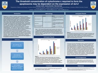
Methodology
!"#$%&#'% (#%)*+,
!"#$%&'()*+,#+*--(#./,0#1234,&55*6#7+-48#
5*'*#+9'(,%:+,#
8"#2;<=#/(#:(*6#,9#(9%,#,0*#+*--(#>&(*6#
9'#123#*?@%*((/9'#/',9#,0*#,0%**#6*(/%*6#
@9@:-&,/9'(#
A'9+B9:,#7+-48
C"#<D,9+0%9E*#<#./--#>*#&66*6#/'#F&%D/'5#
&E9:',(#,9#+*--(#>D#+*--:-&%#E/+%9/'G*+,/9'#
,*+0'/H:*(#/',9#,0*#+D,9(9-#9)#*&+0#+*--#
@9@:-&,/9'
<*--(#9F*%4*?@%*((/'5#7+-48
I"#;@9@,9(/(#./--#>*#H:&',/)/*6#>D#)-9.#
+D,9E*,%D
<9',%9-#J./-64,D@*#7+-48K
L"!4#C"L#E5MEN#9)#+D,9+0%9E*#<!
Table 1: 100-150 cells will be used for each treatment with cytochrome
c1
. Apoptosis will be quantified using fluorescence-labelled inhibitors of
caspases (FLICA) applied with plasma membrane permeability marker
potassium iodine (PI)2
. Cells will be sorted using flow cytometry hourly to
distinguish between viable cells (FLICA -/ PI-) and late apoptotic/necrotic
cells (FLICA +/ PI+)2
.
Anticipated Results
Anticipated Results
Discussion
If we were to vary the concentrations of bcl-2 in cells with varying levels
of cytosolic cytochrome c, we expect to find significant variation in the
frequencies of apoptosis across these trials. These expected observa-
tions suggest that there is a possible correlation between bcl-2 concen-
tration and the frequency of apoptosis in lymphoma and leukemia pa-
tients cells. We expect to find that in cells expressing low levels of bcl-2,
the frequency of apoptosis will be relatively high and require lower levels
of cytosolic cytochrome c. Conversely, expect to find that in cells over-
expressing bcl-2, apoptosis is largely inhibited with higher levels of cyto-
solic cytochrome c required to induce apoptosis. These results suggest
that bcl-2 may play a cytosolic role in apoptosis by interacting with cyto-
chrome c in the formation of the apoptosome complex.
Figure 1:
% Apoptotic cells should increase proportionately with increasing
concentrations of cytochrome C, and cells expressing the most bcl-2
should have the lowest % Apoptotic cells. bcl-2 is not restricted
exclusively to the mitochondria as there is some localization in the Golgi
body and inner plasma membrane3
. bcl-2 can target another protein,
Raf-1, to the mitochondrial membrane4
. bcl-2 may be able to similarly do
so with cytochrome c.
Figure 2:
On subsequent measurements (cytochrome C dosage unchanged) cells
should become increasing apoptotic. This increase in cell death can be
explained by the reliance of cytochrome C-induced apoptosis on
caspase activation. More caspase activation will occur as time passes
which will be proportionate to cell death.
References
1. Li, F. et al. “Cell-specific Induction of Apoptosis by Microinjection of Cytochrome C.” Journal of Biological Chemistry. 272 (1997): 30299-30305.
Print.
2. Wlodkowic, D. et al. “Flow cytometry-based apoptosis detection.” Methods in Molecular Biology. 559 (2009): 19-32. Print.
3. Pozarowski, P. et al. “Interactions of Fluorochrome-Labelled Caspase Inhibitors With Apoptotic Cells: A Caution in Data Interpretation.” Journal
of the International Society for Advancement of Cytometry. 55A (2003): 50-60. Print.
4. Wang, H. et al. “Bcl-2 Targets the Protein Kinase Raf-1 to Mitochondria.” Cell. 87 (1996): 629-638.
Acknowledgements
Limitations
We would like to thank Chi-Chao (Jack) Liu at the Child & Family Re-
search Institute for his consistent encouragement and mentoring and
UBC Undergraduate Research Opportunities (URO) for this opportunity
for learning and to present.
This experiment will not be able to determine the exact way the bcl-2 is
able to regulate cytosolic cytochrome C