More Related Content
Similar to STEM Peanut Poster Final (20)
STEM Peanut Poster Final
- 1. 0
0.1
0.2
0.3
0.4
0.5
0.6
0.7
0.8
0.9
1
C 50x 125x 250x 500x 1000x 2000x 4000x BSA
Absorbanceat450nm
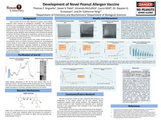
Reagent “P” & “B” modified Ara h2 shows
reduced antibody binding
Reagent P Reagent B
Background
Peanut protein Ara h2 has been observed to produce severe allergic
responses when exposed to endogenous antibodies. Life threatening
conditions such as anaphylaxis could be the result in allergic individuals who
are not treated immediately. Currently, there is little that can be done to
prophylactically treat allergic episodes caused by peanut allergens. Current
research has been focusing on the study of the developing a modified and
safe peanut protein allergoid, which antibodies could ultimately be exposed
to without the threat of inducing an anaphylactic episode by the patient.
This idea has been hypothesized from the successful clinical outcome of
urushiol vaccine for poison ivy treatment.
Purification of this specific protein from whole roasted peanuts is
accomplished mainly by Soxhlet extraction followed by chromatography
columns based on ionic charge and hydrophobicity. The purified protein is
modified and reengineered to generate allergoids by modifying allergen
epitope surface using specific aldehydes and aryl carboxylic acids as well as
specifically designed cross-linkers. The modified proteins has demonstrated
a reduced allergenicity of Ara h2 to Ara h2 antibody shown on ELISA and
Western Blot analyses.
Development of Novel Peanut Allergen Vaccine
Thomas F. Anguella1, Savan V. Patel1, Amanda McCollick1, Laura Wolf2, Dr. Dayalan G.
Srinivasan2, and Dr. Catherine Yang1*
1Department of Chemistry and Biochemistry 2Department of Biological Sciences
Purification of Ara h2
Results and Discussions
Absorbance after Q-Sepharose Column
Chromatography
Ara h2 Modified by Coupling Caffeic
Acid
Ara h2 Coupled
w/ Caffeic Acid Control (Native Ara h2)
Western Blot
17 kDa
Reaction Mechanisms
Caffeic Acid (CA)
References
Conclusion/Future Research
Ara h2 Modified by Coupling with
Reagent “P”
Ara h2 Modified by Coupling with
Reagent “B”
1. Chung SY, Kato Y, & Champagne ET (2005). Polyphenol oxidase/caffeic acid may reduce the
allergenic properties of peanut allergens. J. Sci Food Agric., 85(15):2631-2637.
2. Koppleman SJ, Vlooswijk RA, Knipples LM, Hessing M, vam Reijsen EF, Bruijnzeel-Koomen CA
(2001). Quantification of major peanut allergens Ara h 1 and Ara h 2 in the peanut varieties
Runner, Spanish, Virginia, and Valencia, bred in different parts of the world. Allergy, 56:32-137.
3. Silva CJSM, Sousa F, Gubitz G & Cavaco-Paulo A (2004). Chemical modifications on proteins
using gluteraldehyde. Food Technol Biotechnol, 42(1), 51-56
We have successfully carried out both comprehensive purification and
modification of the peanut protein Ara h2 with multiple protein
modification reagents under varying conditions. The allergenicity of the
modified protein has been illustrated and identified with a significant
decrease in IgE responsiveness when evaluated in-vitro using SDS-PAGE,
ELISA and Western Blot.
Future studies will be focused on the characterization of the allergoids
by Circular Dichroism and to evaluate the extent of reactivity by performing
tests that measure for availability of reactive side chains. Further solubility
of the allergoids will be engineered for modulating the overall solubility in
physiological fluid before vaccine efficacy test in preclinical trials.
kDa
150
100
75
50
35
25
15
Q Phenyl
Ara h2
Sepharose Column Chromatography
30
35
40
45
50
55
60
0 500 1000 1500 2000 2500 3000 3500 4000
%intensity
Ratio of CA to native Ara h2 (242 µg/mL)
Reduced density of native Ara h2 shows evidence of
allergoid
-0.1
0
0.1
0.2
0.3
0.4
0.5
0.6
0.7
0.8
0.9
0.001 0.01 0.1 1 10 100 1000 10000 100000
Absorbanceat450nm
Concentration (ng/ml)
Ara h2 modified with CA is detected less than native
Ara h2
Native Ara h2
BSA (negative control)
x125
Ara h2 Coupled
w/ reagent B
Graph displays absorbance at 595 nm for each fraction after the Q-
Sepharose column. This purification profile demonstrates Ara h 2 separated
from other protein through Q-Sepharose column chromatography. The blue
shaded area gave Ara h2 protein indication eluted from the column.
Caffeic Acid is understood to act as a vehicle for the chemical modification
of Ara h 2 under basic conditions where the formation of o-quinone is
essential for the binding of the protein to caffeic acid and ultimately the
polymerization of Ara h 2.
Modification with caffeic acid was prepared with Ara h 2 in
varying ratio from 62.5 to 4000 mM and incubated at pH 10.5,
at 37° C for 2 days with rotation. The modification resulted in
varied degrees of polymerization smeared SDS-PAGE gel. SDS-
PAGE of Reagent “P” shows dimerization enhancement as
concentration of modification reagent increases. As the ratio of
modification reagent vs Ara h2 increases, the disappearance of
the native Ara h2 has been observed in SDS-PAGE of CA. Ara h
2 modifications were carried out using "Reagent P” and
“Reagent B” and protein cross-linking was observed in the
form of a dimer using SDS-PAGE. Appearance of the formation
of dimer was amplified at higher reagent concentrations.
-10%
10%
30%
50%
70%
90%
110%
0 500 1000 1500 2000 2500 3000 3500 4000 4500
PercentIntensity
Ratio of Reagent "P" to Protein
Varying intensities of both native Ara h2 and modified Ara
h2 with reagent "P"
Native Ara h2 Disappearance
Dimerization
Appearance of Cross-link
0
0.1
0.2
0.3
0.4
0.5
0.6
0.7
0.01 0.1 1 10 100 1000 10000 100000
Absorbanceat450nm
Concentration (ng/ml)
Ara h2 modified with reagent "B" is detected less
than native Ara h2
Native Ara h2
BSA (negative control)
2000x
4000x
Ara h 2 protein epitope surface was modified using
caffeic acid and "Reagent B" which was evident
after performing a Western Blot assay for the two
separate modifications. It is observed that the
allergenicity of modified Ara h 2 is significantly
decreased due to likely chemical altering of the
protein’s epitope surface. Control Ara h2 and
modified Ara h2 extract were delivered to 12-40%
Tris gel, 0.5ug solubilized protein per lane. Proteins
were separated by SDS-PAGE, transferred to
nitrocellulose membrane, and probed with
primary and secondary antibodies identical to
those described in ELISA. Immunoblot was
incubated with Pierce© Western Blot
chemiluminescent substrate and detected with a
CCD camera imaging system.
0
0.1
0.2
0.3
0.4
0.5
0.6
0.7
0.01 0.1 1 10 100 1000 10000 100000
Absorbanceat450nm
Concentration (ng/ml)
Ara h2 modified with reagent "P" is detected less
than native Ara h2
Native Ara h2
BSA (negative control)
2000x
4000x
ELISA was prepared using 25mM Tris buffer at pH 10.5 using a ratio of 1:3,000 - 10,000 dilutions. pAb rabbit anti Ara h2 were used to determine binding of cross-linked protein with
endogenous Ara h 2 antibody in vitro. Secondary antibodies from goat were conjugated with HRP in order to observe primary IgG binding using UV-Vis at 450nm. Antibody binding for Ara h
2 separately exposed to caffeic acid, reagent “P”, and reagent “B” displayed significant decrease in IgG antibody binding.
The resulting SDS-PAGE gels were used to determine level of modification displayed via measuring the
density intensity through a computer program. It can be observed that as the cross-link increases with
increasing concentration of reagents, the Ara h2 band intensity changes.