
Recommended
More Related Content
What's hot
What's hot (20)
Similar to Bedside teaching vvf
Similar to Bedside teaching vvf (20)
More from KhusbuLama
Recently uploaded
Recently uploaded (20)
Bedside teaching vvf
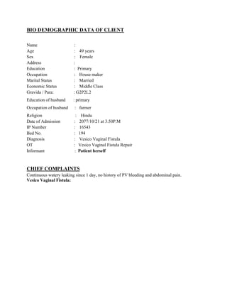
- 1. BIO DEMOGRAPHIC DATA OF CLIENT Name : Age : 49 years Sex : Female Address : Education : Primary Occupation : House maker Marital Status : Married Economic Status : Middle Class Gravida / Para: : G2P2L2 Education of husband : primary Occupation of husband : farmer Religion : Hindu Date of Admission : 2077/10/21 at 3:50P.M IP Number : 16543 Bed No. : 194 Diagnosis : Vesico Vaginal Fistula OT : Vesico Vaginal Fistula Repair Informant : Patient herself CHIEF COMPLAINTS Continuous watery leaking since 1 day, no history of PV bleeding and abdominal pain. Vesico Vaginal Fistula:
- 2. Definition: Vesico Vaginal Fistula (VVF) is an abnormal communication between the urinary bladder and the vagina that results in the continuous involuntary discharge of urine into the vaginal vault. Frequency: In developing countries, the predominant cause of vesicovaginal fistula is prolonged obstructed labor (97%). Etiology: • Obstetrical • Gynecological Obstetrical causes: • Ischemia. • Traumatic causes: it includes instrumental vaginal delivery (especially forceps delivery or during destructive operations) and abdominal operations like hysterectomy for ruptured uterus or Cesarean section. Gynecological causes: • Operative procedures like anterior colporrhaphy, abdominal hysterectomy. • Traumatic: the anterior vaginal wall and the bladder may get trauma following fall on a pointed object, fracture of pelvic bones or due to retained forgotten pessary. • Malignancy: Advanced carcinoma of cervix, vagina or bladder when spread produced fistula. • Radiation: its effect used to treat cervical cancer causes ischemic necrosis which may lead to fistula. • Infective: the infective disease like vaginal tuberculosis, lymphogranuloma venereum In my patient, the cause is operative procedure as abdominal hysterectomy was performed three months back Risk factors: it includes pelvic and vaginal surgery, previous pelvic inflammatory disease (PID), ischemia, diabetes, arteriosclerosis, carcinoma, endometriosis, anatomic dysfunction by uterine myomas and infection particularly, postoperative cuff abscess. Types: Depending on the site of the fistula, the VVF is classified into following types: 1. Juxtacervical fistula: The fistula is between the supratrigonal region of the bladder and the vagina.
- 3. 2. Midvaginal fistula: the fistula is between the base (trigone) of the bladder and vagina. 3. Juxtaurethral fistula: The fistula is between the neck of the bladder and vagina. My patient had juxtracervical fistula. Sign and symptoms: Diagnostic test: Book Picture Patient picture The most common complaint is constant urinary drainage per vagina, although small fistula can present with intermittent wetness that is positional in nature. Present Patient also complain of recurrent cystitis, perineal skin irritation due to constant wetness, vaginal fungal infection and rarely pelvic pain. Present When large vvf is present, patient may not void at all and simply have continuous leakage of urine into the vagina. Present VVF following hysterectomy or other surgical procedures may present upon the removal of the urethral catheter or 1 to 3 weeks later with urinary drainage per vagina. Present VVF resulting from radiation therapy may not be present for months to years following completion of radiation. My patient had not undergone radiation. Speculum examination: A sim’s speculum in sims position gives the information about the size, site and number of fistulas. Laboratory studies: • Upon examination of the vaginal vault, any fluid collection can be tested for urea, creatinine or potassium concentration to determine the likelihood of a diagnosis of VVF.
- 4. • Urine collection for culture and sensitivity test and patient with positive results should be treated prior to surgery. Imaging studies: An intravenous urogram (IVU) is necessary to exclude urethral injury or fistula. If suspicion is high for a urethral injury or fistula and the IVU findings are negative, retrograde pyelography should be performed at the time of cystoscopy examination under anesthesia. Confirmative test: The patient is placed in sims or knee chest position and while examining the anterior vaginal wall, bubbles of air are seen through the small tiny fistula when women coughs. • Dye test: A speculum is introduced and the anterior vaginal wall is swabbed dry. When methylene blue solution is introduced into the bladder by a catheter, a dye will be seen coming out through the opening. • A metal catheter placed through external urinary meatus into the bladder, when comes out through the fistula, confirms the VVF and ensures the patency of the urethra. • Three swab tests: This test also differentiates ureterovaginal and urethrovaginal fistula. In this test three cotton swabs are placed in the vagina- one at a vault, one at a middle and one just above the introitus. The methylene blue is instilled into the bladder through a rubber catheter and a patient is asked to walk for about 15 minutes. She then asked to lie down and swabs are removed for inspection. If upper most swab soaked with urine but unstained with dye and the lower two fistula swabs remain dry then ureterovaginal fistula is confirmed. If upper and lower swabs remain dry but the lower swab stained with dye then vesicovaginal fistula is confirmed. If the upper two swabs remain dry but the lower swab stained with dye then urethrovaginal fistula is confirmed. Management: Prevention: • Adequate antenatal care is to be extended to screen out ‘at risk’ mothers likely to develop obstructed labor like contracted pelvis, malpresentation, history of previous difficult labor, hydrocephalus, etc or early detection of obstructed or prolonged labor. • Emptying the bladder frequently before and after the delivery. • Avoid bladder injuries during pelvic surgeries.
- 5. Medical Therapy: Conservative management or non-surgical management: • If the VVF is diagnosed within the first few days of surgery, a transurethral or suprapubic catheter should be placed and maintained for up to 30 days. Small fistulae (< 1 cm) may resolve or decrease during this period, if the caution is used to ensure proper continuous drainage of the catheter. Sometimes, this prolonged catheterization only increases the risk of infection and offers no increased benefit to fistula care. • Fulguration of the fistula followed by catheter drainage has been shown to have some efficacy as well in small (<5cm), uncomplicated fistula. • Adjuvant measures (such as fibrin glue) have been used. Medications: • Tab cefexime 200mg PO BD • Tab metronidazole 400 mg PO TDS • Tab pantop 40mg PO BD • Tab fortiflex 1 tab OD Surgical therapy: • Success rates approach 90% to 98% regardless of surgical approach. • Adherence to basic surgical principles is essential to achieve success in the repair of all urinary fistula. • No single approach is applicable to all VVF. Transabdominal, transvaginal, trans vesical approaches can be used. Types of repair: • The vaginal approach • The abdominal approach • The laparoscopic approach • Electrocautery • Fibrin glue • Endoscopic closure using fibrin glue with or without adding bovine collagen