
Recommended
More Related Content
Similar to lecture 9
Similar to lecture 9 (20)
Recently uploaded
Recently uploaded (20)
lecture 9
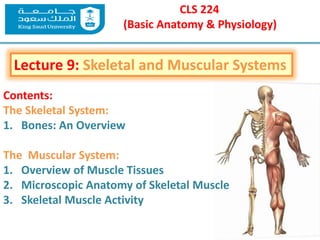
- 1. CLS 224 (Basic Anatomy & Physiology) Lecture 9: Skeletal and Muscular Systems Contents: The Skeletal System: 1. Bones: An Overview The Muscular System: 1. Overview of Muscle Tissues 2. Microscopic Anatomy of Skeletal Muscle 3. Skeletal Muscle Activity
- 3. Objectives: •List the functions of the skeletal system. •Identify the subdivisions of the skeleton as axial or appendicular. •Name the four main classifications of bones. Bones: An Overview
- 4. Functions of Bones: Support of the body Protection of soft organs Movement due to attached skeletal muscles Storage of minerals and fats Blood cell formation (hematopoiesis)
- 5. The Skeletal System Parts of the skeletal system a. Bones (skeleton) b. Joints c. Cartilages d. Ligaments Divided into two divisions 1. Axial skeleton 2. Appendicular skeleton
- 7. Bones of the Human Body The skeleton has 206 bones Classification of Bones: Two basic types of bone (osseous) tissue 1. Compact bone Homogeneous 2. Spongy bone Small needle-like pieces of bone Many open spaces
- 8. Classification of Bones A. Long bones Typically longer than wide Have a shaft with heads at both ends Contain mostly compact bone Examples: Femur, humerus B. Short bones Generally cube-shape Contain mostly spongy bone Examples: Carpals, tarsals
- 9. Classification of Bones C. Flat bones Thin and flattened Usually curved Thin layers of compact bone around a layer of spongy bone Examples: Skull, ribs, sternum D. Irregular bones Irregular shape Do not fit into other bone classification categories Example: Vertebrae and hip
- 10. Classification of Bones on the Basis of Shape
- 11. Gross Anatomy of a Long Bone 1. Diaphysis Shaft Composed of compact bone 2. Epiphysis Ends of the bone Composed mostly of spongy bone Structure of Bone
- 12. 3. Periosteum Outside covering of the diaphysis Fibrous connective tissue membrane 4. Sharpey’s fibers Secure periosteum to underlying bone 5. Arteries Supply bone cells with nutrients Structure of Bone Gross Anatomy of a Long Bone
- 13. Structure of Bone Gross Anatomy of a Long Bone 5. Articular cartilage Covers the external surface of the epiphyses Made of hyaline cartilage Decreases friction at joint surfaces 6. Medullary cavity Cavity of the shaft Contains yellow marrow (mostly fat) in adults Contains red marrow (for blood cell formation) in infants
- 15. Bone Markings Surface features of bones Sites of attachments for muscles, tendons, and ligaments Passages for nerves and blood vessels Categories of bone markings Projections and processes – grow out from the bone surface Depressions or cavities – indentations Structure of Bone
- 16. Microscopic Anatomy of Bone 1. Osteon (Haversian System) A unit of bone 2. Central (Haversian) canal Opening in the center of an osteon Carries blood vessels and nerves 3. Perforating (Volkman’s) canal Canal perpendicular to the central canal Carries blood vessels and nerves Structure of Bone
- 17. Microscopic Anatomy of Bone Copyright © 2003 Pearson Education, Inc. publishing as Benjamin Cummings
- 18. Microscopic Anatomy of Bone Structure of Bone 4. Lacunae Cavities containing bone cells (osteocytes) Arranged in concentric rings 5. Lamellae Rings around the central canal Sites of lacunae 6. Canaliculi Tiny canals Radiate from the central canal to lacunae Form a transport system
- 19. Microscopic Anatomy of Bone Structure of Bone
- 20. Microscopic Anatomy of Bone Structure of Bone Types of Bone Cells Osteocytes Mature bone cells Osteoblasts Bone-forming cells Osteoclasts Bone-destroying cells Break down bone matrix for remodeling and release of calcium Bone remodeling is a process by both osteoblasts and osteoclasts
- 21. Changes in the Human Skeleton In embryos, the skeleton is primarily hyaline cartilage During development, much of this cartilage is replaced by bone Cartilage remains in isolated areas Bridge of the nose Parts of ribs Joints
- 22. Bone Growth Epiphyseal plates allow for growth of long bone during childhood New cartilage is continuously formed Older cartilage becomes ossified Cartilage is broken down Bone replaces cartilage Bones are remodeled and lengthened until growth stops Bones change shape somewhat Bones grow in width
- 23. Long Bone Formation and Growth
- 24. Long Bone Formation and Growth
- 26. Objectives: •Describe similarities and differences in the structure and function of the three types of muscle tissue, and indicate where they are found in the body. •Define muscular system. •Define and explain the role of the following: endomysium, perimysium, epimysium, tendon, and aponeurosis. The Muscular System
- 27. Function of Muscles Produce movement Maintain posture Stabilize joints Generate heat
- 28. Characteristics of Muscles Muscle cells are elongated (muscle cell = muscle fiber) Contraction of muscles is due to the movement of microfilaments All muscles share some terminology Prefix myo refers to muscle Prefix mys refers to muscle Prefix sarco refers to flesh
- 29. The Muscular System Muscles are responsible for all types of body movement Three basic muscle types are found in the body: 1. Skeletal muscle 2. Cardiac muscle 3. Smooth muscle
- 30. Skeletal Muscle Characteristics Most are attached by tendons to bones Cells are multinucleate Striated – have visible banding Voluntary – subject to con-scious control Cells are surrounded and bundled by connective tissue
- 31. Cardiac Muscle Characteristics Has striations Usually has a single nucleus Joined to another muscle cell at an intercalated disc Involuntary Found only in the heart
- 32. Smooth Muscle Characteristics Slide 6.6 Copyright © 2003 Pearson Education, Inc. publishing as Benjamin Cummings Has no striations Spindle-shaped cells Single nucleus Involuntary – no conscious control Found mainly in the walls of hollow organs
- 34. Connective Tissue Wrappings of Skeletal Muscle Endomysium – around single muscle fiber Perimysium – around a fascicle (bundle) of fibers Epimysium – covers the entire skeletal muscle Fascia – on the outside of the epimysium
- 35. Skeletal Muscle Attachments Slide 6.5 Epimysium blends into a connective tissue attachment 1. Tendon – cord-like structure 2. Aponeuroses – sheet-like structure Sites of muscle attachment 1. Bones 2. Cartilages 3. Connective tissue coverings
- 36. Microscopic Anatomy of Skeletal Muscle Cells are multinucleate Nuclei are just beneath the sarcolemma Sarcolemma – specialized plasma membrane Sarcoplasmic reticulum – specialized smooth endoplasmic reticulum
- 37. Myofibril Bundles of myofilaments Myofibrils are aligned to give distrinct bands I band = light band A band = dark band Microscopic Anatomy of Skeletal Muscle Sarcomere Contractile unit of a muscle fiber
- 38. Organization of the sarcomere a. Thick filaments = myosin filaments Composed of the protein myosin Has ATPase enzymes Microscopic Anatomy of Skeletal Muscle b. Thin filaments = actin filaments Composed of the protein actin
- 39. Myosin filaments have heads (extensions, or cross bridges) Myosin and actin overlap somewhat Microscopic Anatomy of Skeletal Muscle At rest, there is a bare zone that lacks actin filaments Sarcoplasmic reticulum (SR) – for storage of calcium
- 41. Properties of Skeletal Muscle Activity Irritability (excitability) – ability to receive and respond to a stimulus Contractility – ability to shorten when an adequate stimulus is received Extensibility: ability to be stretched Elasticity – ability to recoil to resting length
- 42. Nerve Stimulus to Muscles Skeletal muscles must be stimulated by a nerve to contract Motor unit One neuron Muscle cells stimulated by that neuron
- 43. Nerve Stimulus to Muscles Neuromuscular junctions – association site of nerve and muscle Synaptic cleft – gap between nerve and muscle Nerve and muscle do not make physical contact Area between nerve and muscle is filled with interstitial fluid
- 44. Transmission of Nerve Impulse to Muscle Neurotransmitter – chemical released by nerve upon arrival of nerve impulse The neurotransmitter for skeletal muscle is acetylcholine Neurotransmitter attaches to receptors on the sarcolemma Sarcolemma becomes permeable to sodium (Na+) Sodium rushing into the cell generates an action potential Once started, muscle contraction cannot be stopped
- 45. The Sliding Filament Theory of Muscle Contraction Activation by nerve causes myosin heads (cross- bridges) to attach to binding sites on the thin filament Myosin heads then bind to the next site of the thin filament This continued action causes a sliding of the myosin along the actin The result is that the muscle is shortened (contracted)
- 46. The Sliding Filament Theory of Muscle Contraction
- 47. References: