Download as PDF, PPTX
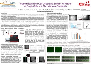

This document describes an image recognition cell dispensing system called On-chip SPiS that allows for damage-less plating of single cells and uniform spheroids. On-chip SPiS uses image recognition to identify and dispense single cells or spheroids into multi-well plates with over 90% accuracy. It is shown to dispense single PC-9 cells and uniform spheroids without damage. The system is also used along with a cell sorter called On-chip Sort to prepare monodisperse spheroid populations for more accurate drug efficacy evaluation compared to unsorted spheroids that show more variable responses.


