Call Girls Service In Shyam Nagar Whatsapp 8445551418 Independent Escort Service
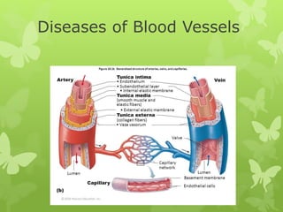
Diseases of blood vessels
1. Diseases of Blood Vessels
Dr. Jyoti Priyadarshini Shrivastava
Associate Professor
Department of Pathology
Gajra Raja Medical College
2. Monckeberg’s Arteriosclerosis
Medial Calcific Sclerosis
Johann Georg Monckeberg in 1903
Site: Media of Large and medium sized arteries.
Location: Extremities and Genital Tract
Etiology:
1.Age related Degenerative changes
Dystrophic calcification
2. Pseudoxanthoma elasticum
3.Idiopathic arterial calcification of Infancy
10. DEFINITION
A localised permanent abnormal dilatation
of a blood vessel occurring due to
congenital or acquired weakening or
destruction of the vessel wall.
12. Sites of Aneurysm
•Major artery from the heart (the aorta)
•Brain (cerebral aneurysm)
•Behind the knee in the leg (popliteal artery aneurysm)
•Intestine (mesenteric artery aneurysm)
•Artery in the spleen (splenic artery aneurysm)
33. Dissecting Aneurysm and Cystic Medial Necrosis
Dissecting Haematoma in which
blood enters the separated
(dissected ) wall of the vessel and
spreads for varying distance
longitudinally
Males
50-70 yrs
Females: Pregnancy
34. TYPES
DeBakey Classification-3 types
Stanford Cassification
1.Type A dissection—The tear begins in the ascending aorta
and progresses throughout the vessel, often extending as
far as the arteries in the leg.
2.Stanford Type B dissection—The tear is located only in the
descending aorta, but may extend into the abdomen.
38. Morphology
No significant dilatation
k/a Dissecting Haematoma
Site:Arch of Aorta
Characteristic: Sharpely incised
Transverse/Oblique
Intimal tear
3-4 cm.
Extends below Into AA and proximally into valves
40. The grade of medial alterations :
cystic medial necrosis,
elastin fragmentation,
fibrosis
medionecrosis
41. Histopathological image of dissecting aneurysm of thoracic aorta in
a patient without evidence of Marfan's trait. The damaged aorta
was surgically removed and replaced by artificial vessel. Victoria
blue & HE stain.
46. Hemangioma
A capillary hemangioma (also known as an Infantile
hemangioma,
Site:Face
Liver,Spleen,Kidney
Gross:Few mm to Few cm.
Histologically:Well defined
UnEncapsulated
Thin walled vessels
47.
48.
49. Cavernous Hemangioma
Cavernous hemangioma, cavernous
angioma, cavernoma, or cerebral
cavernous malformation (CCM)
(when referring to presence in the
brain) is a type of blood vessel
malformation or hemangioma,
where a collection of dilated blood
vessels form a benign tumor.
54. Hemangioendothelioma
True tumour of Endothelial cells.
Intermediate b/w Hemangioma and Hemangiosarcoma
Skin and subcutaneous tissue
They usually grow slowly and can sometimes spread to
other tissues in the body (metastasize).
Vasoproliferative lesion
Gross: well defined, grey red
55.
56.
57. References
Robbins Textbook of Pathology,10th edn.
Textbook of Pathology,Harsh Mohan ,6th edn.
Textbook of Pathology,1st edn.,Vinay Kamal
Wikipedia