1. UNIVERSITY OF HELSINKI
FACULTY OF MEDICINE
BACKGROUND Left atrial
appendage (LAA) of the adult heart contains a
vast number of cardiac and myeloid progenitor
cells.1 The resident myeloid progenitor
population expresses an array of pro-
regenerative paracrine factors.2 Cardiac
constructs have been shown to inhibit
deleterious remodeling of the heart by physical
support. Because of these two aspects, LAA
could be an ideal tissue to be used as a
transplant.
METHODS LAAs from adult mTomato
mice were transplanted to the recipient 129X1-
SvJ mice simultaneously as myocardial
infarction (MI) was performed, a decellularized
LAA patch was implanted to the control group.
Cardiac ultrasound was performed and mice
were sacrificed at 1, 2 and 8 weeks time points.
Hearts were analyzed by 3D fluorescent imaging
(iDisco) and immunohistochemistry. Echo data
was analyzed using Vevo LAB Vevostrain and
3D image data using Imaris software.
Jussi Leinonen1
Päivi Leinikka1
Esko Kankuri1*
Ronen Beeri2*
Eero Mervaala1*
1Department of Pharmacology, University of Helsinki, Finland
2Cardiovascular Research Center, Hadassah-Hebrew
University Medical Center, Israel
*Equal contribution
LEFT ATRIAL APPENDAGE TRANSPLANT
integrates to the left ventricle after a
myocardial infarction initiating
functional recovery
A= MI+LAA patch (n=6) B= MI+Decellularized patch (n=6)
A
B
C
D
Patch
size
( mm²)
0
3
6
Mean ± SD
A= MI+LAA patch (n=6) B= M
2w
4w
6w
7w
8w
0,00 0,20 0,40 0,60 0,80 1,00
0,01
0,02
0,28
0,28
1,00
0,01
0,03
0,35
0,24
0,92
0,00
0,06
0,24
0,05
0,59
EF Strain (L) Strain (C)
A vs B (t-test p-values)
Patch
size
( mm²)
0
3
6
Mean ± SD
2w
4w
6w
7w
8w
0,00 0,20 0,40 0,60 0,80 1,00
0,59
0,23
0,47
0,61
0,44
0,98
0,32
0,31
0,63
0,42
LVd LVs
Troponin C
Tomato
Troponin C
Tomato
F4/80
Troponin C
Tomato
F4/80
Troponin C
Tomato
F4/80
Troponin C
Tomato
Troponin C
Tomato
Troponin C
Tomato
1 week Volume (µm^3)
Infarct size (YELLOW) 3961 x 10^6
Migrated cells (BLUE) 10 x 10^6
Patch 1w (RED) 1747 x 10^6
Migrated cells/Infarct 0,27 %
2 weeks Volume (µm^3)
Infarct size 3889 x 10^6 (YELLOW)
Migrated cells 119 x 10^6 (BLUE)
Patch 2w 1191 x 10^6 (RED)
Migrated cells/Infarct 3,1 %
1 week after MI 2 weeks after MI
Patch 2w/1w 68 %
Migrated 2w/1w 1100 %
A= MI+LAA patch (n=6) B= MI+Decellularized patch (n=6) C= MI (n=3) D= Sham (n=3)
EF Strain (L) Strain (C) Strain rate (L) Strain rate
p-values
0,00
0,11
0,22
0,33
0,44
0,56
0,67
0,78
0,89
1,00
2w 4w 6w 7w
Strain (R) Strain rate (R) LVd LVs
A vs B (t-test p-values)
A B C D A B C D A B C
A B C D A B C D
A B C D A B C D A B C D
A B C D
Patch size (area)
0
1,5
3
4,5
6
Mean
A B
1
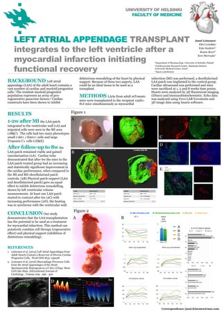
RESULTS
1-2w after MI the LAA patch
integrated to the ventricular wall (1A) and
migrated cells were seen in the MI area
(1B&C). The cells had two main phenotypes:
small c-kit+ / Emr1+ cells and large
Troponin C+ cells (1D&E).
After follow-up to 8w the
LAA patch remained viable and gained
vascularization (2A). Cardiac echo
demonstrated that after 6w the mice in the
LAA patch treated group had an increasing
and statistically significant improvement in
the cardiac performance, when compared to
the MI and MI+decellularized patch
controls. (2B) Physical patch-support (LAA
and decellularized patch) gave an equal
effect to inhibit deleterious remodelling,
shown by left ventricular volume
measurements. At least one LAA patch
started to contract after 6w (2C) with
increasing performance (2D), the beating
was in synchrony with the ventricular wall.
F4/80
Troponin C
Tomato
Figure 1
Figure 2
A
B
C
A B
C
D
E
D 8w
CONCLUSION Our study
demonstrates that the LAA transplantation
has the potential to be used as a treatment
for myocardial infarction. This method can
putatively combine cell therapy (regenerative
effect) and physical support (inhibition of
deleterious remodeling).
REFERENCES
1. Leinonen et al. (2013) Left Atrial Appendages from
Adult Hearts Contain a Reservoir of Diverse Cardiac
Progenitor Cells. PLoS ONE 8(3): e59228
2. Leinonen et al. (2016) Macrophage Precursor Cells
from the Atrial Appendages of the Heart
Spontaneously Reprogram to a C-kit+/CD45- Stem
Cell-Like State. International Journal of
Cardiology , Volume 209 , 296 - 306
7w
Correspondence: jussi.leinonen@mac.com