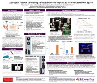
1. Tools for delivering interbody cages are designed to engage
rigid materials made of titantium, stainless steel, or PEEK.
Bone graft delivery tools deliver flowable tissue-derived
materials, such as bone particles mixed with bone marrow
aspirate.
0
20
40
60
80
100
120
140
160
180
1 2 3 4
Plunger Loading Time Suture Loading Time
Plunger Procedure Time Suture Procedure Time
Time
(s)
Experiment #
Loading & Procedure Time
Test Methods and Results
Design 1: Plunger-Push Tool
• Design has hollow core and
curved tip to hold the
biomaterial in place
• Curvature mimics the shape of
the anterior wall, allowing more
accurate biomaterial
deployment
• A plunger is guided through the
hollow core and pushes the
biomaterial into the vertebral
disc space
• Plunger is made of a high
performance blend rubber
plastic tubing
A Surgical Tool for Delivering an Osteoinductive Implant to Intervertebral Disc Space
Tiffany Law1,2, Joshua Liebowitz1, Ryan McNaughton1, Howard Seeherman2, Christopher Wilson2
1Boston University, Department of Biomedical Engineering, Boston, MA 02215
2Bioventus, LLC, Boston University Business Innovation Center, Boston, MA 02215
Acknowledgements
Funding support and laboratory space for this project was provided by Bioventus, LLC. We
would like to acknowledge Heitor Mourato and Glenn Thayer of Boston University’s Scientific
Instrumentation Facility (SIF), Bob Sjostrom and David Campbell of the Engineering Product
Innovation Center (EPIC) for their machining mentorship and Doug Fredericks, University of
Iowa, for helpful discussions.
As of 2009, over 400,000 Americans received spinal
fusion surgeries annually.1 Procedures, such as
Transforaminal Lumbar Interbody Fusion (TLIF) and
Posterior Lumbar Interbody Fusion (PLIF), are used
to treat back pain arising from degenerative disc
disease or spondylolisthesis. It involves inserting
autologous bone graft and an interbody cage
between vertebral bodies to achieve spinal fusion.
Delivery of osteoinductive growth factors, such as
bone morphogenetic protein, improves rate of fusion
and limits the need for revision surgery. Such factors
are delivered on sponge-like biomaterial with porous
architecture. Preservation of the sponge’s structure
is crucial for maximal efficacy. Literature reviews,
patent databases, and corporate websites, revealed
no previously-described tools designed to deliver an
intact sponge-like implant to the intervertebral disc
space.
Active Healing Through Orthobiologics
Candidate Designs
The designs discussed have been protected with a provisional patent application2 and are ultimately intended for human
use; however, the scope of this student project will not expand into clinical testing. Initial human factors assessments
occurred in November 2015, but a final human factors assessment will be made following the final fabrication of the high
fidelity prototype. The goal of the human factors testing is to confirm the device will be suitable for both minimally invasive
or open surgical approaches.
(a) Bone graft is removed from the annulus
using a rotate cuter
(b) A distractor is used to measure the
distance between vertebral bodies
(c) An interbody cage filled with bone graft is
inserted into the disc space
(a) (b) (c)
Design 2: Suture-Snare Tool
• Design has a hollow core
• The end of the tool is a cap
with two holes
• The suture exits through one
hole and into the adjacent
hole, wrapping around the
biomaterial
• The ends are tied to anchors
on the handle
• Post proper insertion and
placement, the suture is cut
and removed
• Material is Ethicon 4-0 Coated
Vicryl Suture (not shown here)
• Designs were drawn using SolidWorks
• Initial prototypes were generated by 3D printing with ABS plastic
• Functional prototypes fabricated by machining and assembly of polycarbonate components
• Various measurements were collected to test the functionality of the different designs using a standard custom test bed
Medtronic’s Threaded
Inserter inserts the
interbody cage into the
intervertebral disc space.
Benvenue’s Multi-
Dimensional Degree In-Situ
Expandable Implant delivers
a flexible interbody cage
Commercialization considerations:
• Decrease the unit variable cost by creating an
injection mold for each device component
• Use of polycarbonate: Inexpensive and FDA
compliant
Safety considerations:
• Device will follow FDA Class II regulations
• Autoclave sterilization is applicable
• Prototype will undergo animal testing to validate
device
• Preservation of the carrier size and shape is
essential to implant performance
Surgical tool has to abide by the following criteria:
1. Secure grip on the biomaterial scaffold prior to
insertion.
2. Ability to fit through a small incision along the
annulus fibrosis.
3. Reliably position biomaterial scaffold against
the anterior wall of the disc space while
preserving architecture of the implant.
4. Deliver biomaterial scaffold with minimal
absorption of BMP to the surgical tool.
5. Intuitive user design such that the surgical tool
is simple to use.
6. Compatible with common sterilization
practices.
Pinnacle’s InFill
system injects
morselized bone graft
into the disc space
Experimental Test Bed:
Material Interaction with Protein During Delivery
A) Initial test apparatus prior to experimentation. Implant is placed for
reference. B) Plunger-Push model experiment in progress.
C) Magnified view of the intervertebral disk space during experiment.
D) Successful placement of the biomaterial against the anterior wall
of the annulus fibrosis.
Testing was performed with a sample size of 4. On average, the plunger-
push design saw a reduced percentage change in mass and a reduced
number of biomaterial granules lost than the suture-snare design. The
results indicate the plunger-push design allowed the biomaterial carrier to
retain more of the BMP throughout delivery and preserve the integrity of
the carrier better than the suture-snare design.
Testing was performed with a sample size of 4. The plunger-
push design was faster than the suture-snare design for both
loading and overall procedure times. A user learning curve was
observed with the suture-snare design and will be monitored in
future testing. Future iterations will incorporate a more diverse
range of users, each with an increased number of trials.
Fluorescent-labeled protein was loaded onto the
biomaterial and testing was performed to observe the
amount of residual protein present in the tool post-delivery.
The pictures show fluorescing residual protein under blue
light. Visual inspection revealed no indication of residual
protein. Quantitative analysis using guanidine extraction
also corroborated no significant results.
images courtesy of Medtronic’s CAPSTONE PEEK Spinal System Technique
images courtesy of Medtronic’s CAPSTONE PEEK Spinal System Technique
images courtesy of www.benevenue.com
Positive Control Design Trials
Discussion
Introduction Design Criteria
Prior Art
Device
A B C
Plunger
Implant*
Intervertebral
disc space
Working
Window
Example of a successful delivery
Incision
Window
Plunger-Push Design
Suture-Snare Design
D
*Implant is made out of
400-800um ceramic
granules in collagen matrix
0
1
2
3
4
Plunger-Push Suture-Snare
# of
Granules
Ceramic Shed from Implant
0%
2%
4%
6%
8%
10%
Plunger-Push Suture-Snare
Percent
Loss
Change in Implant Mass
References
1Rajaee, S. S., L. E. A. Kanim, and H. W. Bae. "National Trends in Revision Spinal Fusion in the USA:
Patient Characteristics and Complications." The Bone & Joint Journal 96-B.6 (2014): 807-16.
2Law T, Liebowitz J, McNaughton R, Wilson C and Seeherman H. “Device and Method for Placement of
Osteoinductive Implants during Spinal Fusion.”