VIP Call Girls Noida Jhanvi 9711199171 Best VIP Call Girls Near Me
Fluid-Balance-Monitoring-Poster.pdf
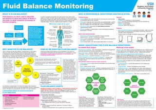
1. Trained Nurses
• Identify patients who need fluid balance monitoring and communicate
via whiteboard behind bed, handover
• Clarify up-to-date plan with medical team and communicate to patient
and visitors, wider MDT including HCAs: SALT, housekeeping, physio
and any colleague who may assist with, provide or remove fluids
• Ensure accuracy using charts and calculating cumulative
measurements 6 hourly
• Escalate promptly to medical team in case of developing imbalance,
deterioration or concern
HCA’s and Student Nurses
• Teamwork, Communicating with MDT to ensure accurate
measurements – eg SALT, housekeeping, physio
• Ensure chart is complete and accurate – use of appropriate equipment
e.g. scales, urometers etc
• Inform trained staff or NIC with changes deterioration or concerns
Doctors
• Daily review:
Indication for monitoring, is it still necessary?
Goal – document with special instructions such as restrictions or
frequency of monitoring
Charts – current balance
Escalation plan or the need for it
Patients
• Must demonstrate capacity (understand information, retain and recall
when asked) concerning their fluid balance monitoring if they are to
complete charts independently.
• Trained nurses to calculate cumulative measurements
Acute illness
Monitoring fluid balance helps monitor
acute illness or with early recognition of
further deterioration. In some illnesses
the fluid may move out of the vascular
system and into extracellular spaces
outside the bloodstream. This fluid is
still in the body but no longer in a useful
space and patients may develop low
urine output due to hypotension and
hypovolaemia. Generally caused by
altered capilliary permeability (leaky blood
vessels) secondary to ischaemia, trauma
or inflammation, conditions include:
• Sepsis
• Bowel obstruction
• Acute pancreatitis (or acute on
chronic)
• Liver failure
• Malnutrition
Fluid Balance Monitoring
An imbalance of electrolytes in the blood can lead to fluid imbalance.
Laboratory blood tests such as urea and electrolytes, glucose, magnesium,
calcium will determine discrepancies and lead to the right treatment.
LABORATORY RESULTS ASSOCIATED WITH
FLUID IMBALANCE
Fluid loss Fluid gain
• Increased serum osmolality
• High urine osmolality and
specific gravity
• Raised haematocrit
• Increased plasma-urea
concentration
• Reduced plasma urea
• Reduced haematocrit
FLUID BALANCE CHARTS
Identifying a postitive (↑input) or negative (↑ output) balance is essential, as
redressing any imbalance is vitally important. As well as aiding assessment,
together with other vital signs it allows us to evaluate and adapt our care,
replacing and restricting fluids appropriately to achieve stability. Maintaining an
accurate fluid chart can present challenges:
Communication with Patient
• Aware of plan, and any restriction to intake?
• Able to use equipment independently?
• Compliant, possibly able to self document?
Communication with MDT?
• MDT aware of monitoring? (SALT, Physio, volunteers)
• Use whiteboard behind bed to convey any specific instructions
Accuracy
• Volumes measured specifically, awareness of volumes of different
drinking vessels
• If patient is using pads these can be weighed with mg converted to ml
WHEN: INDICATIONS FOR FLUID BALANCE MONITORING
WHAT IS FLUID BALANCE?
Fluid balance is a term used to describe
the balance of input and output of fluids in
the body, to allow metabolic processes to
function properly.
In order to maintain homeostasis, the adult human body needs a fluid
intake of 2-3 litres (25-30ml / kg per day), allowing it to keep a balance of
the nutrients, oxygen and water, which are necessary to preserve a stable
healthy internal environment. Output should be roughly equal, though
‘insensible losses’ may give a slightly positive balance on charts.
WHY MONITOR FLUID BALANCE?
Injury or Illness can alter fluid balance. Hypoperfusion of vital organs
may occur with lower circulating volumes caused by dehydration,
or redistribution of within the body during an inflammatory response
post trauma, in Cancer or during Sepsis, requiring fluid replacement.
Alternatively an ‘overload’ may occur as a result of poor cardiac or renal
function, or excessive fluid intake orally or IV.
HOW DO WE MONITOR FLUID BALANCE?
Knowing the signs and symptoms of Fluid Imbalance in the body is a
crucial aspect of hospital care and assessment. It is assessed in 3 ways:
fluid balance charts, physical assessment of fluid balance and
monitoring of blood results.
Fluid intake is regulated by
thirst – which is a natural
response to fluid depletion. As
the osmotic pressure of blood
increases (due to higher ratio
of molecules to H2
O) water is
drawn from cells into blood.
Osmoreceptors in the brain
are dehydrated, and stimulate
release of anti-diuretic hormone
and sensation of thirst. Adrenal
glands produce Aldosterone
– stimulating reabsorbtion of
sodium, and then water, from
the kidneys – less is excreted
Thirst is often a LATE indicator
of hydration, and this response
becomes weaker and more
delayed with increasing AGE.
Physical
assessment
of fluid status
Facial/
oral
assessment
Weight
Urine
output
Thirst
Vital
signs
Overload may present with: tachycardia, hypertension,
increased respiratory rate/effort/noise/moist cough.
Fluid depletion may present with hypotension, postural drop,
a lowered ‘pulse pressure’, rapid, shallow respirations, rapid,
weak thready pulse.
Skin elasticity – ‘tissue turgour’.
• skin is dry and less elastic with dehydration
• presence of oedema indicates overload
Capillary refill time
• good indicator of intravascular pressure/ volume
(and hydration). Blood should return to area
post gentle pressure in less than 2 seconds
• is skin warm, pink?
Jugular/venous pressure
• raised in overload
Mucous membranes
dry/moist – mouth,
tongue, conjuntiva,
saliva – thick, sticky in
depletion or copious
and frothy in overload.
Sunken facial features
particularly around
eyes indicate severe
depletion... or are there
signs of oedema?
If serial weights same
time each day
What does water do for you?
Forms saliva
(digestion)
Needed by the brain to
manufacture hormones
and neurotransmitters
Regulates body
temperature (sweating
and respiration)
Acts as a shock
absorber for brain
and spinal cord
Converts food to
components needed
for survival - digestion
Helps deliver oxygen
all over the body
Keeps mucosal
membranes moist
Allows body’s cells to
grow, reproduce
and survive
Flushes body waste,
mainly in urine
Lubricates joints
Water is the major
component of most
body parts
Although these fluid
compartments are classed as
separate, water and fluids are
constantly moving between
them all, powered by different
processes such as diffusion
(movement of particles)
osmosis (movement of water)
hydrostatic pressure (gravity
and cardiac function) and
oncotic pressure (proteins)
This fluid consists of water and
electrolytes – particles which
carry an electrical charge – an
imbalance in these can cause
cardiac arrhythmias.
Human body is
55-60% fluid
1/3 is
extracellular
2/3 total
body fluid is
intracellular
80% extracellular
fluid is
interstital
20% extracellular
fluid is in
the plasma
Skin
WHO IS RESPONSIBLE: MONITORING AND ESCALATION
Increased fluid output
Diarrhoea and vomiting – risk of dehydration, malnutrition and significant
electrolyte disturbances including hyperkalaemia
High urine output – polyuria -↑200mls /hr – leads to dehydration if
unmanaged. Common causes: diabetes, resolving AKI, excessive diuretics
High output stoma – increased frequency or ↑1 litre in 24 hrs
Urinary catheter, convene, urostomy or irrigation – volumes must
be measured. Incontinent patients may self-limit input in attempt to
manage problem.
Post-operative patients should be closely monitored
• Large open wounds: output should be estimated if an accurate output
is not possible
• Drains: pleural, wound, ascitic
• Increased ‘insensible losses’: sweating, sustained pyrexia of 38°C or
a sustained respiratory ↑rpm. Each example can lead to a fluid loss of
↑500 mls in any 24 hour period
Reduced urine output
Oliguria – low urine output ↓0.5mls per kilogram per hour. Oliguria can be
an early sign of poor renal perfusion. Most common causes: hypotension
or hypovolaemia. Anuria - absence of urine: ↓100mls over 24 hours.
Acute Kidney Injury (AKI) /Chronic Kidney Disease
Patients with raised creatnine blood levels combined with a low urine
output may have an AKI: the kidneys are not effectively filtering blood,
reabsorbing vital elements and excreting others. Prompt identification of
an AKI is crucial as it can lead to serious complications if left untreated.
Medications which increase risk of AKI (patients on these need fluid
balance monitoring)
• Contrast medium – monitor fluid balance for 24 hrs before and
after procedure
• Chemotherapy – monitor Fluid Balance during therapy
• Antibiotic therapy – many antibiotics can cause renal impairment
(Check BNF). High risk are: Gentamycin, Aciclovir and Vancomycin.
Fluid balance should be monitored throughout therapy and for 24hrs
post last dose
• ACE inhibitors and diuretics - often held in acute kidney injury
Heart failure
Acute Heart Failure (HF) is most commonly caused by cardiac dysfunction due to
muscle damage, valvular dysfunction, or arrhythmias. The heart does not pump
enough blood to meet all the needs of the body, and it can be complex to manage
fluid balance for these patients. In acute new onset HF or acute decompensation of
Chronic HF, renal function, weight and Fluid Balance should be closely and accurately
monitored, to ensure appropriate diuretic therapy or fluid management (NICE 2014).
Unconcious
patients
Patients
with
impaired
swallow
Impaired thirst
reflex - this can
worsen with age and
increase risk
of dehydration
Diagnosis,
or at risk of
malnutrition
Intravenous
fluids/enteral
feeding
NBM/
restricted
diets
Decreased
oral
intake
Paralysis
Delirium
Dementia
Poor vision
Poor memory
Stroke
Loss of
independence
Glass 200mls Beaker 200mls Jug 750mls
Cup 160mls
Clodagh Bannerman 2018