illustration of XRF and TEY STXM for image single particle
•Download as PPTX, PDF•
1 like•122 views

![1202 mccormack[2]](data:image/gif;base64,R0lGODlhAQABAIAAAAAAAP///yH5BAEAAAAALAAAAAABAAEAAAIBRAA7)
Recommended
More Related Content
What's hot
What's hot (20)
Similar to illustration of XRF and TEY STXM for image single particle
Similar to illustration of XRF and TEY STXM for image single particle (20)
illustration of XRF and TEY STXM for image single particle
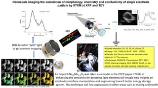
- 1. e SDD detector “split” light to get element mapping XRF 400 600 800 1000 1200 0 10000 20000 30000 40000 Intensity Photon energy (eV) Ni O Mn Fe hv=1100 eV, 50 ms Nanoscale imaging the correlation of morphology, chemistry and conductivity of single electrode particle by STXM at XRF and TEY Spatial resolution: 20, 25, 35, 40, 60 nm ZP Energy: 130 - 2500 eV (E/∆E: 3000 - 10000) Sample: thin films or nano-scale particles under helium or 10-6 Torr vacuum. Techniques: NEXAFS (Transmission, TEY, XRF), 2D/3D chemical imaging, XLD, XMCD, SAXS, In situ devices (humidity, wet cells, heating, magnets etc.). Phys. Chem. Chem. Phys., 2016, 18, 22789--22793 Phys. Chem. Chem. Phys., 2016, 18, 22789--22793 SDD Fe doped LiNi0.5Mn1.5O4 was taken as a model in the PCCP paper. Efforts in enhancing the sensitivity for detecting light elements will enable new insights on electrode interface manipulation and engineering toward better energy storage system. This technique will find applications in other areas such as mining and health.