
Recommended
More Related Content
What's hot
What's hot (20)
Similar to Labor-5 (2).pptx
Similar to Labor-5 (2).pptx (20)
Recently uploaded
Recently uploaded (20)
Labor-5 (2).pptx
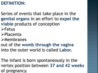
- 1. DEFINITION: Series of events that take place in the genital organs in an effort to expel the viable products of conception Fetus Placenta Membranes out of the womb through the vagina into the outer world is called Labor. The infant is born spontaneously in the vertex position between 37 and 42 weeks of pregnancy.
- 2. Expulsion of a previable live fetus occurs through the same process but in a miniature form and is called mini-labor. Labor is characterized by the presence of regular uterine contractions. Delivery is the expulsion or extraction of a viable fetus out of the womb. It is not synonymous with labor. Delivery can take place without labor as in elective cesarean section. Delivery may be Vaginal(either spontaneous or aided) Abdominal
- 3. NORMAL LABOR (EUTOCIA) Labor is called normal if it fulfills the following criteria. Spontaneous in onset and at term. With vertex presentation. Without undue prolongation Natural termination with minimal aids Without having any complications affecting the health of the mother and/or the baby.
- 4. CAUSES OF ONSET OF LABOR The precise mechanism of initiation of human labor is still obscure. Endocrine, biochemical and mechanical stretch pathways as obtained from animal experiments, however, put forth the following hypotheses. Uterine distension Fetoplacental contribution Estrogen: The probable mechanisms are: — Increases the release of oxytocin from maternal pituitary. ProgesteroneProstaglandinsOxytocin and myometrial oxytocin receptorsNeurological factor:
- 5. CONTRACTILE SYSTEM OF THE MYOMETRIUM The basic elements involved in the uterine contractile systems are: (a) actin, (b) myosin, (c) adenosine triphosphate (ATP), (d) the enzyme myosin light chain kinase (MLCK), and (e) Ca++.
- 6. Uterine muscles have two types of adrenergic receptors—(1) α receptors, which on stimulation, produce a decrease in cyclic AMP (adenosine monophosphate) and result in contraction of the uterus and (2) β receptors, which on stimulation, produce rise in cyclic AMP and result in inhibition of uterine contraction.
- 7. FALSE PAIN: (Synonym: false labor, spurious labor): It is found more in primigravidae than in parous women. It usually appears prior to the onset of true labor pain by 1 or 2 weeks in primigravidae and by a few days in multiparae. Such pains are probably due to stretching of the cervix and lower uterine segment with consequent irritation of the neighboring ganglia. PRELABOR: (Synonym: premonitory stage): The premonitory stage may begin 2–3 weeks before the onset of true labor in primigravidae and a few days before in multiparae. The features are inconsistent and may consist of the following:
- 8. “Lightening” A few weeks prior to the onset of labor especially in primigravidae, the presenting part sinks into the true pelvis. It is due to active pulling up of the lower pole of the uterus around the presenting part. It signifies incorporation of the lower uterine segment into the wall of the uterus. This diminishes the fundal height and hence minimizes the pressure on the diaphragm.
- 9. The mother experiences a sense of relief from the mechanical cardiorespiratory embarrassment. There may be frequency of micturition or constipation due to mechanical factor—pressure by the engaged presenting part. It is a welcome sign as it rules out cephalopelvic disproportion and other conditions preventing the head from entering the pelvic inlet. Cervical changes: A few days prior to the onset of labor, cervix becomes ripe. A ripe cervix is •Soft •80% effaced (<1.5 cm in length) •admits one finger easily •cervical canal is dilatable
- 10. True labor pain is characterized by: Painful uterine contractions at regular intervals Frequency of contractions increase gradually Intensity and duration of contractions increase progressively Progressive effacement and dilatation of the cervix Descent of the presenting part Formation of the “bag of forewaters” Not relieved by enema or sedatives .
- 11. False labor pain is: Dull in nature Confined to lower abdomen and groin Not associated with hardening of the uterus They have no other features of true labor pain as discussed above Usually relieved by enema or sedative !!!Expulsion of cervical mucus plug mixed with blood is called “show”
- 12. Formation of “bag of waters”: Due to stretching of the lower uterine segment, the membranes are detached easily because of its loose attachment to the poorly formed decidua. With the dilatation of the cervical canal, the lower pole of the fetal membranes becomes unsupported and tends to bulge into the cervical canal. As it contains liquor, which has passed below the presenting part, it is called “bag of waters”.
- 13. During uterine contraction with consequent rise of intra-amniotic pressure, this bag becomes tense and convex. After the contractions pass off, the bulging may disappear completely. This is almost a certain sign of onset of labor. However, in some cases the membranes are so well applied to the head that the finding may not be detected.
- 14. STAGES OF LABOR Conventionally, events of labor are divided into three stages: First stage: It starts from the onset of true labor pain and ends with full dilatation of the cervix. It is, in other words, the “cervical stage” of labor. Its average duration is 12 hours in primigravidae and 6 hours in multiparae.
- 15. has 2 phases Latent Active During the latent phase, irregular contractions become progressively better coordinated, discomfort is minimal, and the cervix effaces and dilates to 4 cm. Traditionally, the cervix was expected to dilate about 1.2 cm/hour in nulliparas and 1.5 cm/hour in multiparas.
- 16. Second stage It starts from the full dilatation of the cervix (not from the rupture of the membranes) and ends with expulsion of the fetus from the birth canal. It has got two phases The propulsive phase—starts from full dilatation up to the descent of the presenting part to the pelvic floor. The expulsive phase is distinguished by maternal bearing down efforts and ends with delivery of the baby. Its average duration is 2 hours in primigravidae and 30 minutes in multiparae.
- 17. PHYSIOLOGY OF NORMAL LABOR During pregnancy there is marked hypertrophy and hyperplasia of the uterine muscle and the enlargement of the uterus. At term, the length of the uterus measures about 35 cm including cervix. The fundus is wider both transversely and anteroposteriorly than the lower segment. The uterus assumes pyriform or ovoid shape. The cervical canal is occluded by a thick, tenacious and mucus plug.
- 18. UTERINE CONTRACTION IN LABOR: Throughout pregnancy there is irregular involuntary spasmodic uterine contractions which are painless (Braxton Hicks) and have no effect on dilatation of the cervix. The character of the contractions changes with the onset of labor. The pacemaker of the uterine contractions is situated in the region of the tubal ostia from where waves of contractions spread downward.
- 19. Probable causes of pain are Myometrial hypoxia during contractions (as in angina) Stretching of the peritoneum over the Fundus Stretching of the cervix during dilatation Stretching of the ligaments surrounding the uterus Compression of the nerve ganglion. Pain of uterine contractions is distributed along the cutaneous nerve distribution of T10 to L1. Pain of cervical dilatation and stretching is referred to the back through the sacral plexus.
- 20. Duration: In the first stage, the contractions last for about 30 seconds initially but gradually increase (60 seconds) in duration with the progress of labor. Thus in the second stage, the contractions last longer than in the first stage. Frequency: In the early stage of labor, the contractions come at intervals of 10–15 minutes. The intervals gradually shorten with advancement of labor until in the second stage, when it comes every 2–3 minutes.
- 22. The second stage begins with the complete dilatation of the cervix and ends with the expulsion of the fetus. This stage is concerned with the descent and delivery of the fetus through the birth canal. Second stage has two phases: Propulsive—from full dilatation until head touches the pelvic floor. Expulsive—since the time mother has irresistible desire to “bear down” and push until the baby is delivered.
- 23. The third stage of labor comprises the phase of placental separation; its descent to the lower segment and finally its expulsion with the membranes. PLACENTAL SEPARATION: At the beginning of labor, the placental attachment roughly corresponds to an area of 20 cm (8") in diameter. There is no appreciable diminution of the surface area of the placental attachment during first stage. During the second stage, there is slight but progressive diminution of the area following successive retractions, which attains its peak immediately following the birth of the baby.
- 24. Mechanism of separation: Marked retraction reduces effectively the surface area at the placental site to about its half. But as the placenta is inelastic, it cannot keep pace with such an extent of diminution resulting in its buckling. A shearing force is instituted between the placenta and the placental site which brings about its ultimate separation. The plane of separation runs through deep spongy layer of decidua basalis so that a variable thickness of decidua covers the maternal surface of the separated placenta.
- 25. There are two ways of separation of placenta Central separation (Schultze): Detachment of placenta from its uterine attachment starts at the center resulting in opening up of few uterine sinuses and accumulation of blood behind the placenta (retroplacental hematoma). With increasing contraction, more and more detachment occurs facilitated by weight of the placenta and retroplacental blood until whole of the placenta gets detached. Marginal separation (Mathews-Duncan): Separation starts at the margin as it is mostly unsupported. With progressive uterine contraction, more and more areas of the placenta get separated. Marginal separation is found more frequently.
- 27. EXPULSION OF PLACENTA: After complete separation of the placenta, it is forced down into the flabby lower uterine segment or upper part of the vagina by effective contraction and retraction of the uterus. Thereafter, it is expelled out either by voluntary contraction of abdominal muscles (bearing down efforts) or by manual procedure
- 28. MECHANISM OF NORMAL LABOR DEFINITION: The series of movements that occur on the head in the process of adaptation during its journey through the pelvis is called mechanism of labor. It should be borne in mind that while the principal movements are taking place in the head, the rest of the fetal trunk is also involved in it, either participating in or initiating the movement.
- 29. The engaging anteroposterior diameter of the head is either suboccipitobregmatic 9.5 cm (3 3/4") or in slight deflexion—the suboccipitofrontal 10 cm (4"). The engaging transverse diameter is biparietal 9.5 cm (3.74"). As the occipitolateral position is the most common, the mechanism of labor in such position will be described.
- 30. The principal movements are: Engagement Descent Flexion Internal rotation Crowning Extension Restitution External rotation Expulsion of the trunk Although the various movements are described separately but in reality, the movements at least some, may be going on simultaneously.
- 31. Engagement: Head brim relation prior to the engagement as revealed by imaging studies shows that due to lateral inclination of the head, the sagittal suture does not strictly correspond with the available transverse diameter of the inlet. Instead, it is either deflected anteriorly toward the symphysis pubis or posteriorly toward the sacral promontory (Fig. 13.12). Such deflection of the head in relation to the pelvis is called asynclitism. Mild degrees of asynclitism are common but severe degrees indicate cephalopelvic disproportion.
- 32. Descent: Provided there is no undue bony or soft tissue obstruction, descent is a continuous process. It is slow or insignificant in first stage but pronounced in second stage. It is completed with the expulsion of the fetus. In primigravidae, with prior engagement of the head, there is practically no descent in first stage; while in multiparae, descent starts with engagement. Head is expected to reach the pelvic floor by the time the cervix is fully dilated. Factors facilitating descent are—(1) uterine contraction and retraction, (2) bearing down efforts and (3) straightening of the ovoid fetal especially after rupture of the membranes.
- 33. Flexion: While some degree of flexion of the head is noticeable at the beginning of labor but complete flexion is rather uncommon. As the head meets the resistance of the birth canal during descent, full flexion is achieved. Thus, if the pelvis is adequate, flexion is achieved either due to the resistance offered by the unfolding cervix, the walls of the pelvis or by the pelvic floor. It has been seen that flexion precedes internal rotation or at least coincides with it. Flexion is essential for descent, since it reduces the shape and size of the plane of the advancing diameter of the head.
- 34. Flexion is explained by the two-arm lever theory—the fulcrum represented by the occipito-allantoid joint of the head, the short arm extends from the condyles to the occipital protuberance, and the long arm extends from condyles to the chin. When resistance is encountered, by ordinary law of mechanics, the short arm descends and the long arm ascends resulting in flexion of the head
- 38. Leopold maneuver (A)The uterine fundus is palpated to determine which fetal part occupies the fundus. (B) Each side of the maternal abdomen is palpated to determine which side is fetal spine and which is the extremities. (C) The area above the symphysis pubis is palpated to locate the fetal presenting part and thus determine how far the fetus has descended and whether the fetus is engaged. (D) One hand applies pressure on the fundus while the index finger and thumb of the other hand palpate the presenting part to confirm presentation and engagement.
- 40. CLINICAL COURSE OF FIRST STAGE OF LABOR The first symptom to appear is intermittent painful uterine contractions followed by expulsion of bloodstained mucus (show) per vaginam. Only few drops of blood mixed with mucus is expelled and any excess should be considered abnormal.
- 41. PAIN: Pains are felt more anteriorly with simultaneous hardening of the uterus. Initially, pains are not strong enough to cause discomfort and come at varying intervals of 15–30 minutes with duration of about 30 seconds. But gradually the interval becomes shortened with increasing intensity and duration so that in late first stage the contraction comes at intervals of 3–5 minutes and lasts for about 45 seconds.
- 42. DILATATION AND EFFACEMENT OF THE CERVIX: Progressive anatomical changes in the cervix, such as dilatation and effacement, are recorded following each vaginal examination. Cervical dilatation relates with dilatation of the external os and effacement is determined by the length of the cervical canal in the vagina. The anterior lip of the cervix is the last to be effaced. In primigravidae, the cervix may be completely effaced, feeling like a paper although not dilated enough to admit a fingertip.
- 43. Cervical dilatation is expressed either in terms of fingers—1, 2, 3 or fully dilated or better in terms of centimeters (10 cm when fully dilated). It is usually measured with fingers but recorded in centimeters. One finger equals to 1.6 cm on average. Simultaneously, effacement of the cervix is expressed in terms of percentage, i.e. 25%, 50% or 100% (cervix less than 0.25 cm thick). The term “rim” is used when the depth of the cervical tissue surrounding the os is about 0.5–1 cm.
- 44. Partograph Friedman first devised it. Partograph is a composite graphical record of cervical dilatation and descent of head against duration of labor in hours. It also gives information about fetal and maternal condition, which are all recorded on a single sheet of paper Cervical dilatation is a sigmoid curve and the first stage of labor has got two phases Latent phase Active phase
- 46. Latent phase of labor is defined as the period between the onset of true labor pain and the point when the cervical dilatation becomes 3–4 cm. Normal duration of latent phase of labor in a Primigravida is about 20 hours (average 8.6 hours) Multipara 14 hours (average 5.3 hours) in a. Cervical dilatation averaging only 0.35 cm/h
- 47. The active phase has got three components. Acceleration phase with cervical dilatation of 3–4 cm. Phase of maximum slope of 4–9 cm dilatation. Phase of deceleration of 9–10 cm dilatation.
- 48. Dilatation of the cervix at the rate of 1 cm/h in primigravidae and 1.5 cm in multigravidae beyond 4 cm dilatation (active phase of labor) is considered satisfactory.
- 49. STATUS OF THE MEMBRANES: Membranes usually remain intact until full dilatation of the cervix or sometimes even beyond in the second stage. However, it may rupture any time after the onset of labor but before full dilatation of cervix—when it is called early rupture. When the membranes rupture before the onset of labor, it is called premature rupture. Rarely, spontaneous rupture may not take place at all, allowing the baby to be “born in a caul”.
- 50. CLINICAL COURSE OF SECOND STAGE OF LABOR Second stage begins with full dilatation of the cervix and ends with expulsion of the fetus. PAIN: The intensity of the pain increases. The pain comes at intervals of 2–3 minutes and lasts for about 1 minutes. It becomes successive with increasing intensity in the second stage.
- 51. BEARING-DOWN EFFORTS: It is the additional voluntary expulsive efforts that appear during the second stage of labor (expulsive phase). It is initiated by nerve reflex (Ferguson Reflex) set up due to stretching of the vagina by the presenting part. In majority, this expulsive effort start spontaneously with full dilatation of the cervix.
- 52. Abdominal assessment of progressive descent of the head (using fifth formula) Progressive descent of the head can be usefully assessed abdominally by estimating the number of “fifths” of the head above the pelvic brim (Crichton). The amount of head felt suprapubically in finger breadth is assessed by placing the radial margin of the index finger above the symphysis pubis successively until the groove of the neck is reached. When one-fifth above, only the sinciput can be felt abdominally and nought-fifth represents a head entirely in the pelvis with no poles felt abdominally.
- 53. Advantages over “station of the head” in relation to ischial spines 1. It excludes the variability due to caput and molding or by a different depth of the pelvis. 2. The assessment is quantitative and can be easily reproduced. 3. Repeated vaginal examinations are avoided.
- 55. CLINICAL COURSE OF THIRD STAGE OF LABOR Third stage includes separation, descent and expulsion of the placenta with its membranes. PAIN: For a short time, the patient experiences no pain. However, intermittent discomfort in the lower abdomen reappears, corresponding with the uterine contractions. BEFORE SEPARATION: Per abdomen—Uterus becomes discoid in shape, firm in feel and nonballottable. Fundal height reaches slightly below the umbilicus. Per vaginam: There may be slight trickling of blood. Length of the umbilical cord as visible from outside remains static.
- 56. AFTER SEPARATION: It takes about 5 minutes in conventional management for the placenta to separate. Per abdomen 1. Uterus becomes globular, firm, and ballottable. 2. The fundal height is slightly raised as the separated placenta comes down in the lower segment and the contracted uterus rests on top of it. 3. Slight bulging in the suprapubic region due to distension of the lower segment by the separated placenta.
- 57. Per vaginum 4. Slight gush of vaginal bleeding. 5. Permanent lengthening of the cord is established. This can be elicited by pushing down the fundus when a length of cord comes outside the vulva, which remains permanent even after the pressure is released. Alternatively, on suprapubic pressure upward by the fingers, there is no indrawing of the cord and the same lies unchanged outside the vulva.
- 58. EXPULSION OF PLACENTA AND MEMBRANES: The expulsion is achieved either by voluntary bearingdown efforts or more commonly aided by manipulative procedure. The afterbirth delivery is soon followed by slight to moderate bleeding amounting to 100–250 mL. MATERNAL SIGNS: There may be chills and occasional shivering. Slight transient hypotension is not unusual.
- 59. MANAGEMENT OF NORMAL LABOR VAGINAL EXAMINATION IN LABOR First vaginal examination should be done by a senior doctor to be more reliable and informative. The examination is done with the patient lying in dorsal position. PRELIMINARIES Toileting—Hands and forearms should be washed with soap and running water, a scrubbing brush be used for the finger nails. The procedure should take at least 3 minutes.
- 60. Sterile pair of gloves is donned Vulval toileting is performed. Vulva should once more be swabbed from before backward with antiseptic lotion like 10% Dettol or Hibitane 1 in 2,000. The same solution is poured over the vulva by separating the labia minora by the fingers of left hand. Gloved middle and index fingers of the right hand smeared liberally with antiseptic cream like Cetavlon (cetrimide IP 0.5% W/W and hibitane 0.1% W/W) are introduced into the vagina after separating the labia by two fingers of the left hand. Complete examination should be done before fingers are withdrawn. Vaginal examination should be kept as minimum as possible to avoid risks of infection.
- 61. MANAGEMENT OF THE FIRST STAGE General Antiseptic dressing is as described before Encouragement, Emotional support and assurance are given to keep up the morale Constant supervision is ensured. Generally, a woman in early normal labor may not be confined to bed. While in bed she may take the position most comfortable to her. She should avoid dorsal supine position to avoid aortocaval compression.
- 62. Bowel An enema with soap and water or glycerin suppository is traditionally given in early stage.This may be given if the rectum feels loaded on vaginal examination. But enema neither shortens the duration of labor nor reduces the infection rate. Rest and ambulatio If the membranes are intact, the patient is allowed to walk about. This attitude prevents venacaval compression and encourages descent of the head. Ambulation can reduce the duration of labor, need of analgesia and improve maternal comfort. If, however, labor is monitored electronically or analgesic drug (epidural analgesia) is given, she should be in bed.
- 63. Diet There is delayed emptying of the stomach in labor. Low pH of the gastric contents is a real danger if aspirated following general anesthesia when needed unexpectedly. So food is withheld during active labor. Fluids in the form of plain water, ice chips or fruit juice may be given in early labor. Intravenous fluid with ringer solution is started where any intervention is anticipated or the patient is under regional anesthesia.
- 64. Bladder care—Patient is encouraged to pass urine by herself as full bladder often inhibits uterine contraction and may lead to infection. If the woman cannot go to the toilet, she is given a bed pan. Privacy must be maintained and comfort must be ensured. If the patient fails to pass urine especially in late first stage, catheterization is to be done with strict aseptic precautions. Relief of pain—The detail of analgesia in labor is discussed in Chapter 33. For practical purposes, the common analgesic drug used is pethidine 50–100 mg intramuscularly when the pain is well established in the active phase of labor. If necessary, it is repeated after 4 hours. Pethidine is an effective analgesic as well as a sedative. Metoclopramide 10 mg IM is commonly given to combat vomiting due to pethidine. Pethidine crosses the placenta and is a respiratory depressant to the neonate. The drug should not be given if delivery is anticipated within 2 hours
- 65. Assessment of progress of labor and partograph recording. Pulse is recorded every 30 minutes and is marked with a dot (.) in the partograph. Blood pressure is recorded at every 1 hours and is marked with arrows . Temperature is recorded at every 2 hours. Urine output is recorded for volume, protein or acetone. Any drug (oxytocin or other) when given is recorded in the partograph.
- 66. Abdominal palpation—(a) Uterine contractions as regard the frequency, intensity and duration are assessed. The number of contractions in 10 minutes and duration of each contraction in seconds are recorded in the partograph (see p. 465, 606). Partograph is charted every half an hour (see p. 606) as: contraction duration less than 20 seconds (mild); between 20 and 40 seconds (moderate) and more than 40 seconds (strong). (b) Pelvic grip: Gradual disappearance of poles of the head (sinciput and occiput) which were felt previously, (usually occur in labor). Abdominal palpation for descent of the fetal head in terms of fifths felt above the brim is to be used (Figs 13.18B and C). (c) Shifting of the maximal intensity of the fetal heart beat downward and medially.