VIP Mumbai Call Girls Hiranandani Gardens Just Call 9920874524 with A/C Room ...
External Genitalia Anatomy Guide
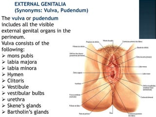
1. EXTERNAL GENITALIA
(Synonyms: Vulva, Pudendum)
The vulva or pudendum
includes all the visible
external genital organs in the
perineum.
Vulva consists of the
following:
mons pubis
labia majora
labia minora
Hymen
Clitoris
Vestibule
vestibular bulbs
urethra
Skene’s glands
Bartholin’s glands
2. MONS VENERIS (MONS
PUBIS): It is the pad of
subcutaneous adipose
connective tissue lying in
front of the pubis and in
the adult female is
covered by hair. The hair
pattern (escutcheon) of
most women is
triangular with the base
directed upwards.
3. LABIA MAJORA: The vulva is
bounded on each side by the
elevation of skin and
subcutaneous tissue which
form the labia majora. They
are continuous where they
join medially to form the
posterior commissure in
front of the anus.
The skin on the outer convex
surface is pigmented and
covered with hair follicle.
The thin skin on the inner
surface has sebaceous glands
but no hair follicle.
4. The labia majora are covered with
squamous epithelium and contain sweat
glands. Beneath the skin, there is dense
connective tissue and adipose tissue. The
adipose tissue is richly supplied with
venous plexus which may produce
hematoma, if injured during childbirth.
The labia majora are homologous to the
scrotum in the male. The round ligament
terminates at its upper border.
5. LABIA MINORA: They are two thin folds of skin, devoid of
fat, on either side just within the labia majora. Except in
the parous women, they are exposed only when the labia
majora are separated. Anteriorly, they divide to enclose
the clitoris and unite with each other in front and behind
the clitoris to form the prepuce and frenulum
respectively. The lower portion of the labia minora fuses
across the midline to form a fold of skin known as
fourchette. It is usually lacerated during childbirth.
Between the fourchette and the vaginal orifice is the
fossa navicularis. The labia minora contain no hair
follicles or sweat glands. The folds contain connective
tissues, numerous sebaceous glands, erectile muscle
fibers and numerous vessels and nerve endings. The labia
minora are homologous to the penile urethra and part of
the skin of penis in males.
6.
7. CLITORIS: It is a small cylindrical erectile body,
measuring about 1.5–2 cm situated in the most
anterior part of the vulva.
It consists of a
glans,
body
two crura.
The clitoris consists of two cylindrical corpora
cavernosa (erectile tissue). The glans is covered
by squamous epithelium and is richly supplied
with nerves. The vessels of the clitoris are
connected with the vestibular bulb and are
liable to be injured during childbirth.
8. Clitoris is homologous to the penis in the
male but it differs in being entirely
separate from the urethra. It is attached
to the under surface of the symphysis pubis
by the suspensory ligament.
9.
10. VESTIBULE: It is a triangular space bounded
anteriorly by the clitoris,
posteriorly by the fourchette
on either side by labia minora.
There are four openings into the vestibule.
(a) Urethral opening: The opening is situated in
the midline just in front of the vaginal orifice
about 1–1.5 cm below the pubic arch. The
paraurethral ducts open either on the
posterior wall of the urethral orifice or
directly into the vestibule.
epithelium.
11. (b) Vaginal orifice and hymen: The vaginal orifice
lies in the posterior end of the vestibule and is of
varying size and shape. In virgins and nulliparae,
the opening is closed by the labia minora, but in
parous, it may be exposed. It is incompletely
closed by a septum of mucous membrane, called
hymen. The membrane varies in shape but is
usually circular or crescentic in virgins. The
hymen is usually ruptured at the consummation
of marriage. During childbirth, the hymen is
extremely lacerated and is later represented by
cicatrized nodules of varying size, called the
carunculae myrtiformes. On both sides it is lined
by stratified squamous
12. (c) Opening of Bartholin’s
ducts: There are two Bartholin
glands (greater vestibular
gland), one on each side. They
are situated in the superficial
perineal pouch, close to the
posterior end of the vestibular
bulb. They are pea-sized and
yellowish white in color.
During sexual excitement, it
secretes abundant alkaline
mucus which helps in
lubrication. The glands are of
compound racemose variety
and are lined by cuboidal
epithelium.
13. Each gland has got a duct which measures
about 2 cm and opens into the vestibule
outside the hymen at the junction of the
anterior two-third and posterior one-third
in the groove between the hymen and the
labium minus. The duct is lined by
columnar epithelium but near its opening
by stratified squamous epithelium.
Bartholin’s glands are homologous to the
bulb of the penis in male.
14. (d) Skene’s glands are the largest paraurethral
glands. Skene’s glands are homologous to the
prostate in the male. The two Skene’s ducts may
open in the vestibule on either side of the
external urethral meatus.
15. VESTIBULAR BULB: These are bilateral elongated masses
of erectile tissues situated beneath the mucous
membrane of the vestibule. Each bulb lies on either side
of the vaginal orifice in front of the Bartholin’s gland and
is incorporated with the bulbocavernosus muscle. They
are homologous to the bulb of the penis and corpus
spongiosum in the male. They are likely to be injured
during childbirth with brisk hemorrhage (Fig. 1.2)
16. PERINEUM: The details of its anatomy are described later
in the chapter.
BLOOD SUPPLY:
Arteries—(a) Branches of internal pudendal artery—the
chief being labial, transverse perineal, artery to the
vestibular bulb and deep and dorsal arteries to the
clitoris.
17. (b) Branches of femoral
artery—superficial and
deep external pudendal.
18. Veins—The veins form
plexuses and drain into:
(a) Internal pudendal
vein, (b) vesical or
vaginal venous plexus
and (c) Long saphenous
vein. Varicosities during
pregnancy are not
uncommon and may
rupture spontaneously
causing visible bleeding
or hematoma formation
19. NERVE SUPPLY: The supply is through bilateral
spinal somatic nerves— (a) anterosuperior part is
supplied by the cutaneous branches from the
ilioinguinal and genital branch of genitofemoral
nerve(L1 and L2 ) and the posteroinferior part by
the pudendal branches from the posterior
cutaneous nerve of thigh (S1.2.3). Between
these two groups, the vulva is supplied by the
labial and perineal branches of the pudendal
nerve (S2.3.4).
20. LYMPHATICS: Vulval lymphatics have bilateral drainage.
Lymphatics drain into—(a) superficial inguinal nodes, (b)
intermediate groups of inguinal lymph nodes—gland of
Cloquet and (c) external and internal iliac lymph nodes.
DEVELOPMENT: External genitalia is developed in the
region of the cranial aspect of ectodermal cloacal fossa;
clitoris from the genital tubercle; labia minora from the
genital folds; labia majora from the labioscrotal swelling
and the vestibule from the urogenital sinus