1. 3. Methods
Preparation:
The upper 3 cm of sediment from three sediment cores collected during October 2015 were used to
enrich for Beggiatoa spp. The presence of Beggiatoa spp. filaments in the sediments was confirmed
by microscopy. Homogenized sediment slurry and shredded paper were added to Hungate tubes.
Enrichment conditions:
• MIS groundwater was added to two different heights.
• 8 tubes were loosely covered with a thin piece of aluminum foil, simulating aerobic conditions;
4 were induced to anaerobic conditions by being tightly capped; the remaining 4 had a hypodermic
needle inserted in the middle of the rubber cap, permitting microaerobic growth (Fig. 2).
• As an alternative potential terminal electron acceptor to O2 for SOB, 0.5 mL of 20 µM nitrate
solution was added to 8 tubes from each treatment, to a final concentration of 1 - 5µM.
• Half of the tubes were incubated in the dark without agitation at 8.6oC (in the following referred to
as “cold”) which resembles the temperature of the sinkhole environment. The other half was
incubated in the dark on an orbital shaker (100 RPM) at 20.0˚C (“room temperature”), to gently
move the water column and simulate the groundwater flow environment.
Observation:
The tubes were checked weekly for a month and any growth of sulfide oxidizing bacterial filaments,
namely Beggiatoa spp., was documented.
1. Introduction
• The underwater Middle Island Sinkhole (MIS) is a photic,
sulfidic, and low-oxygen modern system to understand
microbial life on early Earth.
• Members of Beggiatoa, a genus of filamentous sulfide-
oxidizing bacteria (SOB), live in cyanobacterial-dominated
microbial mats in MIS. Beggiatoa spp. can be found in
either freshwater or marine environments, and are key
autotrophs in light-limited environments such as deep-sea
hydrothermal vents and seeps (Preisler et al., 2007).
• The characteristics and activity of MIS Beggiatoa are of
particular interest due to their unique interaction with the
cyanobacteria. The white filaments cover the purple
cyanobacterial layer during the night but migrate
underneath the purple layer during the day (Biddanda et
al., 2012).
2. Research goal
To optimize growth conditions for Middle Island Sinkhole (MIS) sulfide oxidizing bacteria in order to
isolate a pure culture of Beggiatoaspp. for future physiological and ecological studies.
7. Acknowledgements and References
We thanks Matthew Medina and the NOAA Thunder Bay National Marine Sanctuary, including
captains and scuba divers and especially Russ Green, John Bright, Wayne Lusardi, and Phil
Hartmeyer. This work was supported by NSF grant EAR-1637066 to GJD.
Biddanda, Bopaiah A., Stephen C. Nold, Gregory J. Dick, S. T. Kendall, J. H. Vail, S. A. Ruberg, and C. M. Green. 2012. "Rock, water, microbes: underwater
sinkholes in Lake Huron are habitats for ancient microbial life." Nature Education Knowledge 3: 13.
Kinsman-Costello, L. E., Sheik, C. S., Sheldon, N. D., Allen Burton, G., Costello, D. M., Marcus, D., ... & Dick, G. J. .2016. “Groundwater shapes sediment
biogeochemistry and microbial diversity in a submerged Great Lake sinkhole.” Geobiology
Preisler, André, Dirk De Beer, Anna Lichtschlag, Gaute Lavik, Antje Boetius, and Bo Barker Jørgensen. 2007. "Biological and chemical sulfide oxidation in
a Beggiatoa inhabited marine sediment." The ISME journal 1, no. 4: 341-353.
4. Results and Analysis
Understanding growth and activity of Beggiatoa spp. from a low-oxygen environment
Hui Chien Tan1, Judith Klatt2, Sharon Grim3 and Gregory J. Dick1
1,2,3,4Earth and Environmental Sciences, University of Michigan, Ann Arbor, USA
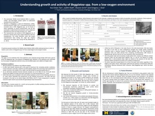
After a month of weekly observations, white filaments were absent in the anaerobic conditions but present in both microaerobic and aerobic conditions. Close inspection
revealed disparity in the growth of white filaments between those in the cold environment and those in the room-temperature environment (Table 1).
A B C D
Figure 3: A. An absence of growth was noted in the anaerobic tubes. B. Muddy yellow mesh growing
near the water level whereas white filament strands were found on the sediments (shown in red rings).
C. Muddy yellow clump potentially representing elemental sulfur. D. White filaments growing around red
tufts along with white ring formation near water surface.
Figure 4: SOB under 40x magnification
5. Discussion and Conclusion
We observed the best growth of SOB, likely Beggiatoa spp., in dark
and microaerobic/aerobic conditions. Though some members of
Beggiatoa are known to use nitrate in sulfide oxidation, we conclude
that nitrate cannot be used efficiently as an alternative terminal
electron acceptor for MIS Beggiatoa. We have four lines of support
for MIS Beggiatoa using oxygen, not nitrate, for sulfide oxidation:
1. We observed migration of SOB filaments in aerobic and
microaerobic treatments, towards the water surface where
atmospheric exchange introduced oxygen.
2. There were insubstantial differences in SOB growth between +/-
nitrate in the aerobic conditions.
3. We did not observe Beggiatoa growth in nitrate-amended,
anaerobic tubes.
4. Low nitrate concentrations in the ambient groundwater (Kinsman-
Costello et al., 2016) support our observation that MIS Beggiatoa
are not cued to use nitrate in their metabolism.
The formation of white rings near the water level provided insight on
the migration pattern of the white mat at MIS. Previous field
observations and laboratory experiments suggest that MIS Beggiatoa
motility is linked to oxygen demand: the oxygenic photosynthetic
cyanobacterial layer likely satisfies their oxygen requirements during
the day, so the SOB reside within and below the cyanobacterial mat;
whereas at night they are the top layer of the microbial mat,
putatively to acquire oxygen from the low-oxygen water column.
• Unlike the tufts of filaments in the tubes found in the cold environment, after two weeks
the color of the tufts at room temperature changed into muddy yellow, putatively
elemental sulfur, a chemical product of sulfide oxidation (Fig. 3 B). The formation of
elemental sulfur was likely derived from competing non-filamentous SOB instead of
Beggiatoa spp. because they do not store elemental sulfur extracellularly.
• The cold growth tubes did not develop muddy yellow tufts; instead, two different colored
filaments (dark red and white) grew together (Fig. 3 C and D). The absence of muddy
yellow tufts in cold-incubated tubes might be related to the presence of the dark red
filaments (likely cyanobacteria) and/or the incubation temperature.
• The white rings were situated about 0.4 cm from the water level surface regardless of the
height of the water column. Microscopy confirmed the presence of filamentous SOB with
intracellular refractive elemental sulfur inclusions, likely Beggiatoa spp., within the white
rings (Fig 4).
We are attempting to isolate Beggiatoa spp. from our enrichments using gradient media that
simulates the natural sediment biogeochemistry. For our first attempt, instead of the targeted
SOB, another species of unknown white rod-shaped bacteria were growing in the gradient
media (Fig. 5). The rod-shaped bacteria likely belong to the genus Thiobacillus. This lead us to
our second attempt where the gradient media consists of autoclaved MIS sediments on a
sulfidic agar plug (Fig. 6). Similar to the presented multivariate experiment, the tubes simulate
aerobic, anaerobic, and microaerobic environments.
Figure 5:
Unknown white
bacteria under
40x magnification
Figure 6: 2nd
attempt gradient
media setup for
culturing
Beggiatoa spp.
Figure 1: The observed migration pattern
of Beggiatoa spp. (Adapted from Biddanda
et al., 2012)
Table 1: Observations of microbial growth in a
multivariate environment
WF = White Filaments (growing on sediments)
WR = White ring (near water level)
YR = Yellow ring (near water level)
DRF = Dark Red Filaments
A B C
Figure 2: The pictures of tubes in different environmental simulations (in order):
A. anaerobic, B. microaerobic, C. aerobic conditions.
Condition Shaken Temp.
Water
column
+ Nitrate 1st Week 2nd Week 3rd Week 4th Week
Aerobic
Yes 20.0oC
2mL
Yes
Faint WR and WF
Tuft in the water column and WF
Muddy yellow clump in the water column and WF
No YR and WF
10mL
Yes No growth of WF Minimal WF White thin layer of WF White mesh with muddy yellow clump and WF
No Growth of WF Muddy YR, white mesh layer and WF
No 8.6oC
2mL
Yes
WF patches with DRF growing on top WR and WF patches with DRF growing on top
No
10mL
Yes
No growth of WF Growth of DRF Thin WR and DRF
No
Anaerobic
Yes 20.0oC
10mL
Yes
No growth of WF
No
No 8.6oC
Yes
No growth of WF but dark red filaments were observed
No
Micro-
aerobic
Yes 20.0oC
2mL
Yes
Faint WR and WF YR with white mesh and WF
No
No 8.6oC
Yes
WF patches with DRF on top Faint WR and WF tufts with DRF on topNo
6. Future Direction