SlideShare a Scribd company logo
1 of 68
Anatomy of EOMs & Ocular Motility
1
By: Dr Henok Samuel (R1)
Moderator: Prof Abebe Bejiga (professor of Ophthalmology, pediatrics and
strabismus surgeon)
Contents
• Introduction
• Recap on Embryology & microscopic anatomy
• Gross Anatomy of EOM
• Origin, Nerve and blood supply
• Action of EOM
• Basic Kinematics, Mechanics
• Ocular Movements
• Fundamental Laws governing ocular motility
2
Why do we move our eyes?
A. To acquire objects for central viewing
Saccadic eye movements
B. To maintain objects in foveal view
Pursuit eye movements
C. To stabilize the world on the retina
3
Extraocular Muscles
4
 EOMS are:
 Striated voluntary muscles
 Among muscles with fastest but also
most sustained contraction
 Get high blood flow exceeded only by
myocardium
 Have got high innervation ratio
Extraocular Muscles
5
 Play a vital role in
 Stereopsis
 Conjugate eye movements
 Maintenance of primary gaze position
 Motor fusion – maintaining corresponding visual elements
within the binocular field on corresponding retinal loci.
 Following of moving objects (smooth pursuit)
 Accomplish rapid changes in fixation (saccades).
Embryology
 The EOMs are derived from three primordial condensations - paraxial and
prechordal mesoderm.
 A pair of premandibular mesodermal condensation – 26th day
 Those muscles innervated by CNIII
 MR, SR, IR & IO .
 Two maxilomandibular condensations – 27th
 Superior oblique .
 Lateral rectus .
 Associated periorbital CT and smooth muscle are derived from the neural crest.
6
Embryology
 Their development begins at 3–4 weeks gestation.
 All of the extraocular muscles and their surrounding tissues are present and in
their final anatomic positions by 6 months gestation.
 The tendon insertions initially extend from the limbus to the equator.
 Continue to change until 18 months to 2 years after birth - 2–3.0 mm narrower in
infants
 At about 1 month, the nerves to the extraocular muscles reach their respective
destinations in the sequence oculomotor, abducens, and trochlear.
7
Microscopic Anatomy of EOMs
 Different from other skeletal muscles by their
 Diameter of its fibers is small
 Contain both slow and fast fibers
 denser connective tissue & blood supply
 Contain an enormous amount of fibroelastic tissue
 connective tissue sheaths are more delicate & richer in elastic fibers
 Have a large nerve to muscle fiber ratio; about 1:5 to 1:10 compared to 1:100
or more in other skeletal muscles
8
Microscopic Anatomy of EOMs
 Components of a muscle fiber
 Sarcolemma: the plasma cell membrane
surrounding each muscle fiber.
 Transverse tubules (T tubules): a series of
invaginations of the sarcolemma into the cell.
 Sarcoplasm: is the cell cytoplasm & contains
normal cellular structures and special muscle
fibers, THE MYOFIBRILS.
9
Microscopic Anatomy of EOMs
 Components of a MYOFIBRIL; two
types
1. Thick myofilaments
2. Thin myofilaments

10
Microscopic Anatomy of EOMs
Twitch single innervated fibers ( thick ):-
 contain little mitochondria ( i.e. anaerobic metabolism ) → rapid
response to the stimulus with rapid contraction of high amplitude &
short duration.
 responsible for saccadic eye movement & help fixation & pursuit
movement.
Tonic multiple innervated fibers ( thin ):-
 contain numerous mitochondria ( i.e. aerobic metabolism ) → slow
response to the stimulus with slow contraction of low amplitude &
long duration.
 responsible for gaze in all positions including 1ry position.
11
Bony orbit
•
12
Extraocular Muscles (EOMs)
 Vertical rectus
- Superior rectus
- Inferior rectus
 Horizontal rectus
- Medial rectus
- Lateral rectus
 Obliques
- Superior oblique
- Inferior oblique
13
Origin of EOMs
 The four recti muscles have their origin on the
common tendinous ring (annulus of Zinn).
 The area enclosed by the tendinous ring is called
the oculomotor foramen
 Superior Oblique originates from the lesser wing
of sphenoid
 Inferior Oblique originates from the maxillary
bone
14
Several blood vessels & nerves pass through
oculomotor foramen
1. The optic nerve
2. Ophthalmic artery
3. Abducens nerve
4. Oculomotor nerve Upper and lower
divisions
5. Nasociliary branch of the ophthalmic 15
Insertion
 Scleral insertions are by tendons
whose fibres are parallel to the
long axis of the muscle.
 The tendon fibres enter the
superficial sclera and quickly
merge into it.
 The EOMs penetrate it ≈10 mm
posterior to their insertions
16
MUSCLE LENGTH OF
TENDON IN mm
MEDIAL
RECTUS
3.7
LATERAL
RECTUS
8.8
SUPERIOR
RECTUS
5.8
INFERIOR
RECTUS
5.5
SUPERIOR
OBLIQUE
25
INFERIOR
OBLIQUE
1
Arterial supply
 Three main sources
1. Muscular branches of ophthalmic artery
Upper (lateral) branch
Lower (medial) branch
1. Infraorbital artery from ECA
2. Lacrimal artery
17
Arterial supply
 The muscular branches give rise to the anterior ciliary
arteries accompanying the rectus muscles; each
rectus muscle has 1–4 anterior ciliary arteries.
 Anterior segment circulation is most dependent on
arteries from vertical rectus muscles and least
dependent on arteries from the lateral rectus muscle
 Surgical manipulation of the rectus muscles
permanently disrupts the anterior ciliary arteries.
18
Venous drainage
19
 Follow the arterial path
 EOMs drain into two
veins:
1) Superior ophthalmic
vein and
2) Inferior ophthalmic
vein
Nerve
20
 Without neural activity, the
visual axes are usually mildly to
moderately divergent.
 The major tonic input to ocular
motility is supplied CNs III, IV,
and VI
Superior rectus
• Origin
• Insertion
• Nerve supply
• Blood supply
21
Inferior rectus
• Origin
• Insertion
• Nerve supply
• Blood supply
22
Medial rectus
• Origin
• Insertion
• Nerve supply
• Blood supply
23
Lateral rectus
• Origin
• Insertion
• Nerve supply
• Blood supply
24
Spiral of tillaux
25
 Imaginary line joining the insertions of the 4 recti and
is an important anatomical landmark when
performing surgery.
Superior Oblique
• Origin
• Insertion
• Nerve supply
• Blood supply
26
Inferior Oblique
• Origin
• Insertion
• Nerve supply
• Blood supply
27
Introduction
• Obliques are inserted behind equator & form an angle of 51°
with visual axis.
28
Orbital Connective Tissue
 Tenon's capsule (fascia bulbi) a layer of delicate
connective tissue that completely envelops the globe -
EOMs penetrate it ≈10 mm posterior to their insertions
 The fascial sheath of the SR muscle closely adheres to the
sheath of LPS of upper lid in front of the equator which
accounts for the cooperation in elevation of the eye.
 The sheath of the IO muscle fuses with the sheath of the
IR and forms the suspensory ligament of lockwood.
29
Pulleys of the EOMs
 Are about 2mm in length & located near the
equator
 Function as a mechanical origins of the EOMs
 Reduce sideslip of the extraocular muscles
during globe rotation and help to determine
the effective direction of pull
30
Pulleys of the EOMs
 They are Composed of
 Elastic fibers
 Collagenous
 The smooth muscles
 The most prominent smooth muscle is the
inframedial orbital muscle extending between
the MR and IR pulleys.
 Anteriorly, these sleeves thin to form slings
known as the intermuscular septum.
31
Muscle Capsule
 Each rectus muscle has a surrounding fascial capsule
 Extends with the muscle from its origin to its insertion
 Its smooth avascular surface allows the muscles to slide
smoothly over the globe.
32
Action of extraocular muscles
Types of Eye Movements
1. Ductions refer to monocular movements of each eye.
2. Versions refer to binocular conjugate movements of both eyes.
3. Vergences refer to binocular disjunctive movements.
33
Uniocular movements
 Ductions – only one eye is open, the other
covered/closed tested by asking the pt. to
follow a target in each direction of gaze.
 Types of ductions:-
1. Adduction
2. Abduction
3. Supraduction
4. Infraduction
5. Incycloduction
6. excycloduction
34
 Three Axes of Eye Rotations
 Fick’s Axis
 Listing Plane
35
Uniocular movements
Binocular movements
36
 Versions:-both eyes open, attempting to fixate a target
& moving in same direction.
 Binocular, simultaneous, conjugate movements in
same direction.
 Abduction of one eye accompanied by adduction of
other eye is called conjugate movements.
Introduction
 Types of versions:-
Dextroversion & laevo version
Supraversion & Infraversion
Dextro elevation & dextro depression
Laevo elevation & laevo depression
37
Introduction
• Torsional movements/righting reflexes:-
When you tilt head to maintain upright image.
• Vergences:-
binocular,simultaneous,disjugate/disjunctive movements (opp
direction)
Convergence– simultaneous adduction
Divergence– outward movement from convergent position
38
Actions of EOMs
• The anterior pole of the globe is the reference point used in the description of any eye
movement
• Eye movements are described based on the movement of the muscle insertion towards its
origin.
• The primary action of a muscle is its major effect when the eye is in the primary position.
• Subsidiary/secondary actions are the additional effects & depend on the position of the eye
• The point at which the center of the muscle or its tendon first touches the globe is called the
tangential point and indicates the direction of the pull.
• Muscles exert force in proportion to their cross-sectional area and length.
• For normal amplitude of rotation (45-50 degree ) 10mm change in muscle length is required
in each direction 39
Positions of Gaze
 Primary position: defined as position of the eye with
 Both head & body erect
 Both eyes are looking straight ahead
 Object of regard is at infinity
 The eye located at the intersection of the sagittal plane of the
head and the horizontal plane passing through the centers of
rotation of bot eyes
40
Positions of Gaze
 Secondary position are rotations around either the vertical axis
or the horizontal axis.
 ADDUCTION
 ABDUCTION
 ELEVATION
 DEPRESSION
41
Positions of Gaze
 Teritiary position: are rotations around both the vertical &
horizontal axis
 DEXTROELEVATION
 DEXTRODEPRESSION
 LEVOELEVATION
 LEVODEPRESSION
42
Positions of Gaze
43
Medial Rectus
 Lies parallel to the sagittal axis & perpendicular to
the vertical axis
 As a result has only one action, which is rotation
around the vertical axis medially => ADDUCTION
44
Lateral Rectus
 Lies parallel to the sagittal axis & perpendicular to
the vertical axis
 Contraction causes rotation in a temporal direction
ABDUCTION
45
Superior Rectus
 Run in line with the orbital axis and are
inserted in front of the equator.
 Forms an angle of 23° with the visual axis.
 With the insertion above the origin and on
the anterior globe, movement around the
horizontal axis causes ELEVATION –
PRIMARY ACTION.
46
 The muscle insertion is lateral to the origin,
so movement around the vertical axis causes
ADDUCTION.
 Its oblique insertion with the nasal side closer
to the limbus than the temporal side on the
superior surface of the globe causes
INTORSION on contraction.
47
Superior Rectus
Inferior Rectus
 Its insertion is below its origin and on the
anterior globe, so movement around the
horizontal axis causes DEPRESSION
48
Inferior Rectus
 The muscle insertion is lateral to the
origin, so movement around the vertical
axis causes ADDUCTION.
 Its oblique insertion on the inferior
surface of the globe causes EXTORSION
on contraction.
49
Superior Oblique
 The oblique insertion on the
posterosuperior lateral aspect of the
globe causes rotation of the eye
around the sagittal axis causing
INTORSION.
50
Superior Oblique
 The insertion is posterior and
inferior to the physiologic origin;
contraction of the muscle pulls
the back of the eye up, and the
anterior pole moves down-
DEPRESSION.

51
Superior Oblique
 Because the insertion is lateral to the
trochlea, contraction pulls the back of
the globe medially, thus moving the
anterior pole laterally – ABDUCTION.

52
Inferior Oblique
 Because the muscle wraps around the lower portion of the
globe and the insertion is superior and lateral to the origin
contraction causes EXTORSION.

53
Inferior Oblique
 Because the insertion is on the
posterior eye and above the
origin, contraction pulls the back
of the eye down, elevating the
front – ELEVATION.

54
Inferior Oblique
 because the insertion on the
back of the eye is pulled toward
the medial side; thus the
anterior pole is moved laterally
in ABDUCTION

55
Actions of the EOMs from secondary position
 HORIZONTAL RECTUS MUSCLES: LR & MR
 If the eye is elevated, contraction of the LR & MR no longer
causes strictly adduction or abduction but also causes a
slight elevation
 if the eye is depressed, contraction causes further
depression
56
VERTICAL RECTUS MUSCLES : SR & IR
57
 When the globe is abducted 23°, the
visual and orbital axes coincide.
 In this position they have no secondary
actions
 The SR can act only as an elevator & IR
only as a depressor
VERTICAL RECTUS MUSCLES : SR & IR
 When the eye is adducted 670 the plane of
the vertical rectus muscles is at a right angle
to the sagittal axis and thus parallel to the
horizontal axis.
 The superior rectus could only act as an
intortor & the inferior rectus could act only as
an extortor.
58
THE OBLIQUE MUSCLES : SO & IO
 The obliques form an angle of 51° with the
visual axis
 So if the eye is adducted 510, the plane of
the oblique muscles becomes parallel to
the sagittal axis and perpendicular to the
horizontal axis.
 Thus contraction of the SO will cause only
depression, and the IO will cause only
elevation
59
THE OBLIQUE MUSCLES : SO & IO
 When the eye is abducted 390, the plane
of the oblique muscles makes a right angle
with the sagittal axis & parallels the
horizontal axis,
 The superior oblique can cause only
intorsion & the IO extorsion
60
Actions of EOMs
61
Fundamental Laws governing Ocular Motility
 Synergists:- refers to muscles having same primary action
in same eye.
Ex:- sup.rectus & inf.oblique----elevators
inf.rectus & sup.oblique-----depressors
 Antagonists:- muscles having opposite action in same eye
Ex:- sup. & inf. Recti
sup. & inf.oblique
62
Fundamental Laws governing Ocular Motility
 Yoke muscle (contralateral synergists):-
Ref. to pair of muscles (one from each eye) which contract
simultaneously during version movements.
Ex :- In dextroversion RLR &LMR
 Contralateral antagonist:- pair of muscle (one from each eye)
having an opposite action.

Ex:-In dextroversion RLR & LLR
63
Fundamental Laws governing Ocular Motility
64
 DONDER’S LAW
 To each positon of line of sight belongs a definite orientation of the vertical and
horizontal retinal meridians relative to the coordinates of space.
 The orientation of the retinal meridians pertaining to a particular position of
globe is achieved irrespective of the path the eye has taken to reach that
position.
 In short, it implies that there is one and only one orientation of the retinal
meridians with each position of the eyes.
Fundamental Laws governing Ocular Motility
 Hering’s Law of Equal Innervation
 Also known as Hering’s law of motor correspondence
 States ‘ equal and simultanous innervation flows from the
brain to a pair of muscles of both eyes (yoke muscles) which
contract simultaneously in different binocular movements.’
 Eg: Right Lateral R and Left Medial rectus: Dextroversion
Both Medial Rectus : Convergence
Right IR and Left Superior Oblique: Dextrodepression
65
Fundamental Laws governing Ocular Motility
 Sherrington’s Law of Reciprocal
Innervation
 ‘During ocular motility, an increased flow of innervation to the
contracting agonist muscle is accompanied by a decreased flow
of innervation to the relaxing antagonist muscle’.
 Eg. During Dextroversion,
Increased innervation – Right LR and Left MR
Decreased innervation – Left LR and Right MR
66
References
 Wolf anatomy of the eye and orbit
 Clinical anatomy and physiology of the visual system
 Duane’s Ophthalmology 2007 edition parts 1 and 2
 Kanski clinical ophthalmology 7thedition chapter18
 BCSC 2019, section 2 and 7
67
68
Thank You!

More Related Content

What's hot

Maddox rod and double maddox rod
Maddox rod and double maddox rodMaddox rod and double maddox rod
Maddox rod and double maddox rodAnuMusyakhwo7
 
Adaptive mechanism of squint
Adaptive mechanism of squintAdaptive mechanism of squint
Adaptive mechanism of squintOm Patel
 
A-V pattern strabismus
A-V pattern strabismusA-V pattern strabismus
A-V pattern strabismusAmrit Pokharel
 
Duanes retraction syndrome ..
Duanes retraction syndrome ..Duanes retraction syndrome ..
Duanes retraction syndrome ..SSSIHMS-PG
 
Binocular refraction techniques, binocular balancing
Binocular refraction techniques, binocular balancing Binocular refraction techniques, binocular balancing
Binocular refraction techniques, binocular balancing Mohammad Arman Bin Aziz
 
Binocular refraction techniques, binocular balancing & binocular
Binocular refraction techniques, binocular balancing & binocularBinocular refraction techniques, binocular balancing & binocular
Binocular refraction techniques, binocular balancing & binocularsabina paudel
 
Specular microscopy
Specular microscopySpecular microscopy
Specular microscopyRuchi sood
 
Cycloplegic retinoscopy
Cycloplegic retinoscopy Cycloplegic retinoscopy
Cycloplegic retinoscopy R J
 
Corneal topography final
Corneal topography finalCorneal topography final
Corneal topography finalanjani kumar
 
Double elevator palsy
Double  elevator  palsyDouble  elevator  palsy
Double elevator palsyVinitkumar MJ
 
Motor adaptation in paretic and nonparetic strabismus
Motor adaptation in paretic and nonparetic strabismusMotor adaptation in paretic and nonparetic strabismus
Motor adaptation in paretic and nonparetic strabismuskopila kafle
 

What's hot (20)

Maddox rod and double maddox rod
Maddox rod and double maddox rodMaddox rod and double maddox rod
Maddox rod and double maddox rod
 
Adaptive mechanism of squint
Adaptive mechanism of squintAdaptive mechanism of squint
Adaptive mechanism of squint
 
A-V pattern strabismus
A-V pattern strabismusA-V pattern strabismus
A-V pattern strabismus
 
Maddox rod n wing
Maddox rod n wingMaddox rod n wing
Maddox rod n wing
 
Duanes retraction syndrome ..
Duanes retraction syndrome ..Duanes retraction syndrome ..
Duanes retraction syndrome ..
 
Binocular refraction techniques, binocular balancing
Binocular refraction techniques, binocular balancing Binocular refraction techniques, binocular balancing
Binocular refraction techniques, binocular balancing
 
Retinoscopy
RetinoscopyRetinoscopy
Retinoscopy
 
Corneal topography...ppt
Corneal topography...pptCorneal topography...ppt
Corneal topography...ppt
 
Binocular refraction techniques, binocular balancing & binocular
Binocular refraction techniques, binocular balancing & binocularBinocular refraction techniques, binocular balancing & binocular
Binocular refraction techniques, binocular balancing & binocular
 
Specular microscopy
Specular microscopySpecular microscopy
Specular microscopy
 
Binocular balancing
Binocular balancing Binocular balancing
Binocular balancing
 
Cycloplegic retinoscopy
Cycloplegic retinoscopy Cycloplegic retinoscopy
Cycloplegic retinoscopy
 
Corneal topography final
Corneal topography finalCorneal topography final
Corneal topography final
 
Retinoscopy
RetinoscopyRetinoscopy
Retinoscopy
 
fixation disparity
fixation disparityfixation disparity
fixation disparity
 
Double elevator palsy
Double  elevator  palsyDouble  elevator  palsy
Double elevator palsy
 
Motor adaptation in paretic and nonparetic strabismus
Motor adaptation in paretic and nonparetic strabismusMotor adaptation in paretic and nonparetic strabismus
Motor adaptation in paretic and nonparetic strabismus
 
Hess chart
Hess chartHess chart
Hess chart
 
Aberrometry
AberrometryAberrometry
Aberrometry
 
Subjective refraction
Subjective refractionSubjective refraction
Subjective refraction
 

Similar to Eoms & ocular motility

ENT and Maxillofacial and Ophtha course.pptx
ENT and Maxillofacial and Ophtha course.pptxENT and Maxillofacial and Ophtha course.pptx
ENT and Maxillofacial and Ophtha course.pptxsamirich1
 
Eom ppt
Eom pptEom ppt
Eom pptLhacha
 
Extraoclar muscles (Anatomy & physiology)
Extraoclar muscles (Anatomy & physiology)Extraoclar muscles (Anatomy & physiology)
Extraoclar muscles (Anatomy & physiology)Sanaa1993
 
Extra ocu.movement by pushkar dhir
Extra ocu.movement by pushkar dhirExtra ocu.movement by pushkar dhir
Extra ocu.movement by pushkar dhirPushkar Dhir
 
Extra ocu.movement by pushkar dhir
Extra ocu.movement by pushkar dhirExtra ocu.movement by pushkar dhir
Extra ocu.movement by pushkar dhirPushkar Dhir
 
ANATOMY AND PHYSIOLOGY OF EXTRAOCULAR MUSCLES.pptx
ANATOMY AND PHYSIOLOGY OF EXTRAOCULAR MUSCLES.pptxANATOMY AND PHYSIOLOGY OF EXTRAOCULAR MUSCLES.pptx
ANATOMY AND PHYSIOLOGY OF EXTRAOCULAR MUSCLES.pptxUttaraBhat
 
ANATOMY AND PHYSIOLOGY OF EXTRAOCULAR MUSCLES.ppt
ANATOMY AND PHYSIOLOGY OF EXTRAOCULAR MUSCLES.pptANATOMY AND PHYSIOLOGY OF EXTRAOCULAR MUSCLES.ppt
ANATOMY AND PHYSIOLOGY OF EXTRAOCULAR MUSCLES.pptANUJA DHAKAL
 
EXTRA OCULAR MUSCLES ANATOMY
EXTRA OCULAR MUSCLES ANATOMYEXTRA OCULAR MUSCLES ANATOMY
EXTRA OCULAR MUSCLES ANATOMYRabia Ammer
 
Extraocular muscles
Extraocular musclesExtraocular muscles
Extraocular musclesOm Patel
 
Anatomy of extraocular muscles and ocular motility
Anatomy of extraocular muscles and ocular motilityAnatomy of extraocular muscles and ocular motility
Anatomy of extraocular muscles and ocular motilityvanya kodali
 
STRABISMUS new-WPS Office.pptx
STRABISMUS new-WPS Office.pptxSTRABISMUS new-WPS Office.pptx
STRABISMUS new-WPS Office.pptxyashabandil155
 
Extraocular muscles.pptx
Extraocular muscles.pptxExtraocular muscles.pptx
Extraocular muscles.pptxSundip Charmode
 
Extraocular muscles.pptx
Extraocular muscles.pptxExtraocular muscles.pptx
Extraocular muscles.pptxshucayb1
 

Similar to Eoms & ocular motility (20)

ENT and Maxillofacial and Ophtha course.pptx
ENT and Maxillofacial and Ophtha course.pptxENT and Maxillofacial and Ophtha course.pptx
ENT and Maxillofacial and Ophtha course.pptx
 
Eom ppt
Eom pptEom ppt
Eom ppt
 
Extraoclar muscles (Anatomy & physiology)
Extraoclar muscles (Anatomy & physiology)Extraoclar muscles (Anatomy & physiology)
Extraoclar muscles (Anatomy & physiology)
 
Eye Muscles Anatomy
Eye Muscles AnatomyEye Muscles Anatomy
Eye Muscles Anatomy
 
Extra ocu.movement by pushkar dhir
Extra ocu.movement by pushkar dhirExtra ocu.movement by pushkar dhir
Extra ocu.movement by pushkar dhir
 
Extra ocu.movement by pushkar dhir
Extra ocu.movement by pushkar dhirExtra ocu.movement by pushkar dhir
Extra ocu.movement by pushkar dhir
 
ANATOMY AND PHYSIOLOGY OF EXTRAOCULAR MUSCLES.pptx
ANATOMY AND PHYSIOLOGY OF EXTRAOCULAR MUSCLES.pptxANATOMY AND PHYSIOLOGY OF EXTRAOCULAR MUSCLES.pptx
ANATOMY AND PHYSIOLOGY OF EXTRAOCULAR MUSCLES.pptx
 
ANATOMY AND PHYSIOLOGY OF EXTRAOCULAR MUSCLES.ppt
ANATOMY AND PHYSIOLOGY OF EXTRAOCULAR MUSCLES.pptANATOMY AND PHYSIOLOGY OF EXTRAOCULAR MUSCLES.ppt
ANATOMY AND PHYSIOLOGY OF EXTRAOCULAR MUSCLES.ppt
 
strabismus
strabismusstrabismus
strabismus
 
EXTRA OCULAR MUSCLES ANATOMY
EXTRA OCULAR MUSCLES ANATOMYEXTRA OCULAR MUSCLES ANATOMY
EXTRA OCULAR MUSCLES ANATOMY
 
Extraocular muscles
Extraocular musclesExtraocular muscles
Extraocular muscles
 
EXTRAOCULAR MUSCLES.pptx
EXTRAOCULAR MUSCLES.pptxEXTRAOCULAR MUSCLES.pptx
EXTRAOCULAR MUSCLES.pptx
 
Anatomy of extraocular muscles and ocular motility
Anatomy of extraocular muscles and ocular motilityAnatomy of extraocular muscles and ocular motility
Anatomy of extraocular muscles and ocular motility
 
STRABISMUS new-WPS Office.pptx
STRABISMUS new-WPS Office.pptxSTRABISMUS new-WPS Office.pptx
STRABISMUS new-WPS Office.pptx
 
EXTRA OCULAR MUSCLES
EXTRA OCULAR MUSCLES EXTRA OCULAR MUSCLES
EXTRA OCULAR MUSCLES
 
2 eye bony orbic and ball
2 eye bony orbic and ball2 eye bony orbic and ball
2 eye bony orbic and ball
 
Extraocular muscles.pptx
Extraocular muscles.pptxExtraocular muscles.pptx
Extraocular muscles.pptx
 
Extraocular muscles.pptx
Extraocular muscles.pptxExtraocular muscles.pptx
Extraocular muscles.pptx
 
Oculomotor nerve
Oculomotor nerveOculomotor nerve
Oculomotor nerve
 
Orbit
OrbitOrbit
Orbit
 

Recently uploaded

Russian Call Girls in Bangalore Manisha 7001305949 Independent Escort Service...
Russian Call Girls in Bangalore Manisha 7001305949 Independent Escort Service...Russian Call Girls in Bangalore Manisha 7001305949 Independent Escort Service...
Russian Call Girls in Bangalore Manisha 7001305949 Independent Escort Service...narwatsonia7
 
Call Girls Service Jaipur Grishma WhatsApp ❤8445551418 VIP Call Girls Jaipur
Call Girls Service Jaipur Grishma WhatsApp ❤8445551418 VIP Call Girls JaipurCall Girls Service Jaipur Grishma WhatsApp ❤8445551418 VIP Call Girls Jaipur
Call Girls Service Jaipur Grishma WhatsApp ❤8445551418 VIP Call Girls Jaipurparulsinha
 
VIP Call Girls Pune Vani 9907093804 Short 1500 Night 6000 Best call girls Ser...
VIP Call Girls Pune Vani 9907093804 Short 1500 Night 6000 Best call girls Ser...VIP Call Girls Pune Vani 9907093804 Short 1500 Night 6000 Best call girls Ser...
VIP Call Girls Pune Vani 9907093804 Short 1500 Night 6000 Best call girls Ser...Miss joya
 
Call Girls Colaba Mumbai ❤️ 9920874524 👈 Cash on Delivery
Call Girls Colaba Mumbai ❤️ 9920874524 👈 Cash on DeliveryCall Girls Colaba Mumbai ❤️ 9920874524 👈 Cash on Delivery
Call Girls Colaba Mumbai ❤️ 9920874524 👈 Cash on Deliverynehamumbai
 
High Profile Call Girls Jaipur Vani 8445551418 Independent Escort Service Jaipur
High Profile Call Girls Jaipur Vani 8445551418 Independent Escort Service JaipurHigh Profile Call Girls Jaipur Vani 8445551418 Independent Escort Service Jaipur
High Profile Call Girls Jaipur Vani 8445551418 Independent Escort Service Jaipurparulsinha
 
Call Girls Service Bellary Road Just Call 7001305949 Enjoy College Girls Service
Call Girls Service Bellary Road Just Call 7001305949 Enjoy College Girls ServiceCall Girls Service Bellary Road Just Call 7001305949 Enjoy College Girls Service
Call Girls Service Bellary Road Just Call 7001305949 Enjoy College Girls Servicenarwatsonia7
 
Call Girls Service Noida Maya 9711199012 Independent Escort Service Noida
Call Girls Service Noida Maya 9711199012 Independent Escort Service NoidaCall Girls Service Noida Maya 9711199012 Independent Escort Service Noida
Call Girls Service Noida Maya 9711199012 Independent Escort Service NoidaPooja Gupta
 
Housewife Call Girls Hoskote | 7001305949 At Low Cost Cash Payment Booking
Housewife Call Girls Hoskote | 7001305949 At Low Cost Cash Payment BookingHousewife Call Girls Hoskote | 7001305949 At Low Cost Cash Payment Booking
Housewife Call Girls Hoskote | 7001305949 At Low Cost Cash Payment Bookingnarwatsonia7
 
Russian Call Girls Chickpet - 7001305949 Booking and charges genuine rate for...
Russian Call Girls Chickpet - 7001305949 Booking and charges genuine rate for...Russian Call Girls Chickpet - 7001305949 Booking and charges genuine rate for...
Russian Call Girls Chickpet - 7001305949 Booking and charges genuine rate for...narwatsonia7
 
Call Girls In Andheri East Call 9920874524 Book Hot And Sexy Girls
Call Girls In Andheri East Call 9920874524 Book Hot And Sexy GirlsCall Girls In Andheri East Call 9920874524 Book Hot And Sexy Girls
Call Girls In Andheri East Call 9920874524 Book Hot And Sexy Girlsnehamumbai
 
CALL ON ➥9907093804 🔝 Call Girls Hadapsar ( Pune) Girls Service
CALL ON ➥9907093804 🔝 Call Girls Hadapsar ( Pune)  Girls ServiceCALL ON ➥9907093804 🔝 Call Girls Hadapsar ( Pune)  Girls Service
CALL ON ➥9907093804 🔝 Call Girls Hadapsar ( Pune) Girls ServiceMiss joya
 
Call Girls Service Chennai Jiya 7001305949 Independent Escort Service Chennai
Call Girls Service Chennai Jiya 7001305949 Independent Escort Service ChennaiCall Girls Service Chennai Jiya 7001305949 Independent Escort Service Chennai
Call Girls Service Chennai Jiya 7001305949 Independent Escort Service ChennaiNehru place Escorts
 
Low Rate Call Girls Pune Esha 9907093804 Short 1500 Night 6000 Best call girl...
Low Rate Call Girls Pune Esha 9907093804 Short 1500 Night 6000 Best call girl...Low Rate Call Girls Pune Esha 9907093804 Short 1500 Night 6000 Best call girl...
Low Rate Call Girls Pune Esha 9907093804 Short 1500 Night 6000 Best call girl...Miss joya
 
Call Girls Doddaballapur Road Just Call 7001305949 Top Class Call Girl Servic...
Call Girls Doddaballapur Road Just Call 7001305949 Top Class Call Girl Servic...Call Girls Doddaballapur Road Just Call 7001305949 Top Class Call Girl Servic...
Call Girls Doddaballapur Road Just Call 7001305949 Top Class Call Girl Servic...narwatsonia7
 
Call Girl Coimbatore Prisha☎️ 8250192130 Independent Escort Service Coimbatore
Call Girl Coimbatore Prisha☎️  8250192130 Independent Escort Service CoimbatoreCall Girl Coimbatore Prisha☎️  8250192130 Independent Escort Service Coimbatore
Call Girl Coimbatore Prisha☎️ 8250192130 Independent Escort Service Coimbatorenarwatsonia7
 
Aspirin presentation slides by Dr. Rewas Ali
Aspirin presentation slides by Dr. Rewas AliAspirin presentation slides by Dr. Rewas Ali
Aspirin presentation slides by Dr. Rewas AliRewAs ALI
 
Artifacts in Nuclear Medicine with Identifying and resolving artifacts.
Artifacts in Nuclear Medicine with Identifying and resolving artifacts.Artifacts in Nuclear Medicine with Identifying and resolving artifacts.
Artifacts in Nuclear Medicine with Identifying and resolving artifacts.MiadAlsulami
 
Russian Call Girls in Chennai Pallavi 9907093804 Independent Call Girls Servi...
Russian Call Girls in Chennai Pallavi 9907093804 Independent Call Girls Servi...Russian Call Girls in Chennai Pallavi 9907093804 Independent Call Girls Servi...
Russian Call Girls in Chennai Pallavi 9907093804 Independent Call Girls Servi...Nehru place Escorts
 
VIP Call Girls Indore Kirti 💚😋 9256729539 🚀 Indore Escorts
VIP Call Girls Indore Kirti 💚😋  9256729539 🚀 Indore EscortsVIP Call Girls Indore Kirti 💚😋  9256729539 🚀 Indore Escorts
VIP Call Girls Indore Kirti 💚😋 9256729539 🚀 Indore Escortsaditipandeya
 

Recently uploaded (20)

Russian Call Girls in Bangalore Manisha 7001305949 Independent Escort Service...
Russian Call Girls in Bangalore Manisha 7001305949 Independent Escort Service...Russian Call Girls in Bangalore Manisha 7001305949 Independent Escort Service...
Russian Call Girls in Bangalore Manisha 7001305949 Independent Escort Service...
 
Call Girls Service Jaipur Grishma WhatsApp ❤8445551418 VIP Call Girls Jaipur
Call Girls Service Jaipur Grishma WhatsApp ❤8445551418 VIP Call Girls JaipurCall Girls Service Jaipur Grishma WhatsApp ❤8445551418 VIP Call Girls Jaipur
Call Girls Service Jaipur Grishma WhatsApp ❤8445551418 VIP Call Girls Jaipur
 
VIP Call Girls Pune Vani 9907093804 Short 1500 Night 6000 Best call girls Ser...
VIP Call Girls Pune Vani 9907093804 Short 1500 Night 6000 Best call girls Ser...VIP Call Girls Pune Vani 9907093804 Short 1500 Night 6000 Best call girls Ser...
VIP Call Girls Pune Vani 9907093804 Short 1500 Night 6000 Best call girls Ser...
 
Call Girls Colaba Mumbai ❤️ 9920874524 👈 Cash on Delivery
Call Girls Colaba Mumbai ❤️ 9920874524 👈 Cash on DeliveryCall Girls Colaba Mumbai ❤️ 9920874524 👈 Cash on Delivery
Call Girls Colaba Mumbai ❤️ 9920874524 👈 Cash on Delivery
 
High Profile Call Girls Jaipur Vani 8445551418 Independent Escort Service Jaipur
High Profile Call Girls Jaipur Vani 8445551418 Independent Escort Service JaipurHigh Profile Call Girls Jaipur Vani 8445551418 Independent Escort Service Jaipur
High Profile Call Girls Jaipur Vani 8445551418 Independent Escort Service Jaipur
 
Call Girls Service Bellary Road Just Call 7001305949 Enjoy College Girls Service
Call Girls Service Bellary Road Just Call 7001305949 Enjoy College Girls ServiceCall Girls Service Bellary Road Just Call 7001305949 Enjoy College Girls Service
Call Girls Service Bellary Road Just Call 7001305949 Enjoy College Girls Service
 
sauth delhi call girls in Bhajanpura 🔝 9953056974 🔝 escort Service
sauth delhi call girls in Bhajanpura 🔝 9953056974 🔝 escort Servicesauth delhi call girls in Bhajanpura 🔝 9953056974 🔝 escort Service
sauth delhi call girls in Bhajanpura 🔝 9953056974 🔝 escort Service
 
Call Girls Service Noida Maya 9711199012 Independent Escort Service Noida
Call Girls Service Noida Maya 9711199012 Independent Escort Service NoidaCall Girls Service Noida Maya 9711199012 Independent Escort Service Noida
Call Girls Service Noida Maya 9711199012 Independent Escort Service Noida
 
Housewife Call Girls Hoskote | 7001305949 At Low Cost Cash Payment Booking
Housewife Call Girls Hoskote | 7001305949 At Low Cost Cash Payment BookingHousewife Call Girls Hoskote | 7001305949 At Low Cost Cash Payment Booking
Housewife Call Girls Hoskote | 7001305949 At Low Cost Cash Payment Booking
 
Russian Call Girls Chickpet - 7001305949 Booking and charges genuine rate for...
Russian Call Girls Chickpet - 7001305949 Booking and charges genuine rate for...Russian Call Girls Chickpet - 7001305949 Booking and charges genuine rate for...
Russian Call Girls Chickpet - 7001305949 Booking and charges genuine rate for...
 
Call Girls In Andheri East Call 9920874524 Book Hot And Sexy Girls
Call Girls In Andheri East Call 9920874524 Book Hot And Sexy GirlsCall Girls In Andheri East Call 9920874524 Book Hot And Sexy Girls
Call Girls In Andheri East Call 9920874524 Book Hot And Sexy Girls
 
CALL ON ➥9907093804 🔝 Call Girls Hadapsar ( Pune) Girls Service
CALL ON ➥9907093804 🔝 Call Girls Hadapsar ( Pune)  Girls ServiceCALL ON ➥9907093804 🔝 Call Girls Hadapsar ( Pune)  Girls Service
CALL ON ➥9907093804 🔝 Call Girls Hadapsar ( Pune) Girls Service
 
Call Girls Service Chennai Jiya 7001305949 Independent Escort Service Chennai
Call Girls Service Chennai Jiya 7001305949 Independent Escort Service ChennaiCall Girls Service Chennai Jiya 7001305949 Independent Escort Service Chennai
Call Girls Service Chennai Jiya 7001305949 Independent Escort Service Chennai
 
Low Rate Call Girls Pune Esha 9907093804 Short 1500 Night 6000 Best call girl...
Low Rate Call Girls Pune Esha 9907093804 Short 1500 Night 6000 Best call girl...Low Rate Call Girls Pune Esha 9907093804 Short 1500 Night 6000 Best call girl...
Low Rate Call Girls Pune Esha 9907093804 Short 1500 Night 6000 Best call girl...
 
Call Girls Doddaballapur Road Just Call 7001305949 Top Class Call Girl Servic...
Call Girls Doddaballapur Road Just Call 7001305949 Top Class Call Girl Servic...Call Girls Doddaballapur Road Just Call 7001305949 Top Class Call Girl Servic...
Call Girls Doddaballapur Road Just Call 7001305949 Top Class Call Girl Servic...
 
Call Girl Coimbatore Prisha☎️ 8250192130 Independent Escort Service Coimbatore
Call Girl Coimbatore Prisha☎️  8250192130 Independent Escort Service CoimbatoreCall Girl Coimbatore Prisha☎️  8250192130 Independent Escort Service Coimbatore
Call Girl Coimbatore Prisha☎️ 8250192130 Independent Escort Service Coimbatore
 
Aspirin presentation slides by Dr. Rewas Ali
Aspirin presentation slides by Dr. Rewas AliAspirin presentation slides by Dr. Rewas Ali
Aspirin presentation slides by Dr. Rewas Ali
 
Artifacts in Nuclear Medicine with Identifying and resolving artifacts.
Artifacts in Nuclear Medicine with Identifying and resolving artifacts.Artifacts in Nuclear Medicine with Identifying and resolving artifacts.
Artifacts in Nuclear Medicine with Identifying and resolving artifacts.
 
Russian Call Girls in Chennai Pallavi 9907093804 Independent Call Girls Servi...
Russian Call Girls in Chennai Pallavi 9907093804 Independent Call Girls Servi...Russian Call Girls in Chennai Pallavi 9907093804 Independent Call Girls Servi...
Russian Call Girls in Chennai Pallavi 9907093804 Independent Call Girls Servi...
 
VIP Call Girls Indore Kirti 💚😋 9256729539 🚀 Indore Escorts
VIP Call Girls Indore Kirti 💚😋  9256729539 🚀 Indore EscortsVIP Call Girls Indore Kirti 💚😋  9256729539 🚀 Indore Escorts
VIP Call Girls Indore Kirti 💚😋 9256729539 🚀 Indore Escorts
 

Eoms & ocular motility

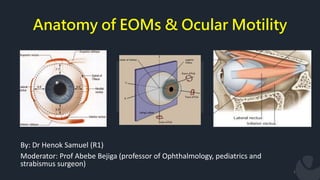
  • 1. Anatomy of EOMs & Ocular Motility 1 By: Dr Henok Samuel (R1) Moderator: Prof Abebe Bejiga (professor of Ophthalmology, pediatrics and strabismus surgeon)
  • 2. Contents • Introduction • Recap on Embryology & microscopic anatomy • Gross Anatomy of EOM • Origin, Nerve and blood supply • Action of EOM • Basic Kinematics, Mechanics • Ocular Movements • Fundamental Laws governing ocular motility 2
  • 3. Why do we move our eyes? A. To acquire objects for central viewing Saccadic eye movements B. To maintain objects in foveal view Pursuit eye movements C. To stabilize the world on the retina 3
  • 4. Extraocular Muscles 4  EOMS are:  Striated voluntary muscles  Among muscles with fastest but also most sustained contraction  Get high blood flow exceeded only by myocardium  Have got high innervation ratio
  • 5. Extraocular Muscles 5  Play a vital role in  Stereopsis  Conjugate eye movements  Maintenance of primary gaze position  Motor fusion – maintaining corresponding visual elements within the binocular field on corresponding retinal loci.  Following of moving objects (smooth pursuit)  Accomplish rapid changes in fixation (saccades).
  • 6. Embryology  The EOMs are derived from three primordial condensations - paraxial and prechordal mesoderm.  A pair of premandibular mesodermal condensation – 26th day  Those muscles innervated by CNIII  MR, SR, IR & IO .  Two maxilomandibular condensations – 27th  Superior oblique .  Lateral rectus .  Associated periorbital CT and smooth muscle are derived from the neural crest. 6
  • 7. Embryology  Their development begins at 3–4 weeks gestation.  All of the extraocular muscles and their surrounding tissues are present and in their final anatomic positions by 6 months gestation.  The tendon insertions initially extend from the limbus to the equator.  Continue to change until 18 months to 2 years after birth - 2–3.0 mm narrower in infants  At about 1 month, the nerves to the extraocular muscles reach their respective destinations in the sequence oculomotor, abducens, and trochlear. 7
  • 8. Microscopic Anatomy of EOMs  Different from other skeletal muscles by their  Diameter of its fibers is small  Contain both slow and fast fibers  denser connective tissue & blood supply  Contain an enormous amount of fibroelastic tissue  connective tissue sheaths are more delicate & richer in elastic fibers  Have a large nerve to muscle fiber ratio; about 1:5 to 1:10 compared to 1:100 or more in other skeletal muscles 8
  • 9. Microscopic Anatomy of EOMs  Components of a muscle fiber  Sarcolemma: the plasma cell membrane surrounding each muscle fiber.  Transverse tubules (T tubules): a series of invaginations of the sarcolemma into the cell.  Sarcoplasm: is the cell cytoplasm & contains normal cellular structures and special muscle fibers, THE MYOFIBRILS. 9
  • 10. Microscopic Anatomy of EOMs  Components of a MYOFIBRIL; two types 1. Thick myofilaments 2. Thin myofilaments  10
  • 11. Microscopic Anatomy of EOMs Twitch single innervated fibers ( thick ):-  contain little mitochondria ( i.e. anaerobic metabolism ) → rapid response to the stimulus with rapid contraction of high amplitude & short duration.  responsible for saccadic eye movement & help fixation & pursuit movement. Tonic multiple innervated fibers ( thin ):-  contain numerous mitochondria ( i.e. aerobic metabolism ) → slow response to the stimulus with slow contraction of low amplitude & long duration.  responsible for gaze in all positions including 1ry position. 11
  • 13. Extraocular Muscles (EOMs)  Vertical rectus - Superior rectus - Inferior rectus  Horizontal rectus - Medial rectus - Lateral rectus  Obliques - Superior oblique - Inferior oblique 13
  • 14. Origin of EOMs  The four recti muscles have their origin on the common tendinous ring (annulus of Zinn).  The area enclosed by the tendinous ring is called the oculomotor foramen  Superior Oblique originates from the lesser wing of sphenoid  Inferior Oblique originates from the maxillary bone 14
  • 15. Several blood vessels & nerves pass through oculomotor foramen 1. The optic nerve 2. Ophthalmic artery 3. Abducens nerve 4. Oculomotor nerve Upper and lower divisions 5. Nasociliary branch of the ophthalmic 15
  • 16. Insertion  Scleral insertions are by tendons whose fibres are parallel to the long axis of the muscle.  The tendon fibres enter the superficial sclera and quickly merge into it.  The EOMs penetrate it ≈10 mm posterior to their insertions 16 MUSCLE LENGTH OF TENDON IN mm MEDIAL RECTUS 3.7 LATERAL RECTUS 8.8 SUPERIOR RECTUS 5.8 INFERIOR RECTUS 5.5 SUPERIOR OBLIQUE 25 INFERIOR OBLIQUE 1
  • 17. Arterial supply  Three main sources 1. Muscular branches of ophthalmic artery Upper (lateral) branch Lower (medial) branch 1. Infraorbital artery from ECA 2. Lacrimal artery 17
  • 18. Arterial supply  The muscular branches give rise to the anterior ciliary arteries accompanying the rectus muscles; each rectus muscle has 1–4 anterior ciliary arteries.  Anterior segment circulation is most dependent on arteries from vertical rectus muscles and least dependent on arteries from the lateral rectus muscle  Surgical manipulation of the rectus muscles permanently disrupts the anterior ciliary arteries. 18
  • 19. Venous drainage 19  Follow the arterial path  EOMs drain into two veins: 1) Superior ophthalmic vein and 2) Inferior ophthalmic vein
  • 20. Nerve 20  Without neural activity, the visual axes are usually mildly to moderately divergent.  The major tonic input to ocular motility is supplied CNs III, IV, and VI
  • 21. Superior rectus • Origin • Insertion • Nerve supply • Blood supply 21
  • 22. Inferior rectus • Origin • Insertion • Nerve supply • Blood supply 22
  • 23. Medial rectus • Origin • Insertion • Nerve supply • Blood supply 23
  • 24. Lateral rectus • Origin • Insertion • Nerve supply • Blood supply 24
  • 25. Spiral of tillaux 25  Imaginary line joining the insertions of the 4 recti and is an important anatomical landmark when performing surgery.
  • 26. Superior Oblique • Origin • Insertion • Nerve supply • Blood supply 26
  • 27. Inferior Oblique • Origin • Insertion • Nerve supply • Blood supply 27
  • 28. Introduction • Obliques are inserted behind equator & form an angle of 51° with visual axis. 28
  • 29. Orbital Connective Tissue  Tenon's capsule (fascia bulbi) a layer of delicate connective tissue that completely envelops the globe - EOMs penetrate it ≈10 mm posterior to their insertions  The fascial sheath of the SR muscle closely adheres to the sheath of LPS of upper lid in front of the equator which accounts for the cooperation in elevation of the eye.  The sheath of the IO muscle fuses with the sheath of the IR and forms the suspensory ligament of lockwood. 29
  • 30. Pulleys of the EOMs  Are about 2mm in length & located near the equator  Function as a mechanical origins of the EOMs  Reduce sideslip of the extraocular muscles during globe rotation and help to determine the effective direction of pull 30
  • 31. Pulleys of the EOMs  They are Composed of  Elastic fibers  Collagenous  The smooth muscles  The most prominent smooth muscle is the inframedial orbital muscle extending between the MR and IR pulleys.  Anteriorly, these sleeves thin to form slings known as the intermuscular septum. 31
  • 32. Muscle Capsule  Each rectus muscle has a surrounding fascial capsule  Extends with the muscle from its origin to its insertion  Its smooth avascular surface allows the muscles to slide smoothly over the globe. 32
  • 33. Action of extraocular muscles Types of Eye Movements 1. Ductions refer to monocular movements of each eye. 2. Versions refer to binocular conjugate movements of both eyes. 3. Vergences refer to binocular disjunctive movements. 33
  • 34. Uniocular movements  Ductions – only one eye is open, the other covered/closed tested by asking the pt. to follow a target in each direction of gaze.  Types of ductions:- 1. Adduction 2. Abduction 3. Supraduction 4. Infraduction 5. Incycloduction 6. excycloduction 34
  • 35.  Three Axes of Eye Rotations  Fick’s Axis  Listing Plane 35 Uniocular movements
  • 36. Binocular movements 36  Versions:-both eyes open, attempting to fixate a target & moving in same direction.  Binocular, simultaneous, conjugate movements in same direction.  Abduction of one eye accompanied by adduction of other eye is called conjugate movements.
  • 37. Introduction  Types of versions:- Dextroversion & laevo version Supraversion & Infraversion Dextro elevation & dextro depression Laevo elevation & laevo depression 37
  • 38. Introduction • Torsional movements/righting reflexes:- When you tilt head to maintain upright image. • Vergences:- binocular,simultaneous,disjugate/disjunctive movements (opp direction) Convergence– simultaneous adduction Divergence– outward movement from convergent position 38
  • 39. Actions of EOMs • The anterior pole of the globe is the reference point used in the description of any eye movement • Eye movements are described based on the movement of the muscle insertion towards its origin. • The primary action of a muscle is its major effect when the eye is in the primary position. • Subsidiary/secondary actions are the additional effects & depend on the position of the eye • The point at which the center of the muscle or its tendon first touches the globe is called the tangential point and indicates the direction of the pull. • Muscles exert force in proportion to their cross-sectional area and length. • For normal amplitude of rotation (45-50 degree ) 10mm change in muscle length is required in each direction 39
  • 40. Positions of Gaze  Primary position: defined as position of the eye with  Both head & body erect  Both eyes are looking straight ahead  Object of regard is at infinity  The eye located at the intersection of the sagittal plane of the head and the horizontal plane passing through the centers of rotation of bot eyes 40
  • 41. Positions of Gaze  Secondary position are rotations around either the vertical axis or the horizontal axis.  ADDUCTION  ABDUCTION  ELEVATION  DEPRESSION 41
  • 42. Positions of Gaze  Teritiary position: are rotations around both the vertical & horizontal axis  DEXTROELEVATION  DEXTRODEPRESSION  LEVOELEVATION  LEVODEPRESSION 42
  • 44. Medial Rectus  Lies parallel to the sagittal axis & perpendicular to the vertical axis  As a result has only one action, which is rotation around the vertical axis medially => ADDUCTION 44
  • 45. Lateral Rectus  Lies parallel to the sagittal axis & perpendicular to the vertical axis  Contraction causes rotation in a temporal direction ABDUCTION 45
  • 46. Superior Rectus  Run in line with the orbital axis and are inserted in front of the equator.  Forms an angle of 23° with the visual axis.  With the insertion above the origin and on the anterior globe, movement around the horizontal axis causes ELEVATION – PRIMARY ACTION. 46
  • 47.  The muscle insertion is lateral to the origin, so movement around the vertical axis causes ADDUCTION.  Its oblique insertion with the nasal side closer to the limbus than the temporal side on the superior surface of the globe causes INTORSION on contraction. 47 Superior Rectus
  • 48. Inferior Rectus  Its insertion is below its origin and on the anterior globe, so movement around the horizontal axis causes DEPRESSION 48
  • 49. Inferior Rectus  The muscle insertion is lateral to the origin, so movement around the vertical axis causes ADDUCTION.  Its oblique insertion on the inferior surface of the globe causes EXTORSION on contraction. 49
  • 50. Superior Oblique  The oblique insertion on the posterosuperior lateral aspect of the globe causes rotation of the eye around the sagittal axis causing INTORSION. 50
  • 51. Superior Oblique  The insertion is posterior and inferior to the physiologic origin; contraction of the muscle pulls the back of the eye up, and the anterior pole moves down- DEPRESSION.  51
  • 52. Superior Oblique  Because the insertion is lateral to the trochlea, contraction pulls the back of the globe medially, thus moving the anterior pole laterally – ABDUCTION.  52
  • 53. Inferior Oblique  Because the muscle wraps around the lower portion of the globe and the insertion is superior and lateral to the origin contraction causes EXTORSION.  53
  • 54. Inferior Oblique  Because the insertion is on the posterior eye and above the origin, contraction pulls the back of the eye down, elevating the front – ELEVATION.  54
  • 55. Inferior Oblique  because the insertion on the back of the eye is pulled toward the medial side; thus the anterior pole is moved laterally in ABDUCTION  55
  • 56. Actions of the EOMs from secondary position  HORIZONTAL RECTUS MUSCLES: LR & MR  If the eye is elevated, contraction of the LR & MR no longer causes strictly adduction or abduction but also causes a slight elevation  if the eye is depressed, contraction causes further depression 56
  • 57. VERTICAL RECTUS MUSCLES : SR & IR 57  When the globe is abducted 23°, the visual and orbital axes coincide.  In this position they have no secondary actions  The SR can act only as an elevator & IR only as a depressor
  • 58. VERTICAL RECTUS MUSCLES : SR & IR  When the eye is adducted 670 the plane of the vertical rectus muscles is at a right angle to the sagittal axis and thus parallel to the horizontal axis.  The superior rectus could only act as an intortor & the inferior rectus could act only as an extortor. 58
  • 59. THE OBLIQUE MUSCLES : SO & IO  The obliques form an angle of 51° with the visual axis  So if the eye is adducted 510, the plane of the oblique muscles becomes parallel to the sagittal axis and perpendicular to the horizontal axis.  Thus contraction of the SO will cause only depression, and the IO will cause only elevation 59
  • 60. THE OBLIQUE MUSCLES : SO & IO  When the eye is abducted 390, the plane of the oblique muscles makes a right angle with the sagittal axis & parallels the horizontal axis,  The superior oblique can cause only intorsion & the IO extorsion 60
  • 62. Fundamental Laws governing Ocular Motility  Synergists:- refers to muscles having same primary action in same eye. Ex:- sup.rectus & inf.oblique----elevators inf.rectus & sup.oblique-----depressors  Antagonists:- muscles having opposite action in same eye Ex:- sup. & inf. Recti sup. & inf.oblique 62
  • 63. Fundamental Laws governing Ocular Motility  Yoke muscle (contralateral synergists):- Ref. to pair of muscles (one from each eye) which contract simultaneously during version movements. Ex :- In dextroversion RLR &LMR  Contralateral antagonist:- pair of muscle (one from each eye) having an opposite action.  Ex:-In dextroversion RLR & LLR 63
  • 64. Fundamental Laws governing Ocular Motility 64  DONDER’S LAW  To each positon of line of sight belongs a definite orientation of the vertical and horizontal retinal meridians relative to the coordinates of space.  The orientation of the retinal meridians pertaining to a particular position of globe is achieved irrespective of the path the eye has taken to reach that position.  In short, it implies that there is one and only one orientation of the retinal meridians with each position of the eyes.
  • 65. Fundamental Laws governing Ocular Motility  Hering’s Law of Equal Innervation  Also known as Hering’s law of motor correspondence  States ‘ equal and simultanous innervation flows from the brain to a pair of muscles of both eyes (yoke muscles) which contract simultaneously in different binocular movements.’  Eg: Right Lateral R and Left Medial rectus: Dextroversion Both Medial Rectus : Convergence Right IR and Left Superior Oblique: Dextrodepression 65
  • 66. Fundamental Laws governing Ocular Motility  Sherrington’s Law of Reciprocal Innervation  ‘During ocular motility, an increased flow of innervation to the contracting agonist muscle is accompanied by a decreased flow of innervation to the relaxing antagonist muscle’.  Eg. During Dextroversion, Increased innervation – Right LR and Left MR Decreased innervation – Left LR and Right MR 66
  • 67. References  Wolf anatomy of the eye and orbit  Clinical anatomy and physiology of the visual system  Duane’s Ophthalmology 2007 edition parts 1 and 2  Kanski clinical ophthalmology 7thedition chapter18  BCSC 2019, section 2 and 7 67

Editor's Notes

  1. Although the eye is responsible for transducing patterns of light energy into neuronal signals, it is the brain that is ultimately responsible for visual perception and cognition.
  2. In the retina we have a highly specialized system in higher mammals and humans where there is only small area where there is high density photoreceptor so to see fine detail u need to move your eyes to location u need to analyze with high level of acuity. It involves Acquire object for central viewing bcz its central viewing that allows u to have high acuity. Those accomplish predominantly by saccadic eye movt – which r rapid eye movt fr one location to another. Another impt whn u see like a bird flying…in order to analyze it u track z object – smooth pursuit mechanism which enable u to analyze the world accurately. Another factor is that whn we move abt its imp for eyes to be stable with respect to the world out there. One of the mechanism is Accessory optic system controlling eye visual stimuli and vestibular system.
  3. There are 7 extraocular muscles (EOMs) in the human eye: 4 rectus muscles 2 oblique muscles levator palpebrae superioris muscle All these muscles except LPS work with the surrounding orbital tissues to provide smooth movements of the globes and allow for binocular vision. Seventh muscle called LPS Similar in innervations Physiologically and metabolically different
  4. There are 7 extraocular muscles (EOMs) in the human eye: 4 rectus muscles 2 oblique muscles levator palpebrae superioris muscle All these muscles except LPS work with the surrounding orbital tissues to provide smooth movements of the globes and allow for binocular vision. Seventh muscle called LPS Similar in innervations Physiologically and metabolically different
  5. The EOMs form from paraxial and prechordal mesoderm, following cues from the developing eye as well as from surrounding neural crest mesenchyme Their development begins at 3–4 weeks gestation All of the extraocular muscles and their surrounding tissues are present and in their final anatomic positions by 6 months gestation.
  6. At about 7 weeks the dorsomedial aspect of the superior rectus muscle gives rise to the levator muscle they do not begin development at their origins and sprout toward their respective insertions
  7. Epimysium : a connective tissue sheath surrounding the entire muscle Perimysium: continuous with epimysium infiltrates the muscle & divides it into bundles Endomysium: a delicate connective tissue enclosure that surrounds the individual muscle fiber within the bundle
  8. Although the eye is responsible for transducing patterns of light energy into neuronal signals, it is the brain that is ultimately responsible for visual perception and cognition.
  9. thick myofibrils composed of hundreds of myosin subunits These filaments lie next to each other and form the “backbone” of the myofibril thin myofibrils formed by the protein actin arranged in a double-helical filament & a molecular complex of troponin and tropomyosin lying within the grooves of the double helix
  10. Although the eye is responsible for transducing patterns of light energy into neuronal signals, it is the brain that is ultimately responsible for visual perception and cognition.
  11. Each eye lies within the orbit The medial walls of the orbit are parallel to the midsagittal plane The lateral walls form an angle of ≈ 900 with each other. medial and lateral orbital walls thus form an angle of ≈ 450 with each other. Each orbital axis diverges from the midline by 22.50 and from the axis of the other orbit by 450
  12. Origin Common tendinous ring: “annulus tendinous communis”, the annulus of Zinn  SR and MR closely attached to the dural sheath of the optic nerve at their origin
  13. the annulus of Zinn  SR and MR closely attached to the dural sheath of the optic nerve at their origin It is this attachment of the superior and medial recti to the nerve sheath which is responsible for the characteristic pain which accompanies extreme movements of the globe in retrobulbar neuritis. Superior Oblique originates from the lesser wing of sphenoid
  14. In thyroid orbitopath MR and IR thickens especially ner the orbital apex compressing the optic nerve as it enters the optic canal
  15. They resemble scleral fibres, being of the same tissue, but differ in size and, whereas tendon fibres are mostly longitudinal, the scleral fibres run in many directions Only the cessation of the thick elastic fibres marks the junction of tendon with sclera Medial rectus muscle is suseptable to injury during anterior segment surgery inadvertent removal of the MR is a well known complication of perygyium removal. If integrity of tenon capsule lost during trauma or surgery 10mm posterior to limbus fatty tissue prolapse will result in restrictive adhesion and limit ocular motility
  16. SUPERIOR/LATERAL BRANCH - LR , SO, SR INFERIOR/MEDIAL BRANCH – MR, IO, IR The arteries to the four rectus muscle give rise to the ant ciliary arteries Blood supply to EOM supplies most of ant segment thus simultaneous surgery on 3 recti induces ant segment ischemia External carotid - Maxillary artery - Infraorbital artery Inferior rectus Inferior oblique
  17. These cilliary vesels pass to the episclera of the globe and then supply blood to the anterior segment The long posterior ciliary arteries also supply the anterior segment of the eye with blood via the major arterial circle of the iris. These long posterior ciliary arteries allow collateral blood flow after rectus muscle surgery. The blood vessels that supply the oblique muscles do not carry any circulation to the anterior segment.
  18. Although the eye is responsible for transducing patterns of light energy into neuronal signals, it is the brain that is ultimately responsible for visual perception and cognition.
  19. Except for IO, innervation to each of the EOMs occurs ≈1/3 the distance from the apex IO receives its innervation at ≈ its midpoint All 6 EOMs receive their innervation on the inside surface, except for SO, where branches of CN IV terminate on the upper (outer) surface
  20.  Arises from upper part of annulus o zinn.  Below the attachment of levator M.  Continuous with attachment of med.,&lat. Recti  Pierces tenon’s capsule &it is inserted into sclera 7.7 mm from superior limbus.  Length 48 mm;width 9mm.  N.supply:-sup.divison of oculomotor N.  B.supply:-lat. Muscular branch of ophthalmic A
  21.   Shortest of all recti  Arises from lower part of optic foramen.  Attached to sclera at 6.5 mm from inferior limbus  Lies b/w globe and inf.oblique.  Also attached to fascial sheath of lower lid.  Length 40mm;width 9mm  N.supply:-branch of inf divison of oculomotor N.  B.supply:-medial muscular branch of ophthalmic A
  22.   Largest ocular M& stronger than lateral rectus.  Arise from medial & inferior sides of optic foramen  Passing along medial wall of orbit ;inserts 5.5mm from nasal limbus.  Length 40mm;thicker than other EOM.  N.supply:-inf.divison of oculomotor N.  B.supply:-medial muscular branch of ophthalmic A
  23.   Arises from annular tendon.  Pierces tenon’s capsule &inserts in sclera at 6.9 mm from temporal limbus.  Length 48mm;2/3 of cross sectional area of MR.  N.supply:-Abducent N enters lR on its ocular aspect,just post.to its mid point.
  24. The insertions are located progressively further away from the limbus in a spiral pattern. the medial rectus insertion is closest. Superior rectus is farthest
  25.  Longest& thinnest EOM.  Arises from common origin at the apex of orbit; superomedial to optic foramen.  Runs forward to trochlea(cartilaginous ring at upper&inner angles of orbit)  After threading through this it becomes tendinous  It changes its direction completely and runs over the globe under SR to attach above & lat, to posterior pole. N.supply:-Trochlear N(4) after dividing into 2-3 branches enters muscle superiorly.  B.supply:-superior muscular branch of ophthalmic A. The trochlea redirects the tendon inferiorly, posteriorly, and laterally, with the tendon forming an angle of 51° with the visual axis in primary position
  26.   Only EOM not arising from apex of orbit  It arises anteriorly from lower & inner orbital walls near lacrimal fossa.  Running below inf.rectus& attaches below&lat. to post.pole of globe.  N.supply:-Inf.divison of oculomotor N.  B.supply:-Infraorbital &medial muscular branches of ophtalmic A
  27. Although the eye is responsible for transducing patterns of light energy into neuronal signals, it is the brain that is ultimately responsible for visual perception and cognition.
  28. Apart from serviing as cavity, support and protection of the globe, they play an important role in control of eye movement, reducing retraction
  29. They are Composed of Elastic fibers which provide reversible extensibility collagenous pulley ring forms the fulcrum of the pulley that inflects the EOM path The smooth muscles provide modulatory force on the pulleys Pulley displacement can clinically mimic muscle dysfunction, and orbital imaging may be needed to distinguish it accurately from a palsy. The pulling direction of each EOM is thus defined by the line segment connecting the EOM's scleral insertion to the respective pulley
  30. Although the eye is responsible for transducing patterns of light energy into neuronal signals, it is the brain that is ultimately responsible for visual perception and cognition.
  31. Although the eye is responsible for transducing patterns of light energy into neuronal signals, it is the brain that is ultimately responsible for visual perception and cognition.
  32. Although the eye is responsible for transducing patterns of light energy into neuronal signals, it is the brain that is ultimately responsible for visual perception and cognition.
  33. Although the eye is responsible for transducing patterns of light energy into neuronal signals, it is the brain that is ultimately responsible for visual perception and cognition.
  34.  center of rotation, located 13.5 mm behind the apex of the cornea & 1.3mm behind the equatorial plane this point varies in ametropia, is slightly more posterior in myopia, and is slightly more anterior in hyperopia  3 types of rotation: Listing Plane is an imaginary coronal plane passing through the center of rotation of globe 1. Rotation around fick vertical axis Z—side to side 2. “ “ fick horizontal axis X– up&down 3. “ “ fick antero posterior axis– torsion Torsions or cyclorotations are rotations around the sagittal axis and are described in relation to a point at the 12-o’clock position on the superior limbus.
  35. Although the eye is responsible for transducing patterns of light energy into neuronal signals, it is the brain that is ultimately responsible for visual perception and cognition.
  36. Although the eye is responsible for transducing patterns of light energy into neuronal signals, it is the brain that is ultimately responsible for visual perception and cognition.
  37. Although the eye is responsible for transducing patterns of light energy into neuronal signals, it is the brain that is ultimately responsible for visual perception and cognition.
  38. ; When the front of the eye moves up, the back moves down When the front of the eye moves right, the back of the eye moves left tangential point is called physiological insertion
  39. Although the eye is responsible for transducing patterns of light energy into neuronal signals, it is the brain that is ultimately responsible for visual perception and cognition.
  40. Although the eye is responsible for transducing patterns of light energy into neuronal signals, it is the brain that is ultimately responsible for visual perception and cognition.
  41. Although the eye is responsible for transducing patterns of light energy into neuronal signals, it is the brain that is ultimately responsible for visual perception and cognition.
  42. The anterior pole of the globe is the reference point used in the description of any eye movement; When the front of the eye moves up, the back moves down When the front of the eye moves right, the back of the eye moves left Eye movements are described based on the movement of the muscle insertion towards its origin. The primary action of a muscle is its major effect when the eye is in the primary position. Subsidiary/secondary actions are the additional effects & depend on the position of the eye The point at which the center of the muscle or its tendon first touches the globe is called the tangential point and indicates the direction of the pull Muscles exert force in proportion to their cross-sectional area The power of the muscle is proportional to its length For normal amplitude of rotation (45-50 degree ) 10mm change in muscle length is required in each direction
  43. Although the eye is responsible for transducing patterns of light energy into neuronal signals, it is the brain that is ultimately responsible for visual perception and cognition.
  44. Although the eye is responsible for transducing patterns of light energy into neuronal signals, it is the brain that is ultimately responsible for visual perception and cognition.
  45. Although the eye is responsible for transducing patterns of light energy into neuronal signals, it is the brain that is ultimately responsible for visual perception and cognition.
  46. Although the eye is responsible for transducing patterns of light energy into neuronal signals, it is the brain that is ultimately responsible for visual perception and cognition.
  47. Although the eye is responsible for transducing patterns of light energy into neuronal signals, it is the brain that is ultimately responsible for visual perception and cognition.
  48. Although the eye is responsible for transducing patterns of light energy into neuronal signals, it is the brain that is ultimately responsible for visual perception and cognition.
  49. Although the eye is responsible for transducing patterns of light energy into neuronal signals, it is the brain that is ultimately responsible for visual perception and cognition.
  50. Although the eye is responsible for transducing patterns of light energy into neuronal signals, it is the brain that is ultimately responsible for visual perception and cognition.
  51. Although the eye is responsible for transducing patterns of light energy into neuronal signals, it is the brain that is ultimately responsible for visual perception and cognition.
  52. Although the eye is responsible for transducing patterns of light energy into neuronal signals, it is the brain that is ultimately responsible for visual perception and cognition.
  53. Although the eye is responsible for transducing patterns of light energy into neuronal signals, it is the brain that is ultimately responsible for visual perception and cognition.
  54. Although the eye is responsible for transducing patterns of light energy into neuronal signals, it is the brain that is ultimately responsible for visual perception and cognition.
  55. As the position of the globe changes, the relationship between the muscle origin and insertion changes relative to the axes contraction of a muscle has a different effect than when the eye is in primary position
  56. Although the eye is responsible for transducing patterns of light energy into neuronal signals, it is the brain that is ultimately responsible for visual perception and cognition.
  57. Although the eye is responsible for transducing patterns of light energy into neuronal signals, it is the brain that is ultimately responsible for visual perception and cognition.
  58. Although the eye is responsible for transducing patterns of light energy into neuronal signals, it is the brain that is ultimately responsible for visual perception and cognition.
  59. Although the eye is responsible for transducing patterns of light energy into neuronal signals, it is the brain that is ultimately responsible for visual perception and cognition.
  60. Both obliques have same tertiary action because inserted behind the center of rotation,  pull post. pole of globe medially  when they contract ant.portion of eye so it causes abduction Both recti have same tertiary action bcz they inserted anterior to centre of rotation  pull ant.portion of globe medially so it causes adduction • In adduction, obliques are the prime vertical movers • In abduction, recti are the prime vertical movers,
  61. Although the eye is responsible for transducing patterns of light energy into neuronal signals, it is the brain that is ultimately responsible for visual perception and cognition.
  62. Although the eye is responsible for transducing patterns of light energy into neuronal signals, it is the brain that is ultimately responsible for visual perception and cognition.
  63. Orientation depends solely on amount of elevation or depression and lateral rotation of the globe.
  64. Major physiologic principle involved in the binocular motor co-operation of the eyes  Applicable to all normal ocular movements including vergence and involuntar movemens  Exception:  Assymmetric convergence
  65. Implies that the state of tension in the agonist exerts a regulatory influence on the state of tension in the antagonist and vice versa.
  66. Although the eye is responsible for transducing patterns of light energy into neuronal signals, it is the brain that is ultimately responsible for visual perception and cognition.
  67. Although the eye is responsible for transducing patterns of light energy into neuronal signals, it is the brain that is ultimately responsible for visual perception and cognition.