EXTRAOCULAR MUSCLES.pptx
•
1 like•41 views

extraocular muscles in a simplest and easy way for the students of eye care ophthalmology and optometry on fingertips .

Recommended
More Related Content
Similar to EXTRAOCULAR MUSCLES.pptx
Similar to EXTRAOCULAR MUSCLES.pptx (20)
More from Hayatabad medical complex peshawar khyber pukhtukhwa pakistan
More from Hayatabad medical complex peshawar khyber pukhtukhwa pakistan (6)
Recently uploaded
Recently uploaded (20)
EXTRAOCULAR MUSCLES.pptx
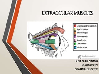
- 1. EXTRAOCULAR MUSCLES BY: Shoaib Khattak BS optometry Pico HMC Peshawar
- 2. Contents • Orbital muscles • Introduction to EOM • Anatomy of EOM • Extra Ocular Muscles • Blood supply • Nerve supply • Functions of EOM • Wholesome
- 4. Introduction to extra ocular muscles • The extraocular muscles are located with in the orbit but are extrinsic and separate from the eyeball itself. • There are six extraocular muscles(4 rectus and 2 oblique) that control the movement of eyeball and one muscle(LPS) that control eyelid elevation.
- 5. Anatomy • The EOM are striated muscles. • They contain slow fibres that creates a graded contraction on the exterior surface and fast muscle fibres that produces rapid movement on anterior surface. • The muscle fibres contain high content of mitochondria and oxidative enzymes .
- 6. • They also contains high amount of glycogen and glycolated enzymes . • EOM contains palisade endings which are believed to act as a sensory receptors that passes signals to CNS to maintain muscle tension .
- 7. SUPERIOR RECTUS • This muscle originates from the superior portion of annulus of zinn. • Courses forward from approx. 42 mm along the dorsal aspect of the globe . • Forms an angle of 23 * with the axis of the globe. • Superiorly it is in close contact with levator muscle . • The muscle is inserted at 7.7 mm from the limbus with a width of 11mm .
- 8. • Function • Its primary function is elevation • Secondary function is intorsion • Tertiary function is adduction • Nervesupply • The superior rectus is supplied by superior division of oculomotor nerve . • Bloodsupply • Superior rectus primarily supplied by superior muscular branch of ophthalmic artery and the two anterior cilliary arteries .
- 9. INFERIOR RECTUS • The IR muscle arises from the inferior portion of annulus of zinn. • Courses forward for approximately 42 mm along the ventral aspect of the globe . • Insertion is about 6.5 mm from the limbus . • Width of the insertion is around 10mm.
- 10. • Nervesupply • The IR muscle is innervated by the inferior division of oculomotor nerve . • Bloodsupply • Blood supply is primarily from the inferior muscular branch of ophthalmic artery + infraorbital artery . • Function • Primary ; depression • Secondary ; extorsion • Tertiary ; adduction
- 12. MEDIAL RECTUS • The MR originatesat the orbitalapex from he medialportionof annulus of zinnin close contactwiththe optic nerve . • It coursesforward for approx.40mm alongthe medialaspectof the globe . • Onlythe last5mmof the MR are in contactwiththe eyeball. • MR is inserted around5.5mm from the limbus withthe widthof 10.50mm.
- 13. • Nervesupply • The MR muscle is innervatedby the inferiordivisionof oculomotor nerve. • Bloodsupply • The MR muscle receiveits bloodsupply fromthe inferior muscularbranchof ophthalmicartery. • Function • Its functionis to adductthe eye towards nose .
- 14. LATERAL RECTUS • The lateral rectus muscle arises from upper and lower portions of annulus of zinn. • Courses forward for approx. 40mm along the lateral aspect of the globe and crosses the insertion of inferior oblique muscle . • It penetrates the tenon’s capsule at 15mm from insertion. • The last 7-8 mm of the muscle is in contact with the eye. • The LR muscle is inserted around 7mm from the limbus with a width of about 9.5mm.
- 15. • Nerve supply • The LR muscle is innervated by VI cranial nerve the abducens nerve. • Bloodsupply • The LR muscle receive its blood supply from the inferior muscular branch of ophthalmic artery . • The LR also receive blood supply from lacrimal artery. • Function • Its function is to move the eyes in lateral direction i.e abduction of the eyeball.
- 16. SUPERIOR OBLIQUE • The SO muscle arises from the orbital apex from periosteum of the body of sphenoid bone. • Courses forward approx. 40mm along the medial wall of the orbit to the trochlea ( a v-shaped fibrocartilage attached to the frontal bone ). • The muscle is encased in synovial sheath. • Form an angle of about 51* with the visual axis . • Passes beneath the superior rectus and inserts on the upper temporal quadrant of the globe .
- 17. • Nervesupply • The SO muscle is innervatedby the trochlearnervethatenters the muscleon its surface roughly12 mm from its origin. • Bloodsupply • The SO muscle is supplied by superiormuscularbranchof the ophthalmicartery. • Function • Primary; intorsion • Secondary; depression • Tertiary; abduction
- 18. INFERIOR OBLIQUE • The IO muscle arisesfrom the floorof the orbitfrom the periosteumcoveringthe anteromedial portionof the maxillary bone. • It is the onlymusclethat’s notoriginatedfrom the annulusof zinn. • Courseslaterallyand posteriorlyapprox. 37mm. • Formsan angleof 51* withthe visualaxis. • Insertedunderthe lateralrectus justanteriorto the macular area.
- 19. • Nervesupply • The IO muscle is suppliedby the inferiordivisionof oculomotor nerve. • Bloodsupply • Inferioroblique is supplied by the ophthalmicartery and infraorbital artery,these arise from the internalcarotidand maxillaryarteries,respectively. • Function • Primary; extorsion • Secondary; elevation • Tertiary; abduction
- 20. LEVATOR PALPEBRAE SUPERIORIS • The LPS muscle arisesfrom undersurface of the lesserwingof sphenoidbone . • InsertedintoSuperior tarsalplate,skinof uppereyelid • As an extraocular muscle,levator palpebraesuperioris indirectly facilitates movementsof the eye by elevatingand retractingthe uppereyelid . • It allows unhinderedupwardgaze. • As a facialmuscle,it contributesto the arrayof facial expressions.
- 21. • Nervesupply • The LPS receivesits nerve supplyfrom superiordivisionof Oculomotornerve (CN III). • Bloodsupply • Receive its bloodsupplyfrom Ophthalmicarteryand supraorbital artery. • Function • The LPS muscle helpsin Elevatingthe upper eyelid
- 22. Summary
- 23. Muscle Course or length Origin Insertion Angle Nerve supply Blood supply Function Medial rectus 40 mm Annulus of zinn 5.5mm to limbus 90 Lower CNIII Inferior muscular branch adduction Lateral rectus 40mm Annulus of zinn 6.9mm to limbus 90 CNVI Superior muscular branch Abductio n Superior rectus 40mm Annulus of zinn 7.7mm to limbus 23 Upper CNIII Superior muscular branch Elevation, intorsion, adduction Inferior rectus 40mm Annulus of zinn 6.5mm to limbus 23 Lower CNIII Inferior muscular branch Depressio n,extorsio n,adducti on Superior oblique 32mm Orbital apex above annulus of zinn Post to equator of eyeball 51 CNIV Superior muscular branch Intorsion, depressio n,abducti on Inferior oblique 37mm Behind lacrimal Macular area 51 Lower CNIII Inferior muscular Extort,ele vates,abd