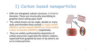
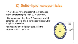
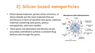
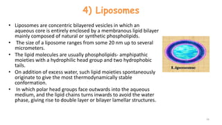
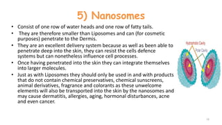
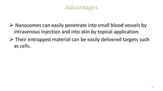
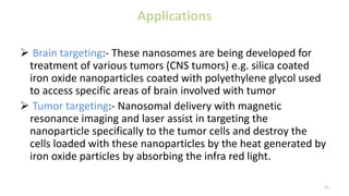
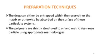
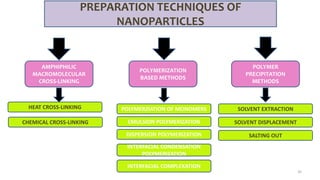
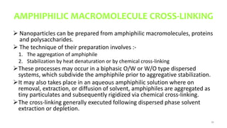
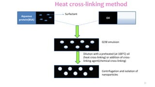
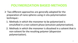
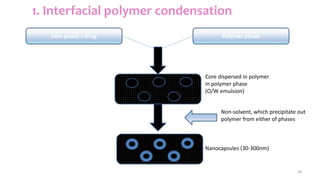
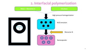
3. NANOPARTICLE
INTRODUCTION:- nano derives from the Greek word “nanos”, which means
dwarf or extremely small.
• Nanoparticles are particles between 1-100 nanometers(nm) in size with a
surrounding interfacial layer.
• Nanoparticles are sub-nano ionized colloidal structures composed of
synthetic or semi-synthetic polymers”.
• The interfacial layer is an integral part of the nanoparticles, fundamentally
affecting all of its properties.
• The interfacial layer typically consists of ions, inorganic and organic
molecules.
• The drug is dissolved, entrapped, encapsulated or attached to a
nanoparticle matrix.
3
4. • Nanoparticles are not simple molecules itself and therefore composed of three
layers –
The surface layer, which may be functionalized with a variety of small
molecules, metal ions, surfactants and polymers.
The shell layer, which is chemically different material from the core in all
aspects.
The core, which is essentially the central portion of the NP and usually refers
the NP itself.
4
5. Properties
• NPs possess unique physical and chemical properties due to their high surface
area and nanoscale size.
• Their optical properties are reported to be dependent on the size, which imparts
different colors due to absorption in the visible region their reactivity, toughness.
• The other properties are also dependent on their unique sizes, shape and
structure.
• Some of the important properties are:-
• Electronic and optical properties
• Magnetic properties
• Mechanical properties
• Thermal properties
5
7. 1) Carbon based nanoparticles
• CNPs are elongated, tubular structure, 1–2 nm in
diameter. These are structurally resembling to
graphite sheet rolling upon itself
• The rolled sheets can be single, double or many
walls and therefore they named as single-walled
(SWNTs), double-walled (DWNTs) or multi-walled
carbon nanotubes (MWNTs), respectively.
• They are widely synthesized by deposition of
carbon precursors especially the atomic carbons,
vaporized from graphite by laser or by electric arc
on to metal particles.
7
8. Advantages
High electrical conductivity
High mechanical strength
Ultra-light weight
High thermal conductivity
Metallic or semi-metallic behaviour
High surface area
8
9. Applications
Cell specifity
Vaccine delivery
Gene delivery
Transport of peptides, nucleic acids and other drug molecules
Reduced toxicity and increase the efficacy
Gene silencing
In diagnostics
9
10. 2) Solid-lipid nanoparticles
• A solid-lipid NP is characteristically spherical
with diameter ranging from 10 to 1000 nm.
• Like polymeric NPs, these NPs possess a solid
core made of lipid and a matrix contains soluble
lipophilic molecules.
• Surfactants or emulsifiers stabilized the
external core of these NPs.
10
11. Advantages
Excellent biocompatibility
Improve stability of pharmaceuticals
High and enhanced drug content
Easy to scale up and sterilize
Enhanced bioavailability of entrapped bioactive compounds
Chemical protection of labile incorporated compounds
11
12. Applications
In cancer therapy
In proteins & peptides delivery
For targeted brain drug delivery
For parasitic diseases
Ultrasonic drug & gene delivery
treatment of malaria
dermatological preparations
agriculture applications
12
13. 3) Silicon based nanoparticles
• Silicon-based materials, porous silicon and silica, or
silicon dioxide are the most materials that are
architecture in form of calcified nano pores, platinum
materials containing nano pores, porous
nanoparticles, and nano needles.
• Nano pores size (diameter) and density can be
accurately controlled to achieve a constant drug
delivery rate through the pores.
13
14. Advantages
It can have a higher mobility due to the presence of the silicon
crystallites.
Higher dielectric constant then bulk silicon.
One of the most important advantage of nano crystalline
silicon, however is that it has increased stability over a-Si.
14
15. Applications
Act as an artificial growth factor
For antibody delivery
For antibiotics, enzymes and DNA delivery
15
16. 4) Liposomes
• Liposomes are concentric bilayered vesicles in which an
aqueous core is entirely enclosed by a membranous lipid bilayer
mainly composed of natural or synthetic phospholipids.
• The size of a liposome ranges from some 20 nm up to several
micrometers.
• The lipid molecules are usually phospholipids- amphipathic
moieties with a hydrophilic head group and two hydrophobic
tails.
• On addition of excess water, such lipid moieties spontaneously
originate to give the most thermodynamically stable
conformation.
• In which polar head groups face outwards into the aqueous
medium, and the lipid chains turns inwards to avoid the water
phase, giving rise to double layer or bilayer lamellar structures.
16
17. Advantages
Provides selective passive targeting to tumor tissues.
Increased efficacy and therapeutic index.
Increased stability of encapsulated drug.
Reduction in toxicity of the encapsulated agent.
Site avoidance effect (avoids non-target tissues).
Improved pharmacokinetic effects (reduced elimination increased ci
rculation life times).
Flexibility to couple with site specific ligands to achieve active target
17
18. Applications
Chelation therapy for treatment of heavy metal poisoning.
Liposomes as protein carriers in immunology
Sustained or controlled delivery
Diagnostic imaging of tumors
Intracellular drug delivery
Site-specific targeting
Enzyme replacement
Oral drug delivery
Gene therapeutics
18
19. 5) Nanosomes
• Consist of one row of water heads and one row of fatty tails.
• They are therefore smaller than Liposomes and can (for cosmetic
purposes) penetrate to the Dermis.
• They are an excellent delivery system because as well as been able to
penetrate deep into the skin, they can resist the cells defence
systems but can nonetheless influence cell processes.
• Once having penetrated into the skin they can integrate themselves
into larger molecules.
• Just as with Liposomes they should only be used in and with products
that do not contain chemical preservatives, chemical sunscreens,
animal derivatives, fragrance and colorants as these unwelcome
elements will also be transported into the skin by the nanosomes and
may cause dermatitis, allergies, aging, hormonal disturbances, acne
and even cancer.
19
20. Advantages
Nanosomes can easily penetrate into small blood vessels by
intravenous injection and into skin by topical application.
Their entrapped material can be easily delivered targets such
as cells.
20
21. Applications
Brain targeting:- These nanosomes are being developed for
treatment of various tumors (CNS tumors) e.g. silica coated
iron oxide nanoparticles coated with polyethylene glycol used
to access specific areas of brain involved with tumor
Tumor targeting:- Nanosomal delivery with magnetic
resonance imaging and laser assist in targeting the
nanoparticle specifically to the tumor cells and destroy the
cells loaded with these nanoparticles by the heat generated by
iron oxide particles by absorbing the infra red light.
21
22. 6) Niosomes
• Niosome is a class of molecular cluster formed by self-association of non-ionic
surfactants in an aqueous phase. They have ability of loading both hydrophilic and
lipophilic drugs.
• Niosomes are vesicles composed of non-ionic surfactants, which are biodegradable,
relatively nontoxic, more stable and inexpensive, an alternative to liposomes.
• Niosomes behave in vivo like liposomes, prolonging the circulation of entrapped
drug and altering its organ distribution and metabolic stability.
• It is reported that the intercalation of cholesterol in the bilayers decreases the
entrapment volume during formulation, and thus entrapment efficiency.
• However, differences in characteristics exist between liposomes and niosomes,
especially since niosomes are prepared from uncharged single-chain surfactant and
cholesterol, whereas liposomes are prepared from double- chain phospholipids
(neutral or charged).
• The concentration of cholesterol in liposomes is much more than that in niosomes.
As a result, drug entrapment efficiency of liposomes becomes lesser than niosomes
22
23. Advantages
They are osmotically active and stable.
They increase the stability of entrapped drug.
The vesicle suspension being water based offers greater patient
compliance over oil based systems
Since the structure of niosome offers place to accommodate
hydrophilic, lipophilic as a well as amphiphilic drug moieties, they
can be used for a variety of drugs.
The vesciles can act as a depot to release the drug slowly and of
controlled release
Biodegradable, non-immunogenic and biocompatible.
23
24. Applications
It is used as drug targeting
It is used as Anti-Neoplastic treatment i.e. cancer disease, eg.
Methotraxate
Used in Leishmaniasis i.e dermal and mucocutaneous infections eg.
sodium stibogluconate
Used in delivery of peptide drugs
Niosomes as carriers for haemoglobin
It is used in ophthalmic drug delivery eg. Cyclopentane
Transdermal drug delivery system utilizing niosomes
eg.Erythromycine
24
25. 7) Nanocrystals and nanosuspensions
To produce Nanocrystals, the drug powder is dispersed in a surfactant
solution and the obtained suspension undergoes a pearl milling
process for hours upto several days.
To produce Nanosuspensions, the drug is dispersed in an aqueous
surfactant solution by high speed stirring.
The obtained macro-suspension is passed through a high speed
homogenizer leading to the formation of nanosuspension of the poorly
water-soluble drug.
25
27. Applications
It is used for targeting to the mucosa of the gastrointestinal
tract.
Targeting to the cells of nonnuclear phagocytic system(MPS) to
treat infections of MPS.
27
28. 8) Hydrogel Nanoparticles
Hydrogel nanoparticles are formed in water by
self-assemblage and self-aggregation of natural
polymer amphiphiles such as hydrophobized
polysaccharides, like cholesteroyl dextran .
Cholestrol bearing polysaccharides (CHP) self-
aggregate to form a mono-disperse and stable
hydrogel nanoparticles, in which the domains of
the associated cholesterol groups of cholestoryl
polysaccharides provide cross-linking points In a
non-covalent manner.
The size and density of hydrogel nanoparticles
can be controlled by changing the degree of
substitution of cholesterol groups of CHP.
28
29. Advantages
Environment can protect cells and other substances (i.e.drugs,
proteins and peptides)
Timed release of growth factors and other nutrients to ensure
proper issue growth
Good transport properties
Biocompatible
Can be injected
Easy to modify
29
30. Applications
Hydrogels are used for manufacturing contact lenses
Hygiene products
Tissue engineering scaffolds
Drug delivery systems and wound dressings.
30
31. 9) Copolymerized peptide nanoparticles
(CPP)
A novel co polymeric nano particulate drug delivery system,
copolymerized peptide particles(CPP) has been developed as a carrier
for the oral uptake of therapeutic peptides.
It is a drug polymer conjugate which forms its own nano particulate
delivery system in which drug moiety is covalently bound, rather
physically entrapped within the system.
31
32. Advantages
Enhance the oral uptake of peptides for drug delivery
Colloidal carriers can theoretically protect a labile drug from
degradation in the gut, and the systemic delivery of the drug
Enhance circulating half-life, Achieve drug targeting, is the
formation of a drug-polymer conjugate, e.g. conjugation of the
active drug moiety with polyethylene glycol .
32
34. PREPARATION TECHNIQUES
The drug can either be entrapped within the reservoir or the
matrix or otherwise be absorbed on the surface of these
particulate systems.
The polymers are strictly structured to a nano metric size range
particle using appropriate methodologies.
34
36. AMPHIPHILIC MACROMOLECULE CROSS-LINKING
Nanoparticles can be prepared from amphiphilic macromolecules, proteins
and polysaccharides.
The technique of their preparation involves :-
1. The aggregation of amphiphile
2. Stabilization by heat denaturation or by chemical cross-linking
These processes may occur in a biphasic O/W or W/O type dispersed
systems, which subdivide the amphiphile prior to aggregative stabilization.
It may also takes place in an aqueous amphiphilic solution where on
removal, extraction, or diffusion of solvent, amphiphiles are aggregated as
tiny particulates and subsequently rigidized via chemical cross-linking.
The cross-linking generally executed following dispersed phase solvent
extraction or depletion.
36
38. POLYMERIZATION BASED METHODS
Two different approaches are generally adopted for the
preparation of nano spheres using in situ polymerization
technique:-
1. Methods in which the monomer to be polymerized is
emulsified in a non-solvent phase (emulsion polymerization).
2. Methods in which the monomer is dissolved in a solvent that is
non-solvent for the resulting polymer (dispersion
polymerization).
38
39. 39
1. Interfacial polymer condensation
Core phase + Drug Polymer phase
Core dispersed in polymer
in polymer phase
(O/W emulsion)
Non-solvent, which precipitate out
polymer from either of phases
Nanocapsules (30-300nm)
41. POLYMER PRECIPITATION METHODS
In these methods, the hydrophobic polymer or a hydrophobic drug is dissolved
in a particular organic solvent followed by its dispersion in a continuous aqueous
phase, in which the polymer is insoluble.
The external phase also contains the stabilizer.
The polymer precipitation occurs as a consequence of the solvent extraction or
evaporation, which can be brought about by:-
Increasing the solubility of the organic solvent in the external medium by adding
an alcohol(i.e. isopropanol)
By incorporating additional amount water into the ultra emulsion
By evaporation of the organic solvent at room temperature or at accelerated
temperatures or by using vacuum
Using an organic solvent that is completely soluble in the continuous aqueous
phase-nanoprecipitation.
41
43. 43
2. Solvent Displacement Method
Distilled water
Polaxamer 188
Distilled water
Polaxamer 188
Organic solvent
Polymer, Drug
Polar solvent, Oil,
Polymer, Drug
Aqueous phase Aqueous phase
Magnetic
stirring
Nanospheres Nanocapsules
Organic phase Organic phase
44. 44
3. Salting out of Polymer
Organic solvent,
Drug, Polymer
Distilled water,
PVA, MgCl2
Organic phase Aqueous phase
Distilled water
O/W emulsion
Mechanical
stirring
45. EVALUATION
1. Particle size
2. Density
3. Molecular weight
4. Structure and crystallinity
5. Specific surface area
6. Surface charge & electronic mobility
7. Surface hydrophobicity
8. In-vitro release
9. Nanoparticle yield
10.Drug entrapment efficiency
45
46. 1. Particle size
Two main techniques are used to determine the particle size of
nanoparticles.
Photon correlation spectroscopy
Electron microscopy
1)scanning electron microscopy
2)transmission electron microscope
3)freeze fracture techniques
46
47. Photon correlation spectroscopy (PCS): for smaller particles
Laser diffractrometry: for larger particles
Electron microscopy (EM): required coating of conductive
material such as gold & limited to dry sample
Transmission electron microscopy (TEM): easier method &
permits differentiation among nanocapsule & nanoparticle.
47
48. 2. Density
The density of nanoparticles is determined with helium or
using a gas pycnometer .
The value obtained with air and with helium may differ from
each other.
48
49. 3. Molecular weight
It is determined by using GEL PERMEATION
CHROMATOGRAPHY using refractive index
detector.
49
51. 5. Specific surface area
It is determined by Sorptometer
The specific surface area of freeze dried nanoparticles is
measured using Sorptometer.
The residual surfactant reduces the specific surface area.
51
52. 6. Surface charge & electronic mobility
Surface charge of particle can be determined by measuring particle
velocity in electrical field.
Laser Doppler Anemometry technique for determination of
nanoparticles velocity.
Surface charge is also measured as electrical mobility.
Charged composition critically decides bio-distribution of nanoparticle.
Zeta potential can also be obtain by measuring the electron mobility.
52
53. 7. Surface hydrophobicity
Important influence in interaction of nanoparticles with
biological environment.
Several methods have been used
1. Hydrophobic interaction chromatography.
2. Two phase partition
3. Contact angle measurement.
53