(Rocky) Jaipur Call Girl - 09521753030 Escorts Service 50% Off with Cash ON D...
APOPTOSIS.pptx
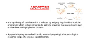
1. APOPTOSIS
• It is a pathway of cell death that is induced by a tightly regulated intracellular
program in which cells destined to die activate enzymes that degrade cells own
nuclear DNA and cytoplasmic proteins.
• Apoptosis is programmed cell death, a normal physiological or pathological
response to specific internal suicidal signals.
2. Morphology
• Cell shrinkage
• Chromatin condensation
• Formation of cytoplasmic blebs and apoptotic bodies
• Phagocytosis of apoptotic cells or cell bodies, usually by macrophages
3. APOPTOSIS
• Physiological or Pathological
• Genetically controlled (Programmed cell
death, ordered disassembly of cells from
within)
NECROSIS
• Always pathological
• Caused by over whelming noxious
stimuli
4. APOPTOSIS
• MORPHOLOGY
• Death of single cell
• Plasma membrane integrity maintained
• Chromatin condensation
• Cell shrinkage
• Lysosomes and other cytoplasmic
organelles are intact
NECROSIS
• MORPHOLOGY
• Death of contiguous cells
• Plasma membrane disruption
• Nuclear swelling and lyses
• Cell swelling
• Lysosomal breakdown
5. • Fragmentation of nucleus and
cytoplasm
• Phagocytosis of apoptotic bodies by
adjacent cells and macrophages
• No inflammatory response
• Cell lysis and disintegration
• Phagocytosis by macrophages
• Inflammatory response present
APOPTOSIS
MORPHOLOGY
NECROSIS
MORPHOLOGY
6. • Apoptosis is responsible for numerous physiologic and pathologic
processes like:
PHYSIOLOGICAL:
• Programmed destruction of cells during embryogenesis
• Hormone dependent involution of the tissues
• Cell deletion in proliferating cell population in lymphoid organ
• Cellular death after completing its function.
7. PATHOLOGICAL:
• Cellular injury produced by a variety of injurious stimuli like radiation, cytotoxic
anticancer drugs, hypoxia.
• Cellular injury and death in certain viral diseases(HIV and adenovirus)
• Cell death in induced by cytoxic T cells in tumors
• Pathological atrophy in parenchymal organs after duct obstruction
8.
9. PROTEIN CLEAVAGE -- caspases (HALLMARK)
ACTIVATE ENDONUCLEASES
NUCLEAR DNA BREAKDOWN
PHAGOCYTIC RECOGNITION
Biochemical features of apoptosis
10. DISTINCTIVE BIOCHEMICAL FEATURES OF APOPTOSIS
• Protein cleavage by cysteine proteases called caspases
• Protein crosslinking by transglutaminase which converts cytoplasmic proteins into
a linked meshwork
• Intranucleosomal cleavage of dna into characteristic ladder pattern of
oligonucleosomal multiples of about 200 base pairs
• Plasma membrane alterations ,such as flipping of the phosphatidylserine from
the inner to the outer surface
16. •CLINICAL SIGNIFICANCE
• Tumours – apoptotic index as a measure of proliferation
• Apoptosis in tumours increases following irradiation & immune
responses.
• Measurement of apoptosis in-vivo/ in-vitro following treatment
may predict effectiveness of therapy.