Characterization of Quasi-Keplerian, Differentially Rotating, Free-Boundary L...
CellWallSeptum
1. Report
Morphogenesis of the Fission Yeast Cell through Cell
Wall Expansion
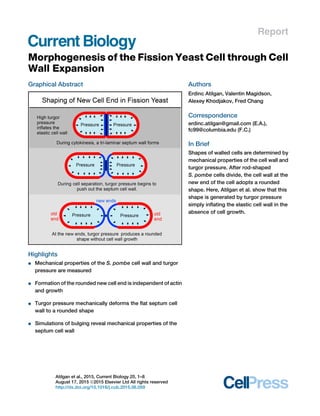
Graphical Abstract
Highlights
d Mechanical properties of the S. pombe cell wall and turgor
pressure are measured
d Formation of the rounded new cell end is independent of actin
and growth
d Turgor pressure mechanically deforms the flat septum cell
wall to a rounded shape
d Simulations of bulging reveal mechanical properties of the
septum cell wall
Authors
Erdinc Atilgan, Valentin Magidson,
Alexey Khodjakov, Fred Chang
Correspondence
erdinc.atilgan@gmail.com (E.A.),
fc99@columbia.edu (F.C.)
In Brief
Shapes of walled cells are determined by
mechanical properties of the cell wall and
turgor pressure. After rod-shaped
S. pombe cells divide, the cell wall at the
new end of the cell adopts a rounded
shape. Here, Atilgan et al. show that this
shape is generated by turgor pressure
simply inflating the elastic cell wall in the
absence of cell growth.
Atilgan et al., 2015, Current Biology 25, 1–8
August 17, 2015 ª2015 Elsevier Ltd All rights reserved
http://dx.doi.org/10.1016/j.cub.2015.06.059
2. Current Biology
Report
Morphogenesis of the Fission Yeast Cell
through Cell Wall Expansion
Erdinc Atilgan,1,* Valentin Magidson,2 Alexey Khodjakov,2 and Fred Chang1,*
1Department of Microbiology and Immunology, Columbia University Medical Center, New York, NY 10025, USA
2Wadsworth Center, New York State Department of Health, Albany, NY 12201, USA
*Correspondence: erdinc.atilgan@gmail.com (E.A.), fc99@columbia.edu (F.C.)
http://dx.doi.org/10.1016/j.cub.2015.06.059
SUMMARY
The shape of walled cells such as fungi, bacteria, and
plants are determined by the cell wall. Models for cell
morphogenesis postulate that the effects of turgor
pressure and mechanical properties of the cell wall
can explain the shapes of these diverse cell types
[1–6]. However, in general, these models await
validation through quantitative experiments. Fission
yeast Schizosaccharomyces pombe are rod-shaped
cells that grow by tip extension and then divide medi-
ally through formation of a cell wall septum. Upon cell
separation after cytokinesis, the new cell ends adopt
a rounded morphology. Here, we show that this
shape is generated by a very simple mechanical-
based mechanism in which turgor pressure inflates
the elastic cell wall in the absence of cell growth.
This process is independent of actin and new cell
wall synthesis. To model this morphological change,
we first estimate the mechanical properties of the
cell wall using several approaches. The lateral cell
wall behaves as an isotropic elastic material with a
Young’s modulus of 50 ± 10 MPa inflated by a turgor
pressure estimated to be 1.5 ± 0.2 MPa. Based upon
these parameters, we develop a quantitative me-
chanical-based model for new end formation that
reveals that the cell wall at the new end expands
into its characteristic rounded shape in part because
it is softer than the mature lateral wall. These studies
provide a simple example of how turgor pressure
expands the elastic cell wall to generate a particular
cell shape.
RESULTS AND DISCUSSION
Morphogenesis of the New End Is Likely to Be a Purely
Mechanical Process
Fission yeast cells serve as an attractive model for eukaryotic
morphogenesis because of their highly regular and simple rod-
shape and growth patterns [7]. These cells have a capsule-like
shape with rounded cell ends, similar to E. coli and many other
rod-shaped cells [1]. S. pombe are approximately 4 mm in diam-
eter and grow by tip extension to 14 mm in length before dividing
medially [8, 9]. Cells are encased in a fibrillar cell wall that is
composed primarily of b-and a-glucans and galactomannan
[10, 11]. For cytokinesis, an actin-based contractile ring guides
the assembly of a cell wall septum, which is composed of a cen-
tral primary septum (PS) disc flanked by two secondary septa
(SS) [9, 12–15]. Following completion of the septum, the PS
and edging are then digested away by endoglucanases for cell
separation [16]. During cell separation, the daughter cells snap
apart abruptly (‘‘rupture’’ event), and then the resultant two
new ends (NEs) adopt a rounded shape within minutes (Fig-
ure 1A; Movie S1). This shape change can be measured by
the amount of bulging from original position of flat septum disc
(s value; Figure 1A). The s value rises and plateaus to
s1 = 1:43 ± 0:25 mm (n = 8 cells) in about 10 min after the rupture
(t = 0). While the old end (OE) begins to grow again soon after the
rupture, NE does not start to elongate till generally about 40 min
later, when cells initiate polarized cell growth at NE in a process
known as ‘‘new end take off’’ (NETO).
An important unresolved question is whether shaping of the
new end is accomplished through active cell wall remodeling
that requires actin and polarity factors, or whether it is formed
by a mechanical process [13, 14]. An initial indication that the
rounded shape might be formed through a purely mechanical
process was seen in laser microsurgery experiments [17]. In cells
with a complete septum, we cut the lateral cell wall in one of the
cellular compartments (Figure 1B; Movie S2). The cut compart-
ment shrank and extruded cellular contents, while the other
cellular compartment stayed intact. Interestingly, the septum
bulged out toward the lysed part of the cell to form a rounded
shape similar to that of a NE. Time-lapse imaging showed that
the deformation occurred extremely quickly (in less than
10 ms) with an outward speed >1 mm/sec (three orders of magni-
tude faster than the normal rate of cell growth). This shape
change occurred similarly in cells treated with the 200 mM latrun-
culin A (LatA) (Figure 1C), which leads to rapid depolymerization
of all F-actin in minutes and cessation of growth [18–20]. Thus,
this bulging of septum is actin independent. Similar deformation
of the septum has also been observed in cell wall mutants
[13, 14]. These initial observations suggest that the larger turgor
pressure in the intact compartment is able to push out the
septum cell wall into a curved shape.
We tested whether a similar mechanism shapes the NE during
cell separation. We propose that before cell separation, the
septum is initially flat because forces from the turgor pressure
on both sides balance each other, but when the cells begin to
separate, turgor pressure pushes out of the cell wall to form
the rounded new cell end. This shape change involves an
approximately 65% increase in the surface area at the new
Current Biology 25, 1–8, August 17, 2015 ª2015 Elsevier Ltd All rights reserved 1
Please cite this article in press as: Atilgan et al., Morphogenesis of the Fission Yeast Cell through Cell Wall Expansion, Current Biology (2015), http://
dx.doi.org/10.1016/j.cub.2015.06.059
3. end. This increase is due to expansion of the new end cell wall,
and not from flow of the lateral cell wall, as indicated by stable
position of birthmarks on the cell wall. To test whether this pro-
cess requires actin, we treated septated cells with LatA and fol-
lowed them by time-lapse imaging. 80% of the cells (n = 30)
divided and separated, with the NEs forming rounded shapes
with similar extent and kinetics as control cells (n = 13 cells; Fig-
ure 1D). We confirmed that the LatA treatment was effective in
inhibiting F-actin and polarized cell growth (Figure S1). We tested
the role of wall synthesis by treating septated cells with the
b-glucan synthase inhibitor caspofungin [21]. At 20 mg/ml caspo-
fungin, cells exhibit five times slower tip growth, and many cells
lyse. However, most septated cells continued to separate, and
those that do formed rounded NEs with a similar extent and time-
scale as untreated cells (n = 12 cells; Figure 1E). Staining with
calcofluor and blankophor, which preferentially labels newly
deposited cell wall [8, 10, 22], further suggests that there is little
or no wall growth on the NE during cell separation. These find-
ings support a model in which turgor pressure simply deforms
the flat cell wall to a rounded shape without wall growth.
The Fission Yeast Cell Wall Behaves as an Isotropic
Elastic Material
To evaluate cell wall shaping mechanism(s) in a quantitative
manner, we next sought to determine the elastic properties of
the cell wall and turgor pressure. To construct a mechanistic
model, we approximate the fission yeast cells as a capsule-
shaped thin elastic shell inflated by internal turgor pressure
(Figure 2A). A pressure difference, DP, stretches the cell wall to
its ‘‘natural state.’’ Without pressure, the tensionless cell wall
is in ‘‘relaxed state.’’ We define the expansion ratio in width
as %RÃ
hðR1 À R0Þ=R03100 and the expansion ratio in length
A
B C
D E
Figure 1. Shaping of the New End Cell Wall and Septum Are Independent of Actin and Wall Synthesis
(A) During cell-cell separation, the process starts with a sudden break in the lateral cell wall (À1 to 0 s) followed by formation of a rounded new cell end (0–15 min).
Movie S1. The plot shows bulging of the NE wall (as measured by the ‘‘s value’’) over time.
(B) Deformation of the septum. In a cell with a complete septum, one of the cellular compartments was lysed by a laser cut to the cell surface (yellow asterisk). The
septum forms a curved shape in %10 ms (within a single frame, arrowhead; see Movie S2).
(C) Septum deformation is actin independent. Similar as in (B), except one compartment of cells (yellow asterisk) was lysed using physical manipulation (see
Supplemental Experimental Procedures). Prior to manipulation, cells were treated with 200 mM LatA (actin inhibitor) or DMSO (control). Cells were stained with
blankophor to visualize the deformed septum.
(D) Cell-cell separation and new end formation are actin independent. Septated cells were treated with 200 mM LatA or DMSO at time 0 and stained with
blankophor at 25 min. Bright-field and fluorescence images at indicated time points are shown.
(E) New end formation and cell separation are independent of wall synthesis. Septated cells were treated with 20 mg/ml caspofungin at time 0 and imaged in bright
field. Scale bars, 4 mm. In this work, all values with errors are mean ± SD.
2 Current Biology 25, 1–8, August 17, 2015 ª2015 Elsevier Ltd All rights reserved
Please cite this article in press as: Atilgan et al., Morphogenesis of the Fission Yeast Cell through Cell Wall Expansion, Current Biology (2015), http://
dx.doi.org/10.1016/j.cub.2015.06.059
4. as %LÃ
hðL1 À L0Þ=L03100. R1 and R0 are the radii in natural
and relaxed state respectively, and similarly for L1 and L0. We
assume that cell wall is an elastic material with Young’s modulus
Yr in circumferential direction, Yl in longitudinal direction, and
that Poisson’s ratio is zero in both directions.
Crude force balance equations yield the connection between
the expansion ratios and the physical parameters of the system
as (Supplemental Experimental Procedures section 1):
R1 À R0
R0
3 100 = %RÃ
y
DPR1
Yrt
3 100
L1 À L0
L0
3 100 = %LÃ
y
DPR1
2Ylt
3 100:
Since the thickness of the wall, t = 0:2 mm, and the radius of the
cell, R1 = 1:92 ± 0:07 mm, are known, DP=Yr and DP=Yl can be
found if one can measure %RÃ
and %LÃ
experimentally.
To measure these expansion ratios, we devised approaches
to release turgor pressure. First, we locally cut the cell wall using
laser microsurgery. Time-lapse imaging showed that cells shrink
rapidly by approximately 17% in width and 9% in length (see
Movie S3 and Figure 2B, left). This corresponds to expansion
ratios of 20% ± 5% and 10 % ± 3%, respectively. We also lysed
cells by (1) micro-manipulation with a glass needle, (2) dehydra-
tion/rehydration, and (3) post-osmotic-shock (see Supplemental
Experimental Procedures). These approaches all produced
A
B
D
C
E
Figure 2. Determining the Mechanical Prop-
erties of the Cell Wall
(A) Model of the fission yeast cell wall as a thin
elastic capsule that is inflated by turgor pressure in
the natural state (intact cell), and shrinks to a
relaxed state when pressure difference is lost.
(B) Left panel shows a bright-field image of an
individual cell before and after lysis by laser
microsurgery (Movie S3). Right panel shows a
ghost cell (cell wall only).
(C) Measurement of expansion ratios. Cells were
lysed by multiple approaches. Dimensions of
individual cells were measured before and after
lysis. Top graph shows expansion ratios of cell
lengths (%LÃ
= ðL1 À L0Þ=L03100) and widths
(%RÃ
= ðR1 À R0Þ=R03100). Bottom graph shows
ratios between expansion ratios. For ghosts, the
length expansion ratio cannot be measured since
the prior geometry of the cells is not known.
(D) Dimensions of individual cells exposed to
various sorbitol concentrations were measured.
Graphs show experimental and theoretical
osmotic response ratios for length (%LhðL1À
LxÞ=Lx3100) and width (%RhðR1 À RxÞ=Rx3100)
where Lx and Rx are dimensions after the
shrinkage. b is the water inaccessible volume
fraction.
(E) Ratio between the response ratios.
Scale bars, 4 mm.
similar expansion ratios (Figure 2C). The
lysed cells, however, retain intracellular
contents that may exert some internal
pressure or force onto the cell wall. There-
fore, we next isolated cell wall ‘‘ghosts’’ that lack intracellular
structures by breaking yeast cells (Figure 2B, right; Figure S2A).
Although we could not measure changes in cell length in these
broken cell walls, the changes in cell width show that ghosts
shrink slightly more than the lysed cells, indicating that the lysed
cells still maintain some low level of internal pressure. The corre-
sponding expansion ratio of the width in ghosts is 24% ± 5%
(Figure 2C).
A significant outcome of our experiments is that the yeast cell
wall is isotropic. For a cylindrical shell, %RÃ
=%LÃ
= 2Yl=Yr. If the
cell wall has the same stiffness in circumferential and longitudinal
directions (i.e., Yr = Yl = Y), then %RÃ
=%LÃ
is predicted to be 2.
Our experimental measurements produced a ratio very close
to 2 (Figure 2C, bottom graph). This behavior is different from
that of the E. coli cell wall, for instance, in which cell wall fibers
are oriented circumferentially and are anisotropic in stiffness
[23, 24].
Taking the expansion ratio of the ghosts for the width (24%),
we found that
DP
Y
=
ð%RÃ
Þt
R1
=
1
40 ± 8
:
This relationship indicates that the value of Young’s modulus is
about 40 times cell turgor pressure (they both carry the same
units). Equations stated for %RÃ
and %LÃ
are derived through
crude force balance relations. To improve the accuracy of our
Current Biology 25, 1–8, August 17, 2015 ª2015 Elsevier Ltd All rights reserved 3
Please cite this article in press as: Atilgan et al., Morphogenesis of the Fission Yeast Cell through Cell Wall Expansion, Current Biology (2015), http://
dx.doi.org/10.1016/j.cub.2015.06.059
5. A
B C E
D
F
G
Figure 3. Modeling New End Formation
(A) Output of simulations in which turgor pressure causes bulging of the new end cell wall. Simulations are shown in which Ys (Young’s modulus of SS) is treated as
a free parameter while DP = 1:5 MPa and Y = 50 MPa. Graph shows the amount of bulging (s1) plotted as a function of Ys=Y for measured average SS thickness
ts = 100 nm.
(legend continued on next page)
4 Current Biology 25, 1–8, August 17, 2015 ª2015 Elsevier Ltd All rights reserved
Please cite this article in press as: Atilgan et al., Morphogenesis of the Fission Yeast Cell through Cell Wall Expansion, Current Biology (2015), http://
dx.doi.org/10.1016/j.cub.2015.06.059
6. result, we have included the higher order terms in our formulas
by applying a continuum mechanical approach (Supplemental
Experimental Procedures section 1), which yields Y=DP = 33 ± 8.
Osmotic Responses Show that Fission Yeast Is Inflated
by High MPa Turgor Pressure
Next, to further characterize the mechanical properties of the
cells, we measured how cells shrink in different doses of sorbitol
in the media (see Supplemental Experimental Procedures). To
estimate the turgor pressure, we sought to determine the
external molarity, c0, required to shrink intact cells to the size
of their ghosts (i.e., the relaxed wall state); see Figure 2D. Sorbi-
tol shift curves show that on average, c0z1:5 M ( = 1.3 M sorbitol
in the growth medium, YE5S). To estimate the pressure from this
result, we applied an osmotic theory that takes into account the
expansion ratios, the water inaccessible volume, and the matrix
potential of the solutions (see Supplemental Experimental
Procedures section 3). The assumptions of the theory are
taken such that it would yield the minimal plausible value for
the pressure. We found the turgor pressure (the osmotic
pressure difference between inside and outside of the cell)
as DP = 1:5 ± 0:2 MPa. Given our previous finding that Y =DP =
33 ± 8, this yields the Young’s modulus (stiffness) of the cell
wall as Y = 50 ± 10 MPa.
The response ratios of cells fit well with the theoretically
calculated osmotic responses (Figure 2D). The data suggest a
water inaccessible volume fraction of b = 0:22. The ratios be-
tween the response ratios (%R=%L) in width and length are
again close to 2, showing that the cell wall material is isotropic
(Figure 2E).
To address a concern that results may be affected by cellular
adaptation mechanisms to osmotic changes, we also analyzed
gpd1D (glycerol-3-phosphate dehydrogenase) mutants, which
are deficient in restoring turgor pressure during osmotic shifts
[25, 26]. The osmotic response ratios of gpd1D mutants (Figures
1D and 1E) were similar to those of wild-type cells.
We also used spheroplast formation as an alternative
approach to estimate c0 because of its importance for applica-
tion of a reliable osmotic theory. Upon removal of the cell wall,
spheroplasts remained intact at external molarities c0 = 1:2À
1:7 M (Figures S2B–S2D). This result is consistent with the
osmotic shift experiment that showed c0z1:5 M (Figure 2D).
Mechanical Forces Can Account for the Shape
of the New End
Having estimated of the properties of the cell wall, we next em-
barked on testing and analyzing the mechanism for NE formation
based upon the effects of turgor pressure on the cell wall. We
developed a finite element simulation program based on non-
linear continuum mechanics principles. Our algorithm relies
upon the deformation of a given surface under a specified force
field (or pressure). The surface is discretized with triangular ele-
ments, and the elastic energies are calculated through stretch as
well as bending terms (Supplemental Experimental Procedures
section 2.1). We validated our program by comparisons to
known analytic solutions (Supplemental Experimental Proce-
dures section 2.2–5) and also verified our previous analytic
work on expansion ratios (Supplemental Experimental Proce-
dures section 2.6). In simulations, we inflate a closed cylindrical
shell (Supplemental Experimental Procedures section 2.7). We
assume that, before cell separation, the septum is at a relaxed
flat geometry, as opposed to the lateral wall, which is expanded
under stress; buckling of the septum in sorbitol-treated cells
supports this assumption (Figure S3A). We assume that the
wall at the NE is also an isotropic material with a constant uniform
elasticity and zero Poisson’s ratio but could have a different
thickness and elasticity. The shapes from simulations are depen-
dent on Y=DP ratio and therefore are independent of DP. Note
that we extract Y through Y=DP, which is found through experi-
mentally measured parameters. Our simulations successfully
yielded the rounded shape of the NE (Figure 3A).
This modeling allowed us to estimate elastic properties of the
NE cell wall simply by the extent of its bulging. Published electron
micrographs show that the thickness of the secondary septum
(ts = 100 ± 20 nm) is about the half that of the lateral wall
(ts=ty0:5) [13, 14, 16, 27–31]. In addition, there may be chemical
and structural differences between the two types of cell walls
[14, 32]. We simulated the NE bulging for varying values of
Young’s modulus of the SS (Ys) by using the values found for
DP and Y (Figure 3A). This plot showed that the best fit to the
measured s1 = 1:42 ± 0:15 mm (n = 30 cells) is at around
Ys=Y = 0:48; SDs in s1 and ts measurements yielded error mar-
gins of 0:3 < Ys=Y < 0:7. This leads to the conclusion that
YsyY=2; cell wall stiffness of the NE is about half of that of the
lateral wall (i.e., twice as soft).
Next, we tested the effect of turgor pressure on the shape of
the NE in simulations and experiments. In simulations, both
cell width and the extent of NE bulging (s value) positively corre-
late with the increasing pressure (Figure 3B). We then compared
these results with effects of altering the turgor pressure in a
series of experiments. First, we used micromanipulation to
measure the intact, lysed, and ghost states of individual cells
(Figure 3C). Compared to the NEs of intact cells, the NEs of
the corresponding ghosts bulged less (smaller s value) and
(B) Simulation of a whole cell at varying values of pressure DP where Y = 50 MPa and Ys = Y=2 = 25 MPa.
(C) Images of a single cell stained with Lectin-TRITC in intact, lysed, and ghost states, which were generated by successive micromanipulations.
(D) Averaged NE shapes (from C; n = 8 cells) were fitted to an ellipse-rectangle combination and compared to simulation outputs (blue dots). Note that the NE in
ghosts does not return to a completely flat geometry (Figure S3B) because of the shrinkage of the lateral cell wall, as also seen in the simulations. The slight
discrepancies (maximum error <0.2 mm) between the ghost profiles could be due to additional structural features between the septum and lateral wall not
included in the simulations [12]. Scale bar, 1 mm.
(E) Effect of sorbitol treatment on NE shape. Fully septated cells were treated with 1.3 M sorbitol at 1 or 15 min (arrows) after the time of rupture (t = 0). Graph
shows curvatures of the OE and NE from ‘‘shift at 15 min’’ experiment and simulations (circles; B).
(F) NE bulging (s values) and cell widths as a function of time after sorbitol treatment. As indicated by their width, cells initially shrink and then re-inflate over 60 min
to their original size without tip growth (Figure S3C).
(G) Relationship between NE bulging and cell width (as an indicator of turgor pressure).
Data from experiments in (C), (E), and (F) are plotted and compared to simulation outputs from (B).
Current Biology 25, 1–8, August 17, 2015 ª2015 Elsevier Ltd All rights reserved 5
Please cite this article in press as: Atilgan et al., Morphogenesis of the Fission Yeast Cell through Cell Wall Expansion, Current Biology (2015), http://
dx.doi.org/10.1016/j.cub.2015.06.059
7. exhibited a flatter shape (as shown by geometric fits with ellip-
ses; Figures 3D and S3B; n = 8 cells).
We further tested the effect of turgor pressure experimentally
by adding sorbitol to the live, dividing cells (Figure 3E). First we
added 1.3 M sorbitol to cells that have just divided (<1 min after
rupture). The cells shrank as expected, and the process of cell
separation halted with the NEs flat (Figure 3E; middle images).
Thus, turgor pressure is needed for cell separation and shaping
of NE. However, in these experiments, one reason why the NEs
are flat could be because the sister cells are still attached by the
PS (see below). Hence, in another set of experiments, to elimi-
nate the effects of the PS, we added the sorbitol just after the
A
C
B
D
E
Figure 4. Modeling Cell Separation and Septum Bulging
(A and B) Images and simulations of cell separation. Experimental images of sister cells stained with blankophor (Movie S5) are compared to outputs from
simulations where P = 1:5 MPa and Y = 50 MPa. The PS is assumed to be an elastic disc with Young’s modulus Yp that gradually decreases in diameter, causing
the gradual deformation of the SS. Lateral cell wall thickness tz200 nm, SS thickness tsz100 nm, and the PS thickness tpz50 nm.
(C) Bulging of the SS (s4) as a function of PS diameter, D. Graph compares experimental data (black dots and red points) with simulations for different values of Yp
and Ys. The best fits to experimental data are simulations with Ys = Y=2 and 0%Yp%Y.
(D) The deformation of the whole septum after laser ablation (as in Figure 1B). Inverse image of a blankophor-stained cell and the output of a simulation.
(E) Comparison of experiments and simulations for NE formation (s1) and whole-septum deformations (s5). The whole-septum simulations are done with
P = 1:5 MPa and Y = 50 MPa; Ys = Y=2 and Yp = 0; Y; 2Y; and 5Y.
6 Current Biology 25, 1–8, August 17, 2015 ª2015 Elsevier Ltd All rights reserved
Please cite this article in press as: Atilgan et al., Morphogenesis of the Fission Yeast Cell through Cell Wall Expansion, Current Biology (2015), http://
dx.doi.org/10.1016/j.cub.2015.06.059
8. NEs have adopted a rounded shape ($15 min after rupture; Fig-
ure 3E, right images). Upon sorbitol treatment, the shape of the
NEs changed from a rounded to a flattened shape. Interestingly,
OEs maintained a rounded shape. Curvature decreased at NEs,
while it increased at OEs (n = 8 cells, bar plot in Figure 3E). These
behaviors were as predicted in simulations that assumed that OE
wall properties are similar to that of the lateral wall (Figures 3B
and 3E). These findings demonstrate that the two ends of fission
yeast cell are mechanically different.
Following the initial shrinkage, the cells in sorbitol gradually re-
inflated over 60 min back to normal dimensions (Figures 3E and
3F). This is likely due to cellular adaptation to osmotic stress,
which gradually restores the relative internal pressure [33].
During this recovery period, the width of the cells increased
with increasing pressure, without any apparent tip growth (Fig-
ure S3C). Thus, these time-lapse images allow us to determine
the resultant cell morphology over a continuous range of turgor
pressure. In Figure 3G, we plot s values versus the width of
the cells (as the measure of turgor pressure) from the sorbitol
shift and the micromanipulation experiments and compared
them to simulations; all show a similar, strong positive correla-
tion. The agreement of our simulations with experimental data
provides quantitative evidence for the role of turgor pressure in
the shaping of NE.
Mechanical Forces Shape the Septum during Cell
Separation and Septum Bulging
During cell separation, the shape of the NE develops gradually
over minutes. An additional factor governing this process is the
PS, which acts as an adhesive that holds the sister cells together.
The PS is a disc about 50 nm thick (one-fourth the thickness of
the lateral wall tpyt=4) [13, 14, 16, 27–31]; however, its mechan-
ical properties are unknown. After initial rupture, the PS gradually
shrinks in diameter (D), and the bulging of the SS (s4 value)
gradually increases as the NE adopts a rounded shape (Figures
4A–4C; Movie S5). In simulations (Supplemental Experimental
Procedures section 2.8), we assumed that the PS is mechani-
cally infinitely strong and stiff in the transverse direction (perpen-
dicular to the disc) but could be elastic in the lateral direction
(direction along surface) with a uniform and constant Young’s
modulus of Yp. We plotted s4 as function of D over a range of
Yp values (Figure 4C). The outputs agreed with the experimental
data when Ys = Y=2 and 0%Yp%Y. These findings suggest that
the PS is an elastic material in the lateral direction with a stiffness
< 2Y and stretches along with the SS during cell separation. In
contrast, simulations with Ys = Y for any value of Yp failed to fit
to experimental data. Thus, these results validate our model
further and explain the evolving morphology of NE during cell
separation.
Finally, we applied our simulation to the laser cut experiments
of fully septated cells (Figures 1B and 4D), in which the turgor
pressure is thought to immediately deform the septum cell
wall. In these cells, the deformation of the septum (s5) was less
than the one of the NE in normally dividing cells (s1) (Figure 4E).
We tested whether this shape difference could be explained by
the difference between single layered wall of the NE versus the
thicker triple-layered wall of the septum. In simulations (Supple-
mental Experimental Procedures section 2.9), we examined
septum deformation at Ys = Y=2 at different values of YP. The
case Yp = Y produced the best fit (Figure 4E), which is consistent
with the estimate from cell-separation experiments (Figure 4C),
providing an independent validation of our results. Thus, our
findings using experiments and simulations in a variety of con-
texts demonstrate that a mechanically based mechanism based
upon turgor pressure and elastic properties of the cell wall can
quantitatively account for shaping of the NE.
In summary, we develop a quantitative model for morphogen-
esis of a rounded cell end in a rod-shaped cell. Our findings
suggest that the rounded shape is produced simply by turgor
pressure inflating the elastic cell wall, even without cell wall
growth. Our measurements of mechanical properties are gener-
ally on the same order of magnitude as previous ones in fission
yeast and other fungi obtained through a variety of approaches
[26, 34, 35]. One advance in this study is in the direct measure-
ments of the relaxed state of the wall that allow us to determine
critical parameters needed to apply a reliable osmotic theory. To
adopt its rounded shape, the cell wall at the new end is 50%
thinner and 50% softer than the lateral wall, and stretches 65%
in area. These mechanical properties of the cell wall may explain
why cell wall mutants lyse primarily at cell separation [36]. Turgor
pressure may not only drive shape changes, but also contribute
to the separation of the sister cells and their movement apart.
Forces from turgor pressure also contribute to growth and
shaping of the growing cell end, but this process is more
complex as it involves addition and remodeling of the cell wall
[1, 3, 26]. Turgor pressure is also thought to drive cell-shape
changes in certain plant cells, such as guard cells that regulate
the opening and closing of stomatal pores [37]. It will be inter-
esting to examine whether similar mechanically based mecha-
nisms are responsible for generating the rounded contours of
new ends after cytokinesis in other cell types. Our studies
illustrate how mechanical as well as molecular agents underlie
cellular morphogenesis.
SUPPLEMENTAL INFORMATION
Supplemental Information includes Supplemental Experimental Procedures,
three figures, and five movies and can be found with this article online at
http://dx.doi.org/10.1016/j.cub.2015.06.059.
ACKNOWLEDGMENTS
We thank the members of F.C.’s lab, K.C. Huang, and Enrique Rojas for sup-
port and discussion. This work was supported by grants NIH GM056836
(to F.C.), NSF Collaborative Research: BIOMAPS-1244441 (to F.C.) and NIH
GM059363 (to A.K.).
Received: January 14, 2015
Revised: April 29, 2015
Accepted: June 22, 2015
Published: July 23, 2015
REFERENCES
1. Chang, F., and Huang, K.C. (2014). How and why cells grow as rods. BMC
Biol. 12, 54.
2. Boudaoud, A. (2003). Growth of walled cells: from shells to vesicles. Phys.
Rev. Lett. 91, 018104.
3. Campa` s, O., and Mahadevan, L. (2009). Shape and dynamics of tip-
growing cells. Curr. Biol. 19, 2102–2107.
Current Biology 25, 1–8, August 17, 2015 ª2015 Elsevier Ltd All rights reserved 7
Please cite this article in press as: Atilgan et al., Morphogenesis of the Fission Yeast Cell through Cell Wall Expansion, Current Biology (2015), http://
dx.doi.org/10.1016/j.cub.2015.06.059
9. 4. Campa` s, O., Rojas, E., Dumais, J., and Mahadevan, L. (2012). Strategies
for cell shape control in tip-growing cells. Am. J. Bot. 99, 1577–1582.
5. Drake, T., and Vavylonis, D. (2013). Model of fission yeast cell shape driven
by membrane-bound growth factors and the cytoskeleton. PLoS Comput.
Biol. 9, e1003287.
6. Dumais, J., Shaw, S.L., Steele, C.R., Long, S.R., and Ray, P.M. (2006). An
anisotropic-viscoplastic model of plant cell morphogenesis by tip growth.
Int. J. Dev. Biol. 50, 209–222.
7. Chang, F., and Martin, S.G. (2009). Shaping fission yeast with microtu-
bules. Cold Spring Harb. Perspect. Biol. 1, a001347.
8. Mitchison, J.M., and Nurse, P. (1985). Growth in cell length in the fission
yeast Schizosaccharomyces pombe. J. Cell Sci. 75, 357–376.
9. Johnson, B.F., Miyata, M., and Miyata, H. (1989). Morphogenesis of fission
yeasts. In Molecular Biology of the Fission Yeast, A. Nasim, P. Young, and
B.F. Johnson, eds. (New York: Academic Press), pp. 469331–469366.
10. Perez, P., and Ribas, J.C. (2004). Cell wall analysis. Methods 33, 245–251.
11. Osumi, M., Sato, M., Ishijima, S.A., Konomi, M., Takagi, T., and Yaguchi,
H. (1998). Dynamics of cell wall formation in fission yeast,
Schizosaccharomyces pombe. Fungal Genet. Biol. 24, 178–206.
12. Robinow, C.F., and Hyams, J.S. (1989). General cytology of the fission
yeasts. In Molecular Biology of the Fission Yeast, A. Nasim, P. Young,
and B.F. Johnson, eds. (Academic Press), pp. 273–330.
13. Corte´ s, J.C., Sato, M., Mun˜ oz, J., Moreno, M.B., Clemente-Ramos, J.A.,
Ramos, M., Okada, H., Osumi, M., Dura´ n, A., and Ribas, J.C. (2012).
Fission yeast Ags1 confers the essential septum strength needed for
safe gradual cell abscission. J. Cell Biol. 198, 637–656.
14. Mun˜ oz, J., Corte´ s, J.C., Sipiczki, M., Ramos, M., Clemente-Ramos, J.A.,
Moreno, M.B., Martins, I.M., Pe´ rez, P., and Ribas, J.C. (2013).
Extracellular cell wall b(1,3)glucan is required to couple septation to acto-
myosin ring contraction. J. Cell Biol. 203, 265–282.
15. Zhou, Z., Munteanu, E.L., He, J., Ursell, T., Bathe, M., Huang, K.C., and
Chang, F. (2015). The contractile ring coordinates curvature-dependent
septum assembly during fission yeast cytokinesis. Mol. Biol. Cell 26,
78–90.
16. Sipiczki, M. (2007). Splitting of the fission yeast septum. FEMS Yeast Res.
7, 761–770.
17. Khodjakov, A., La Terra, S., and Chang, F. (2004). Laser microsurgery in
fission yeast; role of the mitotic spindle midzone in anaphase B. Curr.
Biol. 14, 1330–1340.
18. Chang, F. (1999). Movement of a cytokinesis factor cdc12p to the site of
cell division. Curr. Biol. 9, 849–852.
19. Proctor, S.A., Minc, N., Boudaoud, A., and Chang, F. (2012). Contributions
of turgor pressure, the contractile ring, and septum assembly to forces in
cytokinesis in fission yeast. Curr. Biol. 22, 1601–1608.
20. Pan, K.Z., Saunders, T.E., Flor-Parra, I., Howard, M., and Chang, F. (2014).
Cortical regulation of cell size by a sizer cdr2p. eLife 3, e02040.
21. Martins, I.M., Corte´ s, J.C., Mun˜ oz, J., Moreno, M.B., Ramos, M.,
Clemente-Ramos, J.A., Dura´ n, A., and Ribas, J.C. (2011). Differential ac-
tivities of three families of specific beta(1,3)glucan synthase inhibitors in
wild-type and resistant strains of fission yeast. J. Biol. Chem. 286,
3484–3496.
22. Sawin, K.E., Hajibagheri, M.A., and Nurse, P. (1999). Mis-specification of
cortical identity in a fission yeast PAK mutant. Curr. Biol. 9, 1335–1338.
23. Gan, L., Chen, S., and Jensen, G.J. (2008). Molecular organization of
Gram-negative peptidoglycan. Proc. Natl. Acad. Sci. USA 105, 18953–
18957.
24. Firtel, M., Henderson, G., and Sokolov, I. (2004). Nanosurgery: observation
of peptidoglycan strands in Lactobacillus helveticus cell walls.
Ultramicroscopy 101, 105–109.
25. Aiba, H., Yamada, H., Ohmiya, R., and Mizuno, T. (1995). The osmo-induc-
ible gpd1+ gene is a target of the signaling pathway involving Wis1 MAP-
kinase kinase in fission yeast. FEBS Lett. 376, 199–201.
26. Minc, N., Boudaoud, A., and Chang, F. (2009). Mechanical forces of fission
yeast growth. Curr. Biol. 19, 1096–1101.
27. Morrell-Falvey, J.L., Ren, L., Feoktistova, A., Haese, G.D., and Gould, K.L.
(2005). Cell wall remodeling at the fission yeast cell division site requires
the Rho-GEF Rgf3p. J. Cell Sci. 118, 5563–5573.
28. Ishiguro, J., Shimada, S., Gabriel, M., and Kopecka´ , M. (2001).
Characterization of a fission yeast mutant which displays defects in cell
wall integrity and cytokinesis. Genes Genet. Syst. 76, 257–269.
29. Santos, B., Gutie´ rrez, J., Calonge, T.M., and Pe´ rez, P. (2003). Novel Rho
GTPase involved in cytokinesis and cell wall integrity in the fission yeast
Schizosaccharomyces pombe. Eukaryot. Cell 2, 521–533.
30. Zilahi, E., Miklo´ s, I., and Sipiczki, M. (2000). The Schizosaccharomyces
pombe sep15+ gene encodes a protein homologous to the Med8 subunit
of the Saccharomyces cerevisiae transcriptional mediator complex. Curr.
Genet. 38, 227–232.
31. Sipiczki, M., and Bozsik, A. (2000). The use of morphomutants to investi-
gate septum formation and cell separation in Schizosaccharomyces
pombe. Arch. Microbiol. 174, 386–392.
32. Corte´ s, J.C., Konomi, M., Martins, I.M., Mun˜ oz, J., Moreno, M.B., Osumi,
M., Dura´ n, A., and Ribas, J.C. (2007). The (1,3)beta-D-glucan synthase
subunit Bgs1p is responsible for the fission yeast primary septum forma-
tion. Mol. Microbiol. 65, 201–217.
33. Hohmann, S. (2002). Osmotic stress signaling and osmoadaptation in
yeasts. Microbiol. Mol. Biol. Rev. 66, 300–372.
34. Smith, A.E., Zhang, Z., Thomas, C.R., Moxham, K.E., and Middelberg, A.P.
(2000). The mechanical properties of Saccharomyces cerevisiae. Proc.
Natl. Acad. Sci. USA 97, 9871–9874.
35. Zhao, L., Schaefer, D., Xu, H., Modi, S.J., LaCourse, W.R., and Marten,
M.R. (2005). Elastic properties of the cell wall of Aspergillus nidulans stud-
ied with atomic force microscopy. Biotechnol. Prog. 21, 292–299.
36. Wu, J.Q., Ye, Y., Wang, N., Pollard, T.D., and Pringle, J.R. (2010).
Cooperation between the septins and the actomyosin ring and role of a
cell-integrity pathway during cell division in fission yeast. Genetics 186,
897–915.
37. Levitt, J., and Fischer, R.A. (1969). Stomatal opening: role of potassium
uptake. Science 163, 494.
8 Current Biology 25, 1–8, August 17, 2015 ª2015 Elsevier Ltd All rights reserved
Please cite this article in press as: Atilgan et al., Morphogenesis of the Fission Yeast Cell through Cell Wall Expansion, Current Biology (2015), http://
dx.doi.org/10.1016/j.cub.2015.06.059
![Current Biology
Report
Morphogenesis of the Fission Yeast Cell
through Cell Wall Expansion
Erdinc Atilgan,1,* Valentin Magidson,2 Alexey Khodjakov,2 and Fred Chang1,*
1Department of Microbiology and Immunology, Columbia University Medical Center, New York, NY 10025, USA
2Wadsworth Center, New York State Department of Health, Albany, NY 12201, USA
*Correspondence: erdinc.atilgan@gmail.com (E.A.), fc99@columbia.edu (F.C.)
http://dx.doi.org/10.1016/j.cub.2015.06.059
SUMMARY
The shape of walled cells such as fungi, bacteria, and
plants are determined by the cell wall. Models for cell
morphogenesis postulate that the effects of turgor
pressure and mechanical properties of the cell wall
can explain the shapes of these diverse cell types
[1–6]. However, in general, these models await
validation through quantitative experiments. Fission
yeast Schizosaccharomyces pombe are rod-shaped
cells that grow by tip extension and then divide medi-
ally through formation of a cell wall septum. Upon cell
separation after cytokinesis, the new cell ends adopt
a rounded morphology. Here, we show that this
shape is generated by a very simple mechanical-
based mechanism in which turgor pressure inflates
the elastic cell wall in the absence of cell growth.
This process is independent of actin and new cell
wall synthesis. To model this morphological change,
we first estimate the mechanical properties of the
cell wall using several approaches. The lateral cell
wall behaves as an isotropic elastic material with a
Young’s modulus of 50 ± 10 MPa inflated by a turgor
pressure estimated to be 1.5 ± 0.2 MPa. Based upon
these parameters, we develop a quantitative me-
chanical-based model for new end formation that
reveals that the cell wall at the new end expands
into its characteristic rounded shape in part because
it is softer than the mature lateral wall. These studies
provide a simple example of how turgor pressure
expands the elastic cell wall to generate a particular
cell shape.
RESULTS AND DISCUSSION
Morphogenesis of the New End Is Likely to Be a Purely
Mechanical Process
Fission yeast cells serve as an attractive model for eukaryotic
morphogenesis because of their highly regular and simple rod-
shape and growth patterns [7]. These cells have a capsule-like
shape with rounded cell ends, similar to E. coli and many other
rod-shaped cells [1]. S. pombe are approximately 4 mm in diam-
eter and grow by tip extension to 14 mm in length before dividing
medially [8, 9]. Cells are encased in a fibrillar cell wall that is
composed primarily of b-and a-glucans and galactomannan
[10, 11]. For cytokinesis, an actin-based contractile ring guides
the assembly of a cell wall septum, which is composed of a cen-
tral primary septum (PS) disc flanked by two secondary septa
(SS) [9, 12–15]. Following completion of the septum, the PS
and edging are then digested away by endoglucanases for cell
separation [16]. During cell separation, the daughter cells snap
apart abruptly (‘‘rupture’’ event), and then the resultant two
new ends (NEs) adopt a rounded shape within minutes (Fig-
ure 1A; Movie S1). This shape change can be measured by
the amount of bulging from original position of flat septum disc
(s value; Figure 1A). The s value rises and plateaus to
s1 = 1:43 ± 0:25 mm (n = 8 cells) in about 10 min after the rupture
(t = 0). While the old end (OE) begins to grow again soon after the
rupture, NE does not start to elongate till generally about 40 min
later, when cells initiate polarized cell growth at NE in a process
known as ‘‘new end take off’’ (NETO).
An important unresolved question is whether shaping of the
new end is accomplished through active cell wall remodeling
that requires actin and polarity factors, or whether it is formed
by a mechanical process [13, 14]. An initial indication that the
rounded shape might be formed through a purely mechanical
process was seen in laser microsurgery experiments [17]. In cells
with a complete septum, we cut the lateral cell wall in one of the
cellular compartments (Figure 1B; Movie S2). The cut compart-
ment shrank and extruded cellular contents, while the other
cellular compartment stayed intact. Interestingly, the septum
bulged out toward the lysed part of the cell to form a rounded
shape similar to that of a NE. Time-lapse imaging showed that
the deformation occurred extremely quickly (in less than
10 ms) with an outward speed >1 mm/sec (three orders of magni-
tude faster than the normal rate of cell growth). This shape
change occurred similarly in cells treated with the 200 mM latrun-
culin A (LatA) (Figure 1C), which leads to rapid depolymerization
of all F-actin in minutes and cessation of growth [18–20]. Thus,
this bulging of septum is actin independent. Similar deformation
of the septum has also been observed in cell wall mutants
[13, 14]. These initial observations suggest that the larger turgor
pressure in the intact compartment is able to push out the
septum cell wall into a curved shape.
We tested whether a similar mechanism shapes the NE during
cell separation. We propose that before cell separation, the
septum is initially flat because forces from the turgor pressure
on both sides balance each other, but when the cells begin to
separate, turgor pressure pushes out of the cell wall to form
the rounded new cell end. This shape change involves an
approximately 65% increase in the surface area at the new
Current Biology 25, 1–8, August 17, 2015 ª2015 Elsevier Ltd All rights reserved 1
Please cite this article in press as: Atilgan et al., Morphogenesis of the Fission Yeast Cell through Cell Wall Expansion, Current Biology (2015), http://
dx.doi.org/10.1016/j.cub.2015.06.059](data:image/gif;base64,R0lGODlhAQABAIAAAAAAAP///yH5BAEAAAAALAAAAAABAAEAAAIBRAA7)