
Recommended
Recommended
More Related Content
Similar to Sumon22_Functionality and prophylactic role of probiotics.pdf
Similar to Sumon22_Functionality and prophylactic role of probiotics.pdf (20)
Recently uploaded
Recently uploaded (20)
Sumon22_Functionality and prophylactic role of probiotics.pdf
- 1. Aquaculture Reports 25 (2022) 101220 Available online 26 June 2022 2352-5134/© 2022 The Authors. Published by Elsevier B.V. This is an open access article under the CC BY license (http://creativecommons.org/licenses/by/4.0/). Functionality and prophylactic role of probiotics in shellfish aquaculture Tofael Ahmed Sumon a,1 , Md. Ashraf Hussain b,1 , Md. Afsar Ahmed Sumon c , Won Je Jang d,e , Francisco Guardiola Abellan f , S.M. Sharifuzzaman g , Christopher L. Brown h , Eun-Woo Lee d,i , Chan-Hee Kim j,* , Md. Tawheed Hasan i,k,**,1 a Department of Fish Health Management, Sylhet Agricultural University, Sylhet 3100, Bangladesh b Department of Fisheries Technology and Quality Control, Sylhet Agricultural University, Sylhet 3100, Bangladesh c Department of Marine Biology, King Abdulaziz University, Jeddah 21589, Saudi Arabia d Biopharmaceutical Engineering Major, Division of Applied Bioengineering, Dong-Eui University, Busan, 47340, Republic of Korea e Department of Biotechnology, Pukyong National University, Busan 48513, Republic of Korea f Department of Cell Biology and Histology, Faculty of Biology, University of Murcia, Spain g Institute of Marine Sciences, University of Chittagong, Chittagong 4331, Bangladesh h World Fisheries University Pilot Programme, Pukyong National University, Busan, South Korea i Core-Facility Center for Tissue Regeneration, Dong-Eui University, Busan 47340, Republic of Korea j Major in Aquaculture and Applied Life Sciences, Division of Fisheries Life Sciences, College of Fisheries Sciences, Pukyong National University, Busan, 48513, Republic of Korea k Department of Aquaculture, Sylhet Agricultural University, Sylhet 3100, Bangladesh A R T I C L E I N F O Keywords: Shellfish Probiotics Immunity Disease resistance Feed utilization A B S T R A C T Intensification of aquaculture has led to frequent occurrence of disease outbreaks. To deal with this issue anti biotics are a widely-preferred control strategy, but one that poses risks to the environment and humans, if used indiscriminately. In pursuit of an alternative, probiotics have emerged recently among viable alternatives for health management in aquaculture. The prophylactic use of probiotics in farmed shellfish species, i.e., shrimp, prawn, crab, crayfish, oyster and abalone, has been demonstrated to enhance production, promote the host internal microbiota, resulting in reduced incidence of bacterial, parasitic and even viral (e.g., White Spot Syn drome Virus/WSSV, Yellowhead disease/YHD) diseases. Probiotics can be administered either as feed supple ments or directly into rearing water, the former being generally more effective. Although precise modes of action are unknown, probiotics can deliver some measure of sustainability to shellfish aquaculture in multiple ways, including contributions to pathogen exclusion, better growth, survival and feed utilization, and immune mod ulation. Antiviral mechanisms of probiotics are not well documented, but certain protobionts such as Bacillus and Lactobacillus have been effective in developing disease resistance and in reducing the prevalence of WSSV and YHD in a number of studies. This review discusses recent advances on the role of probiotics in shellfish aqua culture, emphasizing their prophylactic activity against viral diseases. 1. Introduction Shellfish aquaculture has emerged as an important hedge against climate change and declining harvests of wild populations. The world appetite for shellfish is strong, and sustainable cultivation is an integral and economically viable component of sustainable aquaculture. The activity provides employment to hundreds of thousands of skilled and unskilled workers, and provides valuable protein and a healthy profile of unsaturated fats for growing populations (Naylor et al., 2021). Thus, this intensification of shellfish aquaculture has become reliant on cutting-edge technologies like re-circulatory farming systems (or RACs; recirculating aquaculture systems), which effectively address increased threats pathologists to shellfishes (Interaminense et al., 2019; Kumar et al., 2016; Ringø et al., 2020). Efforts to intensify shellfish production have led to excessive use of various antimicrobial agents, the adverse side effects of which have become uncomfortably apparent to both * Corresponding author. ** Corresponding author at: Department of Aquaculture, Sylhet Agricultural University, Sylhet 3100, Bangladesh. E-mail addresses: chkim@pknu.ac.kr (C.-H. Kim), tawheed7788@yahoo.com (Md.T. Hasan). 1 Equal contribution Contents lists available at ScienceDirect Aquaculture Reports journal homepage: www.elsevier.com/locate/aqrep https://doi.org/10.1016/j.aqrep.2022.101220 Received 14 April 2022; Received in revised form 10 June 2022; Accepted 12 June 2022
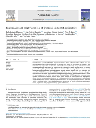
- 2. Aquaculture Reports 25 (2022) 101220 2 producers and consumers. To fight those stressors and their negative impacts, probiotics have become recognized as important substitutes, acting as immunity modulators and increasing resistance against various microbial pathogens (Dawood et al., 2018; Madani et al., 2018; Van Doan et al., 2018). The term probiotic is coined from Greek pro and bios (meaning “for life”), which can be understood further from the revised definition of Merrifield et al. (2010): “a probiotic organism can be regarded as a live, dead or component of a microbial cell, which can be administered via feed or into rearing water, benefiting the host by improving growth performance, feed utilization, immune health status, infectious disease resistance, and stress responses which is achieved at least in part via improving the microbial balance in hosts or ambient environment”. Scientists working with probiotics have noted positive effects on growth (Farsani et al., 2020; Nargesi et al., 2020), activities of digestive en zymes and feed utilization (Makled et al., 2020; Yanbo and Zirong, 2006), immunity elevation by way of immune-gene transcription (Beck et al., 2016; Park et al., 2020), improvement of beneficial gut-microbes and positive modification of intestinal structure (Akter et al., 2019; Duan et al., 2018), and protective actions against diseases (Ramesh et al., 2017; Rengpipat et al., 2000; Van Doan et al., 2018). These pos itive health influences have the additional benefit of being an eco-friendly approach to aquatic environmental management (Cha et al., 2013; Das et al., 2006). The effective use of probiotics can be influenced by some conditions, such as the administration methods, appropriateness of the timing of application, dosages and other physiological factors of the culture spe cies. It is a good general principal that probiotics are most effective when applied preventatively into culture systems before any disease outbreaks occur. Mostly they are designed to be applied as feed additives as opposed to addition into the culture environment, since administration of probiotics orally is considered to be a more practical method (Azad et al., 2005; Gomes et al., 2009; van Hai and Fotedar, 2010). Probiotics have been found to be effective through several modes of action. Major activities of probiotics include i) competitive exclusion of pathogens, ii) stimulation of increased immune responsiveness, iii) promotion of the production of antimicrobial substances and iv) enhancement of growth and survival rates. In addition, the inhibitory effects of probiotics are proven against emerging viral diseases such as white spot syndrome virus (WSSV), yellow head disease virus (YHD) and Taura Syndrome virus (TSV) (Lai et al., 2020a; Thammasorn et al., 2017; Walker and Mohan, 2009). Among established mechanisms of action, immunomodulation ap pears to be actively involved in building resistance against viruses. Probiotics stimulate phagocytic activity, acid phosphatase, lysozymes and cytokines, all of which enhance the immunocompetence of shellfishes, increasing their resistance to viral diseases (Chai et al., 2016; Mamun et al., 2019). In addition, probiotics act as a potential adjuvant as they stimulate cellular and humoral components of shellfish immune systems (Sakai, 1999). Moreover, probiotics were found effective to enhance growth and feed utilization parameters (specific growth rate, final body weight, weight and length gain, feed efficiency and feed conversion ratios etc.), water quality parameters (temperature, dis solved oxygen, pH, nitrate, phosphate, silicate etc.), immune-related gene expressions and much more (Das et al., 2006; Duan et al., 2019; Silva-Aciares et al., 2013; Talib et al., 2017). Aiming to characterize the functionality of probiotics in shellfishes, an extensive search of the Scopus database revealed more than 200 publications focused on the efficiency of probiotics in shellfish, such as prawn, shrimps and lobster species, and citations up to the year 2021 have reached about 6000 (Fig. 1). However, considering those publi cations, this study also focuses on different capacities of probiotics to wards shellfishes. Besides the enhancement of growth, feed utilization, water quality and immune-related parameters, probiotics are known to effectively reduce the viral load and hamper the spread of viral diseases in shellfishes. Future research on shellfish probiotics should focus on finding an organized approach to relevant functions of probiotics on shellfishes such as shrimps, prawns, crabs, crayfishes, lobsters, oysters and abalones. 2. Mode of action of probiotics Over the last two decades, a significant number of investigations have focused on the effects of probiotics as they pertain to disease resistance, growth performance, microbial balance, intestinal health, feed utilization, and water quality in farmed shellfish. These studies have collectively revealed multiple modes and sites of action for pro biotics applied in aquaculture, leading to the proposal of several mechanisms of action in the literature (Dawood et al., 2018; Dawood and Koshio, 2016; Hai, 2015; Hoseinifar et al., 2018; Newaj-Fyzul et al., 2014; A. Wang et al., 2019), including the formation of inhibiting compounds, competition with potential pathogens, bolstered immune response, and improved growth and survival. Probiotic actions have been reported in larval zebrafishes prior to the initiation of feeding, indicating improved yolk absorption and nutrient utilization (Padeniya et al., 2022). These modes of action contribute insights into the diverse range of positive benefits on aquatic species (Fig. 2), which can differ in some cases according to developmental status. For a deeper compre hension of probiotic effectiveness, forthcoming experimentation on the interaction between probiotic and host needs to incorporate both tran scriptomic and proteomic analysis. For a better understanding, we recommend articles on such analysis, such as, Brunt et al. (2008) Fig. 1. The volume of publications in the Scopus database covering the effects of probiotics on shellfish and their citation numbers (Years: 1998–2021). T.A. Sumon et al.
- 3. Aquaculture Reports 25 (2022) 101220 3 (proteomic analysis of Rainbow trout serum after probiotic adminis tration) and Tacchi et al. (2011) (transcriptomic responses of Atlantic salmon to functional feeds). 2.1. Competitive exclusion of pathogens Competitive exclusion has been posed as among possible mecha nisms of probiotic effect against pathogens (Merrifield et al., 2010; Newaj-Fyzul et al., 2014; A. Wang et al., 2019). Probiotics enter into the gastrointestinal (GI) tracts of aquatic organisms in the same way as they do in terrestrial animals, and can disrupt the function of pathogens through the production of antagonistic compounds and by competing for substrates, and resources such as nutrients, physical space, and even oxygen (Fuller, 1989; Hai, 2015) (Fig. 2). Competition for binding re ceptors which might antagonize pathogens and limit their colonization could be the main explanation for this action (Chabrillon et al., 2006; Luis-Villaseñor et al., 2011). The inhibitory role of probiotics on infec tious microbes has been investigated in a number of studies. For instance, LAB was revealed to be inhibitory to Vibrio harveyi and V. parahaemolyticus in Litopenaeus vannamei and Penaeus indicus respectively (Ajitha et al., 2004; Vieira et al., 2007). Similarly, Entero coccus faecium (in Penaeus monodon), Lactococcus lactis subsp. lactis (in L. vannamei), several Lactobacillus species in shrimps were reported to be inhibitory towards many Vibrio spp. such as V. harveyi V. alginolyticus and V. parahaemolyticus (Adel et al., 2017; Karthik et al., 2014; Nguyen et al., 2018; Sivakumar et al., 2012; Swain et al., 2009). It should be noted that not all probiotic candidates colonize the GI tract; some inhabit the skin, gills, and other tissues. In addition, various factors including passive forces, electrostatic interactions, hydrophobic, steric forces, and lipoteichoic acids have all been documented to influence probiotic adherence to binding points (Hoseinifar et al., 2018). Pro biotics appear to have very good potential for utilization as a replace ment to antibiotics for disease preventative purposes. 2.2. Immune modulation One critically important mode of action of probiotics is immuno modulation, which involves enhancing the host’s immunological response. In recent years, a growing degree of attention has been attracted to understanding the underlying mechanism of action of pro biotics on the immune system of shellfish, in response to numerous re ports that probiotics stimulate immune function. In tiger shrimp (Penaeus monodon), dietary Bacillus sp. S11 had a beneficial effect on cellular and humoral immunity, leading to enhanced protection against disease (Rengpipat et al., 2000). Moreover, probiotic application of Clostridium butyricum CBG01 has been demonstrated to augment innate immunity of whiteleg shrimp (L. vannamei), in which significantly increased activities of alkaline phosphatase, acid phosphatase, lyso zyme, and total nitric oxide synthase were observed with elevated res piratory burst activity (Li et al., 2019a). Moreover, increases in other innate and cellular immune parameters such as heamolymph bacteri cidal activity (Chandran et al., 2014; Rengpipat et al., 2000), phagocytic activity (Liu et al., 2014; Y. C. Wang et al., 2019; Zhao et al., 2019), superoxide dismutase (SOD) (Miao et al., 2020; Tseng et al., 2009), total anti-oxidant capacity (Amoah et al., 2020; Duan et al., 2017), total haemocyte count (Dash et al., 2015; Kumar et al., 2013), catalase (Hindu et al., 2018; Miao et al., 2020), phenoloxidase (Rahiman et al., 2010) and prophenoloxidase (proPO) (Kolanchinathan et al., 2017) have been described among effects of probiotics in multiple shellfish species (Fig. 2). Furthermore, numerous studies have demonstrated that adding probiotics to shellfish diets improves the expression of immunological and stress-related genes. including pen-3a (Chai et al., 2016; Tepaa morndech et al., 2019), proPO (Li et al., 2019b; Wu et al., 2014), SOD (Sánchez-Ortiz et al., 2016; Yang et al., 2019), HSP70 (Duan et al., 2018; Miao et al., 2020), lipopolysaccharide and β-1, 3-glucan binding protein (LGBP) (Hao et al., 2014; Interaminense et al., 2019). Fig. 2. Mode of action of probiotic microorganisms in shellfish. A) Competitive exclusion of pathogens like Vibrio sp., Vibrio anguillarum, white spot syndrome virus, yellow head virus, taura syndrome virus etc. B) Development of the immune system is another important mode of action of probiotics in shellfishes, where they increase alkaline and acid phosphatase activities and provide protection against bacterial, viral and parasitic diseases. C) Probiotic also produce many antimicrobial substances, e.g., bacteriocin, nisin, lysozyme enzymes and bactericidal proteins and so on. D) Probiotics like Bacillus, Lactobacillus, Clostridium etc. enhance growth and survival rates of shellfishes through enhancing feed utilizations, increasing digestive enzyme activities and producing vital nutrients. T.A. Sumon et al.
- 4. Aquaculture Reports 25 (2022) 101220 4 2.3. Production of antimicrobial compounds Probiotics are employed as an alternative to antimicrobial agents such as antibiotics and chemicals in shellfish farming (Decamp et al., 2008; Van Hai et al., 2009). Although the specific mechanism by which probiotics exhibit antibacterial properties is still unknown, numerous investigations have suggested that a variety of probiotic species generate bactericidal molecules such as bacteriocin, siderophore and lysozyme enzymes (Hai, 2015; Hoseinifar et al., 2018). Immune systems of shellfishes have been activated by these molecules, increasing their resistance to infections by viruses, bacteria, fungi, and parasites (Fig. 2). Digestion and fermentation of non-digestible fibers and polysaccharides by probiotics led to formation of a group of fermented short- and medium-chain fatty acids which reduce the pH in the intestine, thereby inhibiting pathogenic bacteria (Dawood, 2021; Ma et al., 2009). In addition, common probiotic bacteria like Lactobacillus spp. have been found to inhibit pathogens via the generation of short chain fatty acids, diacetyl, hydro peroxide, and bactericidal proteins (Hai, 2015; Rengpi pat et al., 1998; Verschuere et al., 2000). 2.4. Promotion of growth and survival The activity of digestive enzymes is closely associated with healthy feed intake, digestion ability, growth rates, and the general health of the host species (A. Wang et al., 2019). By augmenting the functionalities of digestive enzymes, probiotics improve feed utilization and growth in shellfish (Newaj-Fyzul et al., 2014). Significantly higher growth rate, feed utilization and protease enzyme activity were observed in whiteleg shrimp following the administration of Bacillus subtilis E20 (Liu et al., 2009). Probiotic Clostridium butyricum has been shown to enhance the growth performance and feed conversion ratio (FCR) of tiger shrimp (Duan et al., 2018). Similarly, a combination of three probiotic strains fed to ornate spiny lobster (Panulirus ornatus) promoted higher weight gain with increased specific growth rate and FCR (V. D. Nguyen et al., 2014). Synthesis of extracellular enzymes such as proteases, lipases and amylases have been reported in response to probiotics, with evidence that increases in digestive enzyme production result in improved digestive function (Adel et al., 2017; Amoah et al., 2020; Seenivasan et al., 2016; Sumon et al., 2018). Additionally, probiotics might enhance the uptake and utilization of nutrients and micronutrients, since the application of probiotics resulted in the production of vital materials such as vitamin B12, fatty acids, and biotin (Hai, 2015; Verschuere et al., 2000; Vine et al., 2006). One net effect is that feeding probiotics to shellfish has been confirmed to boost survival rates. (Zokaeifar et al., 2012) reported higher survival rate of whiteleg shrimp (Litopenaeus vannamei) upon dietary supplementation of two strains of B. subtilis (L10 and G1). Agarivorans albus strain F1-UM and Vibrio sp.-fed abalone (Haliotis rufescens) (Silva-Aciares et al., 2013) and Bacillus-fed mud crab (Scylla paramamosain) (Talib et al., 2017) also had notably higher sur vival rates during challenges with Vibrio parahaemolyticus and Vibrio sp. respectively. 3. Antiviral activity/ability of probiotics The diseases of aquatic animals that have been reported for centuries are mostly either non-infectious or caused by parasites, bacteria, or other pathogens. However, in last few decades the culture of aquatic animals has undergone a series of intensifications, including some extremely intensive methods. Accordingly, the global improvement of aquaculture production systems became associated with various stresses to the aquatic ecosystems and the emergence of a number of new dis eases in fish and shrimp. Shrimps are the largest seafood commodity, contributing around 17% in globally-traded fishery products, and they are often cultured intensively. Diseases have had a disproportionately heavy impact on the shrimp industry, and they are recognized as criti cally important limiting factors (Lakshmi et al., 2013; Walker and Mohan, 2009; Walker and Winton, 2010). The growth of shrimp farming is constrained by diseases but treating and preventing them with ranges of chemicals - particularly antimicrobial agents – has had a catastrophic impact on the industry. The infusion of tons of antibiotics into aquatic environments has resulted in the accumulation of harmful residues in commercial shellfish produced for human consumption. In addition, these antibiotics also devastate the normal microbial communities, including numerous benevolent bacteria, thereby promoting the prolif eration of resistant pathogens and compromising shrimp and fish pro duction (Grave et al., 1999; Lakshmi et al., 2013; Le and Munekage, 2004). The only microbes that can survive such chemical attacks are antibiotic-resistant strains, and consequently this strategy for control has led to the emergence of “superbugs”, or antibiotic-resistant strains of pathogens. As a result, alternatives to antibiotics become a crucial, extremely high-priority industry need. Besides bacterial and parasitic pathogens, a series of novel viral pathogens of shrimp have continued to emerge since the 1970 s, such as monodon baculovirus (1977), hepatopancreatic parvovirus (1983), Yellow Head Virus - YHV (1990), White Spot Syndrome virus - WSSV (1992), Taura Syndrome virus - TSV (1992) and so on (Walker and Mohan, 2009). Viruses evolve extremely fast – a painful lesson that we have learned in the course of the Covid pandemic. The viral life cycle is as short as a single day, enabling mutant strains to become established extremely quickly. Among aquatic pathogenic viruses, WSSV has caused huge economic losses worldwide, and no commercial vaccine to improve adaptive immunity against WSSV is available to date (Pham et al., 2017; Sánchez-Martínez et al., 2007). Recently, a number of sci entists have identified the potential of probiotics to improve resistance of shellfishes against viral diseases like White Spot Syndrome (WSS) and YHV disease (Table 1). In most cases, Bacillus and LABs (including Lactobacillus) have been used experimentally, and many of these LAB species have been found effective to reduce mortality due to WSSV and YHV. Moreover, WSSV prevalence has been significantly reduced after administration of some probiotic microorganisms. Additionally, the immune system of shrimps responded positively to probiotics in nearly all experiments, leading to improved shrimp health (Chai et al., 2016; Partida-Arangure et al., 2013; Thammasorn et al., 2017). For instance, Chai and coworkers (Chai et al., 2016) found the PC465 strain of Bacillus decreased the tran scription of crustin in hemocytes of L. vannamei, which is responsible for inhibiting replication of WSSV and the reduction of mortality in WSSV-challenged shrimps, thereby providing some protection against WSS (Chai et al., 2016). Likewise, Lai and his colleagues found Bacillus amyloliquefaciens to stimulate innate immunity, resulting in reduced copy numbers of WSSV and significantly reduced mortality of WSSV-challenged crayfish (Lai et al., 2020b). In providing resistance to WSSV and prophylaxis treatment, probiotics have also been found effective. For example, the VP28 candidate antigen vaccine using B. subtilis spores decreased the death rate to 33% from 66% of otherwise untreated whiteleg shrimps. In addition, a 75% protection rate was found in the case of tiger shrimps ( A. T. V Nguyen et al., 2014; Pham et al., 2017). To recapitulate, as there are some probiotics that potentially inhibited or reduced or provided resistance to WSSV, this should be subjected to intense scientific pursuit. The inhibitory capacity of pro biotics against WSSV could serve as a model for the management of other emerging viral diseases. To achieve this, a systemic and accurate method of studying antiviral activity of probiotics should be established first. In this context, Lakshmi et al. (2013) suggested some modifications to the cell culture model proposed by Botić et al. (2007), in which probiotic bacteria exclude pathogens through competition for attach ment sites coupled with the stimulation of host cell immune defenses in humans (Isolauri, 2003). The proposed cell culture model could include several antiviral assays, such as the incubation of cell lines with viable probiotic bacteria, or the viruses and bacteria could be administered simultaneously to the cell lines, or the bacteria and virus could be T.A. Sumon et al.
- 5. Aquaculture Reports 25 (2022) 101220 5 co-incubated, or the supernatants containing products of viable pro biotic bacteria could be added in the cell lines as components of the incubation media (Lakshmi et al., 2013). However, the application of probiotics in shellfish culture is a safe and novel tactic. Further experi mental attention should focus on the mechanisms involved in probiotic resistance to viral infections. 4. Effects of probiotics on shellfish 4.1. Whiteleg shrimp Whiteleg shrimp, L. vannamei (also known as Pacific white shrimp) is the most cultured crustacean species, accounting for nearly 53% of total global crustacean production. It is a genetically improved and domesticated species. Production of L. vannamei has substantially surged to 4.156 million tonnes (MT) in 2018, up from 1.13 MT in 2000 (FAO, 2020); alternative favorite species such as Penaeus monodon have not come close to keeping pace with the industry-dominating growth of P. vannamei. Disease occurrence also coupled with increased and intensified production of this species, accounts for significant financial losses for the aquaculture industry. The effect of probiotics on whiteleg shrimp has piqued researchers’ interest in recent years, resulting in the highest number of studies among shellfish. The Scopus database yielded a total of 97 articles on the application of probiotics in L. vannamei as of July 16, 2021 (Fig. 1). Whiteleg shrimp have been treated with a diversity of microorgan isms as probiotics, producing a variety of beneficial effects, including enhancement of growth performance, immune response and protection against various pathogens, including V. harveyi, V. alginolyticus, V. campbellii, V. parahaemolyticus, and WSSV (Table 2). Among the studied probiotic candidates, Bacillus spp., Lactobacillus spp., and Clos tridium spp. are the most extensively utilized species (Butt et al., 2021). In addition, the yeast probiotic Saccharomyces cerevisiae has been shown to be helpful in a number of investigations (Ayiku et al., 2020; Y. C. Wang et al., 2019). These probiotics are derived from a variety of sources, most of which have been cultured from the guts of healthy specimens of the host species (Kewcharoen and Srisapoome, 2019; Kongnum and Hongpattarakere, 2012; Liu et al., 2014). A summary of the effects of probiotics in whiteleg shrimp aquaculture is presented in Table 2. The majority of probiotic investigations on this shrimp species have focused on single-species probiotic treatment (Adel et al., 2017; Duan et al., 2017; Shen et al., 2010; Tseng et al., 2009). Kewcharoen and Srisapoome (2019) fed white shrimp a diet supplemented with host-derived B. subtilis AQAHBS001 for five weeks at different concen trations: 105 , 107 and 109 CFU g–1 . When compared to the control treatment, probiotic-treated shrimp demonstrated significantly improved growth, feed conversion ratios (FCR), and immunological responses as revealed by increased phagocytic activity and clearance efficiency. Furthermore, probiotic found to augment disease resistance of shrimp against Vibrio parahaemolyticus AHPND by upregulating the expression of immune-related genes and increasing microvilli and in testinal wall thickness. In another six–weeks experiment, addition of Lab. plantarum MRO3.12 (2 – 4 ×10⁸ CFU g–1 ) isolated from digestive tract of wild shrimp to the diet improved growth, feed efficiency and survival rate of white shrimp (Kongnum and Hongpattarakere, 2012). Furthermore, when challenged with V. harveyi, probiotic-fed shrimp had a 30% higher survival rate than the control group. Multi-strain probiotics have garnered interest recently owing to their cumulative favorable impact with higher potency against a broader range of pathogenic microorganisms. However, a mixture of probiotics has been shown to yield enhanced benefits to the hosts compared to probiotics administered singularly, although shrimps do not consistently benefit from exposure to such combinations of species. For instance, Cai et al. (2019) evaluated the effect of B. licheniformis, B. flexus and their mixture in L. vannamei postlarvae after feeding probiotic included diet for 21 days. These authors reported that these two strains not only increased the growth, survival, digestive enzyme functions, innate im munity, stress tolerance and disease resistance against V. harveyi, but also elevated the quality of rearing water. Likewise, administration of single or combined Shewanella haliotis D4, B. cereus D7 and Aeromonas bivalvium D15 at 107 CFU g− 1 added to the feed of the whiteleg shrimps for 28 days was demonstrated to modulate non-specific immune pa rameters (Hao et al., 2014). Probiotics-fed shrimp displayed increased activities of respiratory burst, lysozyme, superoxide dismutase and acid phosphatase, as well as upregulated expression of genes such as lipo polysaccharide and β-1,3-glucan binding protein (LHBP), prophenolox idase (proPO) and penaeidin 3 (Pen 3). Some investigations have explored the use of probiotics as water supplements, as opposed to frequent analyses of the dietary application Table 1 Effects of probiotics against viral diseases of shellfish. Probiotics Shellfish species Action against viral diseases References Bacillus Lactic acid Bacteria (LAB) Litopenaeus vannamei Improved host immunity Reduced prevalence of White Spot Syndrome Virus (WSSV) (Partida-Arangure et al., 2013) Bacillus PC465 Litopenaeus vannamei Immune enhancement of shrimp Reduced copy number of WSSV Protection against WSSV (Chai et al., 2016) Bacillus OJ Litopenaeus vannamei Increased immune response and resistance to WSSV with increasing doses (Li et al., 2009) Probiotic mixture of four LAB with extract and powdered forms of plant Litopenaeus vannamei High survival rate and low prevalence of WSSV (Peraza-Gómez et al., 2011, 2009) Lactobacillus plantarum Penaeus vannamei Promoted shrimp health against viruses Reduced copy number of Yellow head virus (YHV) (Thammasorn et al., 2017) Bacillus amyloliquefaciens Procambarus clarkia (crayfish) Regulate innate immunity Reduced copy number of WSSV Significantly reduced mortality of crayfish challenged with WSSV (Lai et al., 2020) Bacillus subtilis with VP28 gene Litopenaeus vannamei Increased survival of shrimp challenged with WSSV (Fu et al., 2011) Bacillus subtilis with VP28 gene Fenneropenaeus chinensis Significant resistance to WSSV (Fu et al., 2010) Bacillus subtilis with VP28 gene Penaeus monodon Found effective against White Spot Syndrome (WSS) (Pham et al., 2017) Bacillus subtilis with VP28 gene Litopenaeus vannamei Found effective against WSS (A.T.V. Nguyen et al., 2014) T.A. Sumon et al.
- 6. Aquaculture Reports 25 (2022) 101220 6 Table 2 Influence of probiotics on growth, feed utilization, immunological and haemato-biochemical parameters, and disease resistance in whiteleg shrimp (Litopenaeus vannamei). Symbol: (→) no change; (↑) increase; (↓) decrease. Probiotics Mode of administration and dosage Duration Effects on Litopenaeus vannamei References Bacillus subtilis Dietary supplementation at 104 , 5 × 10⁴ & 105 CFU g–1 40 days GP WG↑; SR→; HBP ALP & ACP→; IP TAC, SOD, GPx, RB, PO & SL↑, and MDA & CAT→ (Shen et al., 2010) B. subtilis S12 Dietary supplementation at 5 × (109 , 1010 & 1011 ) CFU kg–1 8 weeks GP WG & SGR→; FUP FCR→; IP PcA, SL, SOD, SBA & PO↑, and THC→; SR↑; IDR Vibrio harveyi↑ (Liu et al., 2014) B. subtilis E20 Dietary supplementation at 107 , 108 & 10 CFU kg–1 98 days GP WG↑; FUP FCR↓; SR→; DEA PrA↑ (Liu et al., 2009) B. subtilis E20 Dietary supplementation at 106 , 107 & 108 CFU kg–1 56 days GP FBW, WG & SGR↑; IP THC, RB, PO & SOD↑; IDR Vibrio alginolyticus↑ (Adilah et al., 2022) B. subtilis AQAHBS001 Dietary supplementation at 105 , 107 & 109 CFU g–1 5 weeks GP FBW, WG & SGR↑; FUP FCR↓; SR→; IRGE SOD, SL, proPO & ALF↑; Gut Morphology ↑; IDR V. parahaemolyticus ↑ (Kewcharoen and Srisapoome, 2019) B. subtilis, B. megaterium, B. cereus & B. infantis Dietary supplementation at 108 CFU g–1 6 weeks GP WG, FBW & SGR→; FUP FCR↓; SR↑; IP SOD, CAT & PO↓; DEA PrA, AmA, Lipase & Cellulase↑ (Tamilarasu et al., 2021) B. aryabhattai TBRC8450 Dietary supplementation at 108 CFU g–1 6 weeks IR PO & TAC↑; IRGE Pen-3a, Lec-3, Trx, HSP60 & fer↑, and SP, CAT, SOD, GPx→, and LGBP↓; IDR V. harveyi↑; GutM Vibrio spp.↓, and Bacterial diversity↑ (Tepaamorndech et al., 2019) B. subtilis L10 + B. subtilis G1 Dietary supplementation at 105 & 108 CFU g–1 8 weeks GP FBW, WG & SGR↑; FUP FCR→; SR↑; DEA PrA & AmA↑; IRGE proPO, PE, LGBP & SP↑; IDR V. harveyi↑ (Zokaeifar et al., 2012) B. subtilis L10 + B. subtilis G1 Water supplementation at 105 & 108 CFU mL–1 8 weeks GP FBW, WG & SGR↑; FUP FCR↓; SR↑; DEA PrA & AmA↑; IRGE proPO, PE, LGBP & SP↑; WQP Ammonia, Nitrite & Nitrate ions↓; IDR V. harveyi↑ (Zokaeifar et al., 2014) Bacilli (50% B. subtilis & 50% B. licheniformis) Dietary supplementation at 104 & 108 CFU g–1 60 days GP WG & SGR↑; FUP FCR↓; SR↑; HBP Glu & Cortisol↓, and ALB→; IP SL, THC & Ig↑ (Madani et al., 2018) Photosynthetic bacteria & Bacillus sp. Dietary supplementation at 2, 10 & 20 g kg–1 28 days GP WG↑; DEA PrA, AmA, Lipase & Cellulase↑ (Wang, 2007) B. subtilis & Shewanella algae Dietary supplementation at 106 CFU g–1 60 days GP FBW↑; IRGE proPO, LGBP & HEM↑; IDR V. parahaemolyticus↑ (Interaminense et al., 2019) B. licheniformis MAt32 + B. subtilis MAt43 + B. subtilis subsp. subtilis GAtB1 Dietary supplementation at (1, 2, 4 & 6) × 106 CFU g–1 32 days GP SGR↑, and FBW→; Prevalence WSSV & IHHNV↓; SR→; IRGE proPO, LvToll1 & SOD↑, and HSP70↓, and TGase→ (Sánchez-Ortiz et al., 2016) B. licheniformis, B. flexus, B. licheniformis + B. flexus Dietary supplementation at 2 × 10⁹ CFU g–1 21 days GP FBW & SGR↑; HBP ALP↑; IP MPO & SL↑; SR↑; DEA PrA & Lipase↑, and AmA→; WQP Ammonia & COD↓; ESC Freshwater↑; IDR V. harveyi↑ (Cai et al., 2019) Shewanella haliotis D4, B. cereus D7, Aeromonas bivalvium D15, & their mixture Dietary supplementation at 107 CFU g− 1 28 days GP FBW, WG & SGR↑; IP RB, SL, ACP & SOD↑; IRGE proPO, LGBP & Pen-3a↑; IDR V. harveyi↑ (Hao et al., 2014) Clostridium butyricum Dietary supplementation at 0.25, 0.5 & 1% 56 days GP WG & SGR↑; FUP FCR↓; IP TAC, SL & iNOS↑, and NO→; IRGE Toll, Imd, & HSP70↑; DEA AmA, PrA & Lipase↑; SR→; Gut Morphology Intestine epithelium height↑; Intestine scFA↑; ESC Ammonia↑ (Duan et al., 2017) C. butyricum Dietary supplementation at (2.5, 5 & 10) × 109 CFU g–1 56 days GutM Bacillus, Clostridium, Lachmoclostridium, Lachnospiraceae & Lactobacillus↑, and Desulfovibrio & Desulfobulbus↓; DGE α-amylase, lipase, trypsin, fatty acid-binding protein & fatty acid synthase↑; IRGE proPO, LGBP, SL, Crustin & SOD↑; (Duan et al., 2018) C. butyricum CBG01 (fermentation supernatant [FS]; live cells [LC]; cell-free extract [CE]; spray-dried spores [DS]; mixture of live cells & supernatant [LCS]) Dietary supplementation at 1011 CFU kg–1 of LC, DS, & CE, 120 mL kg–1 (FS), & 1011 CFU kg–1 LC + 120 mL kg–1 FS 42 days GP FBW & SGR↑; FUP FCR↓; SR→; IRGE SOD, SL, proPO, Toll, Imd & Relish↑; Gut Morphology VH & Intestinal wall thickness↑; IDR V. parahaemolyticus↑ (Li et al., 2019a) C. butyricum CBG01 Dietary supplementation at 107 to 1012 CFU kg–1 42 days GP WG & SGR↑; FUP FCR↓; SR→; HBP ALP↑; IP SL, RB, NO, ACP & SOD↑; IRGE Toll, Imd & Relish↑; Gut Morphology VH & Intestinal wall thickness↑; IDR V. parahaemolyticus↑ (Li et al., 2019b) Lactobacillus plantarum Dietary supplementation at 109 CFU mL–1 45 days GP FBW, WG & SGR↑; FUP FCR↓; IRGE proPO, SOD & SL (at stress challenge) ↑; ESC Low salinity↑ (Zheng et al., 2017) Lab. plantarum Dietary supplementation at 109 CFU mL–1 15 days GP FBW, WG & SGR↑; FUP FCR↓; DEA AmA, Lipase & Pepsin↑; Gut Morphology Enterocytes height↑ (Zheng et al., 2018) Lab. plantarum MRO3.12 Dietary supplementation at 2–4 × 10⁸ CFU g–1 6 weeks GP WG & FBW↑; FUP FCR↓; SR↑; IP THC↑; GutM LAB↑; IDR V. harveyi↑ (Kongnum and Hongpattarakere, 2012) Lab. pentosus AS13 Dietary supplementation at 106 , 107 & 108 CFU g–1 28 days GP WG & SGR↑; FUP FCR↓; SR→; DEA PrA & Cellulase↑, and Lipase→; IDR V. vulnificus, V. rotiferianus & V. campbellii↑ (Zheng and Wang, 2017) Lab. pentosus HC-2 Dietary supplementation at 5 × 10⁸ CFU g–1 6 weeks GP WG & FBW→; FUP FER→; SR↑; IRGE Rab, GST, mucin-like-PM, Dorsal & proPO↓, and Relish→; GutM ↑ (Fang et al., 2020) Lab. pentosus BD6, Lab. fermentum LW2, B. subtilis E20, & Saccharomyces cerevisiae P13 Dietary supplementation at 107 , 108 & 109 CFU kg–1 56 days GP WG↑; FUP FER↑; SR→; IP PO, RB, SL, SOD & PcA↑; IDR V. alginolyticus↑ (Y.C. Wang et al., 2019) Pediococcus pentosaceus Dietary supplementation at 106 , 107 & 108 CFU g–1 8 weeks GP WG, FBW & SGR↑; FUP FCR↓; HBP THC↑; IP SL↑; DEA PrA & AmA↑, and Cellulase→; SR↑; GutM Lactobacillus sp. & Bacillus sp.↑, and Vibrio sp.↓; IDR V. anguillarum↑ (Adel et al., 2017) Paenibacillus polymyxa ATCC 842 Dietary supplementation at 106 , 107 & 108 CFU g–1 8 weeks GP WG, FBW & SGR↑; FUP PER↑, and FCR↓; HBP TSP, Alb, Glb, TG & ALP↑, and AST & ALT↓; IP SL, ACP, TAC, SOD & GPx↑, and MDA↓; DEA AmA, Trypsin & Lipase↑; Gut Morphology VH and width & Muscle thickness↑; GutM (Amoah et al., 2020) (continued on next page) T.A. Sumon et al.
- 7. Aquaculture Reports 25 (2022) 101220 7 of probiotics in white shrimp (Wu et al., 2016; Xia et al., 2014; Zokaeifar et al., 2014). Wu et al. (2016) reported that administering probiotic B. subtilis FY99–01 to the water enhanced water quality by lowering pH, nitrite, and soluble reactive phosphorus levels, but had no influence on shrimp growth, survival, or FCR. In contrast, Zokaeifar et al. (2014) observed that supplementation of B. subtilis strains (L10 + G1) in the rearing water of L. vannamei at 105 and 108 CFU mL–1 conferred bene ficial effects to water quality, apparently enhancing growth parameters, gastrointestinal enzyme activities along with elevated immune response and effective inhibition of V. harveyi. 4.2. Tiger shrimp The tiger shrimp, Penaeus monodon a commercially important species primarily in Asia, contributed 9% of total global crustacean production in 2016 (FAO, 2020). The recent development of intensive culture sys tem and environmental deterioration have caused frequent disease outbreaks, for which effective treatment measures are lacking (Duan et al., 2019). Up to 41 research articles have been found in the Scopus database on probiotic application for growth, immunity and disease resistance modulation of this species. Unlike L. vannamei, P. monodon has not been domesticated because breeding is a relatively more time-cosuming and expensive process involving artificial insemination (Muthu and Laxminarayana, 1984). Different types of probiotics (single and multiple) application effects on P. monodon are summarized in Table 3. Most of the supplemented probiotics in P. monodon were isolated from this species’ intestine and Bacillus as well as lactic acid bacteria (LAB) were mostly supplemented (80–120 days) probiotics for shrimp. Single dose of Bacillus S11 and P11 produced increased growth and some degree of resistance to V. harveyi disease (Rengpipat et al., 2000, 1998; Utiswannakul et al., 2011). Ba cillus S11 and P11 are different regarding cell size, physical appearance and optimal growth temperature. Colonization of Bacillus S11 in the shrimp gut acts as competitor against infectious pathogens, thereby interfering with infectious disease outbreaks (Meunpol et al., 2003). Presence of amorphous poly-betahydroxybutyrate (PHB) in Bacillus JL47 is the main causal factor of immune genes transcription as well as protection of Vibrio challenge (Laranja et al., 2017). It should be noted that sometimes shrimp growth performance in re-circulatory aquacul ture systems (RACs) might be slower than outdoor pond culture, due to filtration of important nutritive compounds for water quality manage ment. Aquatic bacteria are abundant in aquaculture environments, and they can play important roles like continuation of geochemical cycles and activity in fish immune systems (Fig. 3). Bacillus strains are commonly used as probiotics in commercial aquaculture because of their spore formation ability and stability in aquatic environments as well as in the intestine. Live and freeze-dried B. subtilis, B. polymyxa, B. megaterium, B. licheniformis and B. thuringiensis improved Bacillus concentration in hepatopancreas and intestine by lowering the Vibrio count (Boonthai et al., 2011). Probiotics like B. cereus (0.4%) (Chandran et al., 2014) and Streptomyces sp. (Das et al., 2006) showed highly favorable outcomes for the modulation of vitally important water quality parameters such as the abundance of dissolved ammonia, alka linity, nitrate, phosphate and the salinity of culture water. Most of the LAB (Lactobacillus) produce bactericidal proteins with demonstrable antimicrobial activity against both gram-positive and gram-negative pathogens. Both Lactobacillus sp. AMET1506 and Lab. acidophilus 04 improved shrimp growth and decreased E. coli (Karthik et al., 2014) and Vibrio spp. (Sivakumar et al., 2012) concentration in GIT, respectively. Probiotics generally increase digestive enzyme activity by the secretion of exoenzymes in the intestine, thereby improving digestion and absorption of ingested food resulting in better growth and FUPs. B. subtilis + B. licheniformis and B. subtilis & Enterococcus sp. improved the level of 4 digestive enzymes, such as protease, amylase, lipase and cellulases (Wang et al., 2020) and only trypsin (Nimrat et al., 2013), respectively. Supplementation of Pseudomonas sp. PM 11 & V. fluvialis PM 17 to culture water (Alavandi et al., 2004) and B. coagulans + B. firmus by diet (Kolanchinathan et al., 2017) upregulated innate immune parameters of shrimp. Streptococcus phocae and Enterococcus faecium exposure resulted in the production of bacteriocin and other antibacterial substances, producing pores on the cell wall of V. parahaemolyticus, Listrea monocytogenes and E. coli and efflux of K+ ions, leading to the mortality of these pathogens. Shrimp fed with S. phocae PI80 and E. faecium MC13 upregulated growth parameters and infection of V. harveyi and V. parahaemoliticus, respectively (Swain et al., Table 2 (continued) Probiotics Mode of administration and dosage Duration Effects on Litopenaeus vannamei References Ruegeria & Pseudoalteromonas↑, and Vibrio, Photobacterium, Tenacibaculum & Shewanella↓; IDR V. parahaemolyticus↑ Arthrobacter sp. CW9 Water supplementation at 105 , 106 & 107 CFU mL–1 24 days GP FBW↑; SR↑; IP PO & PcA↑ (Xia et al., 2014) B. subtilis FY99–01 Water supplementation at 5 × 10⁴ CFU mL–1 84 days FUP FCR→; SR →; WQP pH, Nitrite & Soluble reactive phosphorus↓; Water Microbiota Flavobacteria↑, and α-Proteobacteria & Vibrionaceae↓ (Wu et al., 2016) Bacillus PC465 Dietary supplementation at 107 & 109 CFU g–1 30 days GP WG↑; SR↑; DEA AmA & PrA↑; IRGE Pen-3a, Lec-3, Trx, proPO & Peroxinectin↑; PCI↑; IDR WSSV↑ (Chai et al., 2016) Saccharomyces cerevisiae Dietary supplementation at 1% & 2% 8 weeks GP WG, FBW & SGR↑; FUP FCR↓; HBP TSP, AST, ACP & ALP↑, and Glu & ALT↓, and TG & TC→; IP SL, SOD, PO & CAT↑; Gut Morphology VH and width & Muscle thickness↑; GutM ↑; IDR V. harveyi↑ (Ayiku et al., 2020) B. subtilis, B. subtilis+B. pumilus Dietary supplementation at 2.1 × 10⁷ , 4.4 × 10⁷ , 1.6 × 10⁷ , and 3.5 × 10⁷ CFU g–1 10 weeks GP WG, FBW & SGR↑; FUP FCR↓; IP RB, SOD & GPx↑; (Lee et al., 2021) ACP: Acid phosphatase; Alb: Albumin, ALP: Alkaline phosphatase; ALT: Alanine aminotransferase; AmA: Amylase activity; AST: Aspartate aminotransferase; CAT: Catalase; COD: Chemical oxygen demand; DEA: Digestive enzyme activities; ESC: Environmental stress challenge; FBW: Final body weight; FCR: Feed conversion ratio; FER: Feed efficiency ratio; fer: Ferritin; FUP: Feed utilization parameters; Glb: Globulin; Glu: Glucose; GP: Growth parameters; GPx: Glutathione peroxidase; GutM: Gut microbiota; HBP: Haemato-biochemical parameters; HEM: Hemocyanin; HSP: Heat shock protein; IDR: Infectious disease resistance; Ig: Immunoglobulin; IHHNV: Infectious hypodermal and hematopoietic necrosis virus; Imd: Immune deficiency; iNOS: Inducible nitric oxide synthase; IP: Immunological parameters; IRGE: Immune related gene expression; LAB: Lactic acid bacteria; Lec-3: C-type lectin 3; LGBP: Lipopolysaccharide and β-1,3-glucan binding protein; LvToll: Toll receptor; MDA: Malondialdehyde; MPO: Myloperoxidase; NO: Nitric oxide; PcA: Phagocytic activity; PCI: Probiotic count in the intestine; PE: Peroxinectin; Pen-3a: Penaeidin 3a; PER: Protein efficiency ratio; PO: Phenoloxidase; PrA: Protease activity; proPO: Prophenoloxidase; RB: Respiratory burst (NBT assay); SBA: Serum bactericidal activity; scFA: Short chain fatty acid; SGR: Specific growth rate (%); SL: Serum lysozyme; SOD: Superoxide dismutase; SP: Serine protein; SR: Survival rate; TAC: Total anti- oxidant capacity; TG: Triglyceride; TGase: Transglutaminase; THC: Total hematocyte count; Trx: Thioredoxin; TSP: Total serum protein; VH: Villous height; WG: Weight gain (%); WQP: Water quality parameters; WSSV: White spot syndrome virus. T.A. Sumon et al.
- 8. Aquaculture Reports 25 (2022) 101220 8 2009). Application of C. butyricum improved shrimp digestive enzymes activity along with immune genes transcription (Duan et al., 2019). 4.3. Giant river prawn The giant river prawn, Macrobrachium rosenbergii is a cultured crus tacean for which there is growing and impressive market demand, with worldwide production of 2.34 million tons in 2016, contributing 3% of total global crustacean production (FAO, 2020). Application of pro biotics in prawn aquaculture is gaining favor as an environmentally friendly means of enhancing health (Gatesoupe, 1999; Zhao et al., 2019). Intensive research is underway, focused among other things on dietary supplementation with different species of Lactobacillus and Ba cillus genera, however C. butyricum, E. faecalis and S. cerevisiae have also been administrated. In the Scopus database, 38 articles are available on probiotics application in M. rosenbergii with 542 citations (Fig. 1). Lactobacillus sp. are found in the healthy intestines of prawns and some species found in the gut possess antimicrobial activity in V. harveyi. Dietary application of various Lactobacillus sp. (Ahmmed et al., 2020), sporogenes (Seenivasan et al., 2016, 2014, 2012; Venkat et al., 2004) and plantarum (Dash et al., 2015) for 60–90 days increased growth and feed utilization parameters. Probiotics act as growth promoters when administered as feed supplements for fishes and crustaceans, acceler ating growth and positively modulating feed digestion. Among these, Lactobacillus sp. and Lab. plantarum improved immunological parame ters of prawn, thereby protecting against infections caused by V. harveyi and A. hydrophila, respectively (Table 4). It is difficult to maintain the desired concentration of Lactobacillus in the feed during storage because of their non-spore forming nature, which limits their effectiveness to counteract pathogens. Application of the yeast S. cerevisiae increased digestive enzymes and amino acid production (Seenivasan et al., 2012) and this species, similarly to L. sporogenes, L. acidophilus, is effective enough to inhibit dominant gram-negative microflora in the intestine of M. rosenbergii post-larvae (Venkat et al., 2004). Table 3 Influence of probiotics on growth, feed utilization, immunological and haemato-biochemical parameters, gene expression and disease resistance in tiger shrimp (Penaeus monodon). Symbol: (→) no change; (↑) increase; (↓) decrease. Probiotics Mode of administration and dosage Duration Effects on Penaeus monodon References Bacillus S11 Dietary supplementation at 2 × 10² CFU g–1 100 days GP WG↑; SR↑; WQP Temp, DO, pH, Nitrate & Phosphate→; BC Vibrio↓, and Bacillus S11↑; IDR Vibrio sp. & Vibrio harveyi↑ (Rengpipat et al., 1998) Bacillus S11 Dietary supplementation at 102 CFU g–1 90 days GP WG↑; SR↑; IP THC, PcA, proPO & HBA↑; BC Vibrio↓, and Bacillus S11↑; IDR Bacillus S11 & V. harveyi↑ (Rengpipat et al., 2000) Bacillus P11 Dietary supplementation at 3.9 × 10⁸ - 3.6 × 10⁹ CFU g–1 90 days GP WG↑; FUP FCR↓; SR↑; CM↓; IDR V. harveyi↑ (Utiswannakul et al., 2011) Bacillus sp. JL47 Fed with Artemia nauplii enriched with probiotic 15 days IRGE proPO, TGase & HSP70↑ (Laranja et al., 2017) Bacillus S11 & Ozone (O3) Dietary supplementation at 1010 CFU g–1 30 days SR↑ (Meunpol et al., 2003) Bacillus subtilis, B. polymyxa, B. megaterium, B. licheniformis & B. thuringiensis Dietary supplementation at 9.98 ± 1.53 × 101 CFU mL–1 9.29 ± 2.01 × 101 CFU g–1 120 days GP WG, LG & SGR↑; FUP FER↓; SR↑; BC Bacillus (Hp & It) ↑& Bacillus (CW)→, and Vibrio (Hp, It, & CW) ↓; WQP Temp, pH, Salinity, TDS, Nitrite & nitrate→ (Boonthai et al., 2011) B. coagulans, B. firmus, & B. coagulans + B. firmus Dietary supplementation at 3.6 × 10⁹ , 3.01 × 10⁹ & 3.9 × 10⁹ CFU g–1 14 days GP FBW, LG, WG & SGR↑; FUP FCR↓; SR↑; HBP Protein, Lipid & Carbohydrates (Muscle & Hp)↑; IP THC, RB & proPO↑; BC Vibrio↓ (Kolanchinathan et al., 2017) B. subtilis + B. licheniformis Dietary supplementation at 104 –108 CFU g–1 7 weeks DEA PrA, AmA, Cellulase & Lipase ↑ (Wang et al., 2020) B. cereus Dietary supplementation at 1–5% 90 days GP FBW, WG & SGR↑; FUP FCR↓, and FCE↑; IP THC, proPO, SOD, PPC, RB & SL↑; BC Haemolymph↓ (Chandran et al., 2017) B. cereus Dietary supplementation at 0.1–0.4% 100 g–1 90 days GP FBW & SGR↑; FUP FCR↓, and FCE↑; IP HBA, PPC, THC, ProPO & SL↑; WQP Temp, pH & DO→, and Ammonia & Alkalinity↑ (Chandran et al., 2014) Pseudomonas sp. PM 11 & V. fluvialis PM 17 Water supplementation at 103 CFU mL–1 45 days IP THC, proPO & HBA→ (Alavandi et al., 2004) Lab. acidophilus 04 Dietary supplementation at 105 CFU g–1 30 days GP FBW &WG↑; SR↑; BC Lactobacillus in CW↑, and Vibrio in GIT↓ (Sivakumar et al., 2012) Lactobacillus sp. AMET1506 Dietary supplementation at 106 CFU g–1 30 days GP WG↑; SR↑; BC E. coli & THB↓, and Vibrio & Lactobacillus↑ (Karthik et al., 2014) Clostridium butyricum Dietary supplementation at 109 CFU g–1 56 days GP FBW, WG & SGR↑; FUP FCR↓; SR Nitrite Stress↑; DEA AmA, Lipase & Trypsin↑; IP SOD, CAT & GPx↑; IRGE HSP70 & FER↑ (Duan et al., 2019) B. subtilis & Enterococcus sp. Dietary supplementation at 3 mL probiotic suspension kg–1 84 days GP WG & LG↑, and SGR→; DEA Trypsin↑, and Chymotrypsin↓; IDR V. harveyi↑; (Nimrat et al., 2013) Streptococcus phocae PI80, Enterococcus faecium MC13, & Lactococcus garvieae LC149 & B49 Dietary supplementation at 107 CFU mL–1 100 days GP FBW & SGR (PI80, MC13) ↑, and FBW & SGR (LC149 & B49) ↓; SR↑; IDR V. harveyi (PI80) and V. parahaemoliticus (MC13)↑ (Swain et al., 2009) Streptomyces Dietary supplementation at 2.5–10 g kg–1 25 days GP WG & LG↑; SR↑; WQP Temp, DO, pH, Nitrate, Phosphate & Silicate↑, and Ammonia & Salinity↓; BC THB↑, and Vibrio↓ (Das et al., 2006) B. licheniformis TSK71, B. amyloliquefaciens SK27, B. subtilis SK07, Pseudomonas sp. ABSK55 Water supplementation at 109 CFU mL–1 120 days Immune response↑; IDR V. alginolyticus & V. harveyi↑ (Fernandes et al., 2021) B. coagulans & B. frmus Dietary supplementation at 3.01 × 10⁹ , 3.91 × 10⁹ , 3.65 × 10⁹ , 3.73 × 10⁹ , 3.9 × 10⁹ & 3.81 × 10⁹ CFU g− 1 30 days GP SGR & WG↑; FUP FCR↓; GutM ↑ (Kolanchinathan et al., 2022) AmA: Amylase activity; BC: Bacterial counts, CAT: Catalase; CM: Cumulative mortality; CW: Culture water; DEA: Digestive enzyme activities; DO: Dissolved oxygen; FBW: Final body weight; FCE: Feed conversion efficiency; FCR: Feed conversion ratio; FER: Feed efficiency ratio; FUP: Feed utilization parameters; GIT: Gastroin testinal tract; GP: Growth parameters; GPx: Glutathione peroxidase; GutM: Gut microbiota; HBA: Heamolymph bactericidal activity; HBP: Haemato-biochemical parameters; Hp: Hepatopancreas; HSP: Heat shock protein; IDR: Infectious disease resistance; IP: Immunological parameters; IRGE: Immune related gene expres sion; It: Intestine; LG: Length gain (%); PcA: Phagocytic activity; PPC: Plasma protein concentration; proPO: Prophenoloxidase; RB: Respiratory burst (NBT assay); SGR: Specific growth rate (%); SL: Serum lysozyme; SOD: Superoxide dismutase; SR: Survival rate; TDS: Total dissolved solids; Temp: Temperature; TGase: Trans glutaminase; THB: Total heterotrophic bacteria; THC: Total haemocyte count; WG: Weight gain (%); WQP: Water quality parameters. T.A. Sumon et al.
- 9. Aquaculture Reports 25 (2022) 101220 9 Bacillus is effective in the biocontrol of Vibrio sp. infection, which is consistent with its antagonistic relationship with other bacterial path ogens. In cultured fish and shellfish, this group of probiotics acts as growth promotors by elevating digestive enzymes activities, improving feed utilization (Fig. 3). Leonel Ochoa-Solano and Olmos-Soto (2006) reported the exoenzymes, lipase, amylase and protease production stimulated by Bacillus after dietary administration of these probiotics. Application with different Bacillus species like B. subtilis (Keysami et al., 2012; Keysami and Mohammadpour, 2013), B. coagulans (Gupta et al., 2016), B. coagulans (Kumar et al., 2013) and Bacillus NL110 (Rahiman et al., 2010) improved growth and feed utilization of M. rosenbergii. In the intestine, probiotics cell wall components bind with toll-like re ceptors, inducing the activity of immune enzymes and cytokines (Hasan et al., 2019), resulting in immunity upregulation in fish. Some in vertebrates respond comparably to these LABs; feeding of M. rosenbergii with B. pumilus (Zhao et al., 2019), cereus (Wee et al., 2018) and vireti 01 (Hindu et al., 2018) improved innate immune parameters resulting in infection resistance against A. hydrophila and P. aeruginosa. Pyrrolo [1, 2-a] pyrazine-1,4-dione, hexahydro- of Bacillus bacteria acts as a po tential antioxidant to counter the oxidative damage caused by O2 free radicals (Kiran et al., 2018). Among these above-mentioned probiotics, B. vireti 01, B. coagulans and B. pumilus improved digestive enzyme ac tivity and B. coagulans modulated the intestinal microbial community. However, mixture of B. subtilis and B. licheniformis (Frozza et al., 2021) and B. subtilis (Keysami et al., 2012) showed no effect on growth and water quality parameters of prawn, respectively. Most of these Bacillus probiotics were isolated and identified from M. rosenbergii intestine and supplemented for 60 days. Commercial probiotics, Zymetin (B. mesentericus, C. butyricum, & E. faecalis) supplementation for 240 and 60 days increased total pond production of Macrobrachium sp. prawns (Ghosh et al., 2016) and im munity along with intestinal and the ambient water microbial popula tion (Azad et al., 2019), respectively. Similar concentrations (2 ×10⁹ CFU g–1 ) of C. butyricum (Sumon et al., 2018) and E. faecalis (Khushi et al., 2020) resulted in identical positive modulation in terms of growth, immunity, digestive enzymes and protection against V. harveyi after 60 days feeding of prawn. 4.4. Crab, crayfish and lobster The application of probiotics in the culture of other crustaceans, such as the decapods crab, crayfish, and lobsters, has recently attracted the attention of researchers and the aquaculture sector as a prospective strategy for preventing disease outbreaks and enhancements of pro ductivity (Fig. 3). Nonetheless, there have only been a few investigations on the effects of probiotics on these species, with only 18 studies found Fig. 3. Effect of probiotics on cultured shellfish. Different types of probiotics like Aeromonas, Bacillus, Lactobacillus and Clostridium etc. are applied either through feeding or directly into the waterbody and they propagate the digestion and growth of shellfishes. Besides, immune related genes and other immune parameters also improved following the probiotic administration. Probiotics also assist in improving the water quality parameters by removing toxic substances from the water and improve resistances against many bacterial and viral diseases. T.A. Sumon et al.
- 10. Aquaculture Reports 25 (2022) 101220 10 in the Scopus database (Fig. 1). The outcomes of these experiments are outlined in Table 5. The effects of probiotics on cultured crab have been inadequately investigated. In crab aquaculture, only a few probiotic agents are used: Bacillus, Lactobacillus, Enterococcus and Pediococcus. Available data revealed that these probiotics are capable of enhancing disease resis tance, innate immunity, growth and survival of crab. Yang et al. (2019) administered Enterococcus faecalis Y17 and Pediococcus pentosaceus G11 to mud crab (Scylla paramamosain) at 109 CFU g–1 for 6 weeks and exposed the crab to V. parahaemolyticus. Both strains effectively modu lated the crab’s immune responses, with considerably higher expression of numerous genes in the hepatopancreas and elevated activity of im mune indicators prophenoloxidase, serum lysozyme, and superoxide dismutase in the hemolymph. Moreover, better growth performance and higher survival rate in probiotic-fed crab were reported throughout the challenge test, as compared to the controls. When employing B. subtilis E20, however, there was no significant improvement in feed utilization, growth, or survival rate of mud crab (Yeh et al., 2014). In another study, Boonyapakdee and Bhujel (2020) demonstrated addition of B. licheniformis kmp-9 to the diet of blue swimming crab (Portunus pelagicus) improved the carapace length, weight gain, moulting per centage and survival rate of the crab at the end of 45-day experiment. In a 14-day study, a Lab. plantarum strain was incorporated into the rearing water of P. pelagicus larvae at various concentrations, yielding not only improved water quality but also significantly increased protease and amylase enzyme activity of the crab (Talpur et al., 2013). There is a scarcity of information on the efficiency of probiotics in lobsters since there are realtively few studies on the topic, and also because lobster farming is not particularly widespread. Despite this, available data suggest that probiotic administration improved several features of cultured lobsters, including growth, survival rate, gut microbiota, and stress tolerance (Table 5). Juveniles of the spiny lobster (Panulirus ornatus) were fed B. pumilus B3.10.2B and a multi-strain probiotic containing B. pumilus B3.10.2B, B. cereus D9, and Lab. plan tarum T13 for 60 days, and then challenged with a pathogen, V. owensii (V. D. Nguyen et al., 2014). Juveniles under the probiotic treatments exhibited enhanced growth and improved feed conversion rates, as well as a higher survival rate after pathogen challenge than those in the Table 4 Influence of probiotics on growth, feed utilization, immunological and haemato-biochemical parameters, and disease resistance in giant river prawn (Macrobrachium rosenbergii). Symbol: (→) no change; (↑) increase; (↓) decrease. Probiotics Mode of administration and dosage Duration Effects on Macrobrachium rosenbergii References Zymetin (Bacillus mesentericus, Clostridium butyricum, Enterococcus faecalis) Dietary supplementation at 5 g kg–1 60 days SR↑; IP THC, DHC, PcA & Clearance efficiency↑; GutM↑; Water Microbiota↑; IDR Vibrio spp. & Aeromonas spp.↑ (Azad et al., 2019) C. butyricum Dietary supplementation at 2 × 10⁹ CFU g–1 60 days GP FBW, WG & SGR↑; IP THC & DHC →; DEA PrA & AmA↑; IDR V. harveyi↑ (Sumon et al., 2018) Lactobacillus sp. Dietary supplementation at 2 × 10⁹ CFU g–1 8 weeks GP WG & SGR↑; IP DHC ↑, and THC→; DEA PrA & AmA↑; IDR V. harveyi↑ (Ahmmed et al., 2020) Enterococcus faecalis Dietary supplementation at 2 × 10⁹ CFU g–1 60 days GP WG & SGR↑; IP DHC ↑, and THC→; DEA PrA & AmA↑; IDR V. harveyi↑ (Khushi et al., 2020) Lactobacillus sporogenes, B. subtilis, & Saccharomyces cerevisiae Dietary supplementation at 1, 2, 3 & 4% 90 days GP WG & SGR↑; FUP FCR↓, and PER↑; SR↑; EUP FR, AR, CR, NH3 ER & MR↑ (Seenivasan et al., 2012) Lab. sporogenes, B. subtilis, & S. cerevisiae Dietary supplementation at 1, 2, 3 & 4% 60 days GP WG & SGR↑; SR↑; FUP FCR↓, and PER↑; DEA PrA, AmA & Lipase↑ (Seenivasan et al., 2016) Lab. sporogenes Dietary supplementation at 1, 2, 3 & 4% 90 days GP WG & SGR↑; SR↑; FUP FCR↓, and PER↑; EUP FR, AR, CR & NH3↑ (Seenivasan et al., 2014) Lab. acidophilus & Lab. sporogenes Dietary supplementation at 140 × 1011 & 24 × 107 CFU 100 g–1 60 days GP WG & SGR↑; SR↑; FUP FER & PER↑ (Venkat et al., 2004) Lab. plantarum Dietary supplementation at 107 , 108 & 109 CFU g–1 90 days GP WG & SGR↑; FUP FCR↓, and PER↑; IP THC, PO, RB & clearance efficiency↑; IDR A. hydrophila↑ (Dash et al., 2015) B. pumilus Dietary supplementation at 107 , 108 & 109 CFU g–1 60 days GP WG, SGR & FBW↑; IP RB, CAT, PcA, ACP, NOS & PO↑ and SOD→; DEA PrA & AmA↑, and Lipase→; SR↑ (Zhao et al., 2019) B. subtilis Dietary supplementation at 108 CFU g–1 60 days GP WG & SGR↑; FUP FCR↓; SR↑; WQP Temp, DO, pH & Ammonia→; GutM↓ (Keysami et al., 2012) B. subtilis Dietary supplement at 108 CFU g–1 60 days GP WG & SGR↑; FUP FCR↓; SR↑; IDR A. hydrophila↑ (Keysami and Mohammadpour, 2013) B. coagulans Dietary supplement at 105 , 107 & 109 CFU g–1 60 days GP FBG & SGR↑; FUP PER↑; IP RB & SL↑; SR↑; DEA PrA, AmA & Lipase↑; IDR V. harveyi↑ (Gupta et al., 2016) B. licheniformis Dietary supplementation at 106 , 107 , 108 & 109 CFU g–1 60 days GP WG & SGR↑; FUP FCR↓; IP THC, SOD & PO↑; SR→; GutM TBC & Bacillus spp.↑, and Pseudomonas spp. & Aeromonas spp.↓; IDR V. alginolyticus↑ (Kumar et al., 2013) B. cereus Dietary supplementation at 104 CFU g–1 28 days GP WG & SGR↑; SR→; IP SOD↑, and MDA→; IDR A. hydrophila ↑ (Wee et al., 2018) B. subtilis + B. licheniformis Dietary supplementation at 1.08 × 10⁵ , 2.17 × 10⁵ & 1.08 × 10⁵ CFU g–1 40 days GP WG, FBW & SGR→; FUP FCR→; SR↑; (Frozza et al., 2021) B. vireti 01 Dietary supplementation at 108 CFU g–1 14 days IP SOD, CAT & GSH↑; SR↑; DEA PrA & Lipase↑; IDR P. aeruginosa↑ (Hindu et al., 2018) Bacillus NL110 & Vibrio NE17 Dietary & culture water supplementation at (4.34 & 3.16) × 109 CFU g–1 respectively 60 days GP WG & SGR↑; FUP FCR↓; IP THC, RB & PO↑; SR↑; WQP Nitrate & Ammonia↓ (Rahiman et al., 2010) Zymetin (B. mesentericus, C. butyricum, & E. faecalis) Dietary supplementation at 1.10 × 10⁶ CFU g–1 240 days GP WG & SGR↑; FUP PER↑, and FCR↓; TPP↑ (Ghosh et al., 2016) ACP: Acid phosphatase; AmA: Amylase activity; AR: Absorption rate; CAT: Catalase; CR: Conversion rate; DEA: Digestive enzyme activities; DHC: Differential hae mocyte count; DO: Dissolved oxygen; EUP: Energy utilization parameters; FBW: Final body weight; FC: Feed co-efficiency; FCR: Feed conversion ratio; FER: Feed efficiency ratio; FR: Feeding rate; FUP: Feed utilization parameters; GP: Growth parameters; GPx: Glutathione peroxidase; GSH: Serum glutathione; GutM: Gut microbiota; HSP70: Heat shock protein; IDR: Infectious disease resistance; IRGE: Immune related gene expression; MR: Metabolic Rate; NH3ER: Excretory rate; NOS: Total nitric oxide synthase; PcA: Phagocytic activity; PER: Protein efficiency ratio; PO: Phenoloxidase activity; PrA: Protease activity; RB: Respiratory burst (NBT assay); SGR: Specific growth rate (%); SL: Serum lysozyme; SOD: Superoxide dismutase; SR: Survival rate; TBC: Total bacterial count; Temp: Temperature; THC: Total haemocyte count; TNF: Tumor necrosis factor; TPP: Total pond production; WG: Weight gain (%); WQP: Water quality parameters. T.A. Sumon et al.
- 11. Aquaculture Reports 25 (2022) 101220 11 control group. Similarly, Daniels et al. (Daniels et al., 2013) found that feeding European lobsters (Homarus gammarus) Bacillus spp. at 100 mg L-1 for 12 days improved weight gain, carapace length, and survival, along with increased tolerance to salinity stress. Conversely, the same probiotic was used as a water supplement for 18 days and had no effect on the lobster’s growth performance or tolerance to increased salinity (Middlemiss et al., 2015). Studies on the efficacy of probiotics on crayfish are still in the early stages, as no research on this topic had been carried out until a few years ago. Recently, a few trials have been conducted to evaluate the impacts of dietary probiotics like Bacillus and Lactobacillus on three crayfish species: freshwater crayfish marron (Cherax cainii), red swamp crayfish (Procambarus clarkii), and narrow clawed crayfish (Astacus leptodactylus) (Foysal et al., 2020a, 2020b, 2021, 2019; Lai et al., 2020b; Valipour et al., 2019; Xu et al., 2021). Foysal et al. (2021) investigated the effect of Lab. plantarum on gut health and immunity of marron by feeding probiotic at 109 CFU mL–1 for 56 days. Post-trial data revealed Lab. plantarum significantly improved gut microbiota diversity and morphology, and elevated immune responses, including increased total hemocyte count, lysozyme activity and upregulated expression of several immune-system associated genes in the gut tissues. In another study, the same research group demonstrated similar improvement in overall health of gut and immune responses of marron following the combined administration of Lab. acidophilus and Lab. plantarum (Foysal et al., 2020b). Neither of these studies demonstrated insignificant im pacts of probiotic supplementation on growth parameters of marron. Xu et al. (2021) isolated B. amyloliquefaciens A23 from the intestines of healthy red swamp crayfish. The authors assessed the effects of the probiotic on swamp crayfish by feeding them a diet for 28 days con taining the isolated strain at 107 & 108 CFU g–1 . The findings indicated that this probiotic successfully ameliorated the activities of intestinal and innate immune enzymes of swamp crayfish and conferred protec tion against white spot syndrome virus (WSSV). 4.5. Abalone and oyster Studies of the efficacy of probiotics on molluscan shellfish are sparse when compared to those on crustacean shellfish. Nevertheless, abalone and oyster are the most investigated molluscan species for probiotics applications, with 17 and 9 articles in Scopus, respectively. In Table 6, findings of these researches are summarized. A range of probiotics has reportedly been tested in the aquaculture of various abalone species, including Bacillus, Lactobacillus, Exiguobacte rium, Vibiro, Enterococcus, among others. The majority of these trials employed dietary administration for probiotics treatment and docu mented a multitude of benign responses, e.g., enhanced immunity, greater immune gene expression, increased digestive enzyme activity, and improved growth and survival rates (Fig. 3). For example, autoch thonous Lab. pentosus was incorporated into the diet of abalone (Haliotis discus hannai Ino) for eight weeks at 103 , 105 and 107 CFU g–1 (Gao et al., 2018b). The immune responses of probiotic-fed abalone were signifi cantly enhanced, exhibiting higher PcA, RB, CAT, SOD, ACP, and SL, as Table 5 Influence of probiotics on growth, feed utilization, immunological and haemato-biochemical parameters, and disease resistance in crab, lobster and crayfish. Symbol: (→) no change; (↑) increase; (↓) decrease. Shellfish species Probiotics Mode of administration and dosage Duration Effects on host References Mud crab (Scylla paramamosain) Enterococcus faecalis Y17 & Pediococcus pentosaceus G11 Dietary supplementation at 109 CFU g–1 6 weeks GP FBW, WG & SGR↑; IP CAT, PO, SL, SOD & THC↑; IRGE CAT, SL, proPO & SOD↑; IDR Vibrio parahaemolyticus↑ (Yang et al., 2019) S. paramamosain Bacillus subtilis DCU, B. pumilus BP & B. cereus HL7 Dietary supplementation at 105 CFU g–1 30 days IP RB↑; IRGE CAT, proPO & SOD↑; IDR V. parahaemolyticus↑ (Wu et al., 2014) S. paramamosain B. subtilis E20 Dietary supplementation at 108 , 109 & 1010 CFU kg–1 28 days GP FBW→; FUP FCR→; SR→; IP PO & PcA↑, IDR V. parahaemolyticus↑ (Yeh et al., 2014) Blue swimming crab (Portunus pelagicus) Lab. plantarum PPG-2–10-Talpur Water supplementation at 106 , 5 × 10⁶ & 107 CFU mL–1 14 days SR↑; DEA PrA & AmA↑; WQP Ammonium & pH→; Total bacteria and Vibrio counts in water↓ (Talpur et al., 2013) Blue swimming crab (P. pelagicus) B. licheniformis kmp-9 Dietary supplementation at 1.5, 3 & 6 mL kg–1 45 days GP WG, CL & Moulting ↑; SR↑; FUP FCR ↓; Vibrio count↓ (Boonyapakdee and Bhujel, 2020) Ornate spiny lobster (Panulirus ornatus) Bacillus pumilus B3.10.2B & B. pumilus B3.10.2B + Bacillus cereus D9 + Lactobacillus plantarum T13 Dietary supplementation at 109 CFU mL–1 60 days GP FBW & SGR↑; FUP FCR↓; IDR Vibrio owensii↑ (V. D. Nguyen et al., 2014) European lobster (Homarus gammarus) Bacillus spp. Dietary supplementation at 100 mg L–1 12 days GP FBW, WG & CL↑; SR↑; Vibrio levels ↑; GutM Bacillus↑; ESC Low salinity↑ (Daniels et al., 2013) H. gammarus Bacillus spp. Water supplementation at 3.5 × 10⁷ CFU L–1 18 days GP WG & CL→; ESC Low salinity→; Vibrio like spp. in water↑ (Middlemiss et al., 2015) Marron (Cherax cainii) Lab. plantarum Dietary supplementation at 109 CFU mL–1 56 days GP WG & SGR→; FUP FCR→; IP THC & SL↑; IRGE proPO, SL, ALF, Toll & CTL↑; GutM↑; IFH↑; IDR V mimicus↑ (Foysal et al., 2021) C. cainii Lab. acidophilus + Lab. plantarum Dietary supplementation at 109 CFU mL–1 60 days GP WG→ and SGR↑; FUP FCR↓; IP THC & SL↑; IRGE proPO, cytMnSOD, IL8, IL10, IL17F & IL1β ↑; GutM↑; Microvilli count↑ (Foysal et al., 2020) Red swamp crayfish (Procambarus clarkii) B. amyloliquefaciens Dietary supplementation at 5 g kg–1 72 h IDR WSSV↑; IRGE TLR, CLT & NF-κB↑; IR THC↑ (Lai et al., 2020) Red swamp crayfish (Procambarus clarkii) B. amyloliquefaciens A23 Dietary supplementation at 107 & 108 CFU g–1 28 days DEA AmA, Trypsin & Lipase↑; IR SL, SOD & ACP↑; GutM↑; IDR WSSV↑ (Xu et al., 2021) Narrow clawed crayfish (Astacus leptodactylus) Lab. plantarum Dietary supplementation at 107 , 108 & 109 CFU g–1 97 days GP WG & SGR→; DEA PrA, AmA, ALP & Lipase↑; IR THC, TPP, PO, SL, SOD & CAT↑; GutM↑; SR→ (Valipour et al., 2019) ALF: Anti-lipopolysaccharide factor; AmA: Amylase activity; CAT: Catalase; CL: Carapace length; CTL: C-type lectin; cytMnSOD: cytosolic manganese superoxide dismutase; DEA: Digestive enzyme activities; ESC: Environmental stress challenge; FBW: Final body weight; FCR: Feed conversion ratio; FUP: Feed utilization pa rameters; GP: Growth parameters; GPx: Glutathione peroxidase; GutM: Gut microbiota; IDR: Infectious disease resistance; IFH: Intestinal fold height; IL: Interleukin; IP: Immunological parameters; IRGE: Immune related gene expression; mVH: micro-Villous height; NF-κB: Nuclear factor-κB; PcA: Phagocytic activity; PO: Phenoloxidase; PrA: Protease activity; proPO: Prophenoloxidase; RB: Respiratory burst (NBT assay); SGR: Specific growth rate (%); SL: Serum lysozyme; SOD: Superoxide dismutase; SR: Survival rate; THC: Total hematocyte count; TLR: Toll like receptor; TPP: Total plasma protein; W/CL ratio: Weight and carapace length ratio; WG: Weight gain (%); WQP: Water quality parameters. T.A. Sumon et al.
- 12. Aquaculture Reports 25 (2022) 101220 12 well as elevated expression of HSP70, TPx, CAT, and MnSOD. In addi tion, probiotics increased feed efficiency, overall survival rate and reduced mortality during challenge test with V. parahaemolyticus. In another experiment, Xiaolong et al. (2020) obtained analogous out comes in the same abalone species, as well as substantially increased activity of intestinal enzymes, at the end of a 70-day feeding trial with B. lincheniformis ge6–1. Administration of multi-strain probiotics in farmed abalone have also been reported. Host-derived bacterial strains, Exiguobacterium JHEb1, Vibrio JH1 and Enterococcus JHLDc, were added in combination to the diet of New Zealand abalone H. iris at varied doses. Post-trial data revealed that multi-strain probiotics brought various beneficial effects in the abalone such as significantly improved shell length, wet weight, and survival rate (Hadi et al., 2014), immune response (Grandiosa et al., 2018) and resistance against V. splendidus (Grandiosa et al., 2020). The effects of probiotics on oysters have been studied minimally so far; nearly all of the research that has been performed has focused on growth performance, survival rate, or pathogen resistance. Aguilar- Macías et al. (2010) investigated the survival and growth of juvenile pearl oyster, (Pinctada mazatlanica) fed microalgae supplemented with several probiotics such as Lactobacillus sp. NS6.1, Burkholderia cepacian Y021 + Pseudomonas aeruginosa YC58, and Yarrowia lipolytica 020 for 21 days at 106 CFU mL–1 . When compared to non-probiotic fed oysters, probiotic treated oysters had considerably improved shell length, weight gain, and survival rate. After being fed a diet enriched with the same probiotics for 30 days, Cortez oysters (Crassostrea corteziensis) also demonstrated similar improvements in growth and survival (Cam pa-Córdova et al., 2009). Sohn et al. (2016) administered two probiotic strains, Phaeobacter inhibens S4 and Bacillus pumilus RI06–95, in the rearing water of Eastern oyster (C. virginica) larvae at 104 CFU mL–1 for two weeks. Results revealed that probiotics conferred protection against V. coralliilyticus and Roseovarius crassostreae. Similar probiotic treat ments, on the other hand, had no effect on the growth and survival of larval oysters. Some probiotic candidates appear to have potential protective value in oyster larviculture, promoting resistance to challenges with Vibrio spp. and other pathogens (Karim et al., 2013; Lim et al., 2011). 5. Conclusions and future prospects A sense of urgency has arisen for the transition from the use and overuse of antibiotics to control pathogens in aquaculture. The effec tiveness of antibiotics is counteracted by selection favouring resistant strains, leading to the emergence of dangerously capable pathogens. The generation and proliferation of such pathogens is particularly threat ening in the culture of human food organisms. The dangers of routine antibiotic applications are well-recognized now, and alternatives are critically needed. Probiotics are being marketed to aquaculture pro fessionals, but it is generally true that their effectiveness in any given situation is uncertain. Probiotic organisms decrease the likelihood of the impairment or mortality of culture subjects by pathogens through a poorly understood complex of mechanisms, and more specific Table 6 Influence of probiotics on growth, feed utilization, immunological and haemato-biochemical parameters, and disease resistance in abalone and oyster. Symbol →, no change; ↑, increase; ↓, decrease versus controls. Shellfish species Probiotics Mode of administration and dosage Duration Effects on host References New Zealand abalone (Haliotis iris) Exiguobacterium JHEb1, Vibrio JH1 & Enterococcus JHLDc Dietary supplementation at 3 × 10⁹ CFU g–1 60 days GP WG & Shell length↑; SR↑ (Hadi et al., 2014) H. iris Exiguobacterium JHEb1, Vibrio JH1 & Enterococcus JHLDc Dietary supplementation at 2 × 10⁹ CFU g–1 4 months GP WG & Shell length↑; IR THC, ROS↑ (Grandiosa et al., 2018) H. iris Exiguobacterium JHEb1, Vibrio JH1 & Enterococcus JHLDc Dietary supplementation at 3 × 10⁹ CFU g–1 4 months IDR Vibrio splendidus↑ (Grandiosa et al., 2020) Abalone (Haliotis asinine) Bacillus amyloliquefaciens MA228, Enterobacter ludwigii MA208 and Pediococcus acidilactici MA160 Dietary supplementation at ~ 107 CFU mL− 1 62 days GP FBW, SGR & Shell length ↑; SR→; Bacteria count in water↑ (Amin et al., 2020) Abalone (Haliotis diversicolor) B. stratosphericus A3440, Phaeobacter daeponensis AP1220 and their mixture Dietary supplementation at ~ 108 CFU g–1 180 days GP WG & Shell length↑; DEA Lipase & Trypsin↑; IP ALP, ACP, SOD, CAT & GPx↑; IDR V. harveyi↑ (Zhao et al., 2018) Abalone (H. discus hannai) B. lincheniformis ge6–1 Dietary supplementation at 105 CFU g–1 70 days GP SGR↑; FUP FCE & FI↑; SR↑; DEA PrA, AmA, Lipase & Cellulase↑; IR THC, PcA, RB, SL, ACP, SOD & CAT↑; IRGE CAT, TPx, HSP70, MnSOD↑; IDR V. parahaemolyticus↑ (Xiaolong et al., 2020) Abalone (H. discus hannai Ino) B. lincheniformis Dietary supplementation at 103 , 105 & 107 CFU mL–1 8 weeks GP SGR↑; FUP FCE & FI↑; SR↑; IP THC, PcA, RB, SL, ACP, SOD, CAT & MPO↑; IRGE CAT, TPx, HSP70, MnSOD↑; IDR V. parahaemolyticus↑ (Gao et al., 2018a) Abalone (H. discus hannai Ino) Lactobacillus pentosus Dietary supplementation at 103 , 105 & 107 CFU g–1 8 weeks GP SGR↑; FUP FCE & FI↑; SR↑; IP THC, PcA, RB, SL, ACP, SOD & CAT↑; IRGE CAT, TPx, HSP70, MnSOD↑; IDR V. parahaemolyticus↑ (Gao et al., 2018b) Abalone (H. discus hannai Ino) Shewanella colwelliana WA64 & S. olleyana WA65 Dietary supplementation at 109 CFU g–1 28 days IP THC, SL & RB↑, and SOD, ACP & PcA→; HBP TP↑; IDR V. harveyi↑ (Jiang et al., 2013) Cortes oyster (Crassostrea corteziensis) Lactobacillus sp. NS61, Pseudomonas aeruginosa YC58 + Burkholderia cepacia Y021, Yarrowia lipolytica 020 Dietary supplementation at 5 × 10⁴ CFU mL–1 30 days GP↑; SR↑; IP SOD↑; HBP TP→ (Campa-Córdova et al., 2009) Eastern oyster (C. virginica) Phaeobacter inhibens S4 & B. pumilus RI06–95 Water supplementation at 104 CFU mL–1 2 weeks GP→; SR →; IDR V. coralliilyticus & Roseovarius crassostreae ↑ (Sohn et al., 2016) C. virginica Phaeobacter sp. S4 & B. pumilus RI06–95 Water supplementation at 102 –106 CFU mL–1 24 h SR →; larvae IDR V. tubiashii & R. crassostreae ↑ (Karim et al., 2013) Pearl oyster (Pinctada mazatlanica) Lactobacillus sp. NS6.1, Burkholderia cepacian Y021 + Pseudomonas aeruginosa YC58, Yarrowia lipolytica 020 Dietary supplementation at 106 CFU mL–1 21 days GP WG & Shell length↑; SR↑ (Aguilar-Macías et al., 2010) ACP: Acid phosphatase; AmA: Amylase activity; ALP: Alkaline phosphatase; CAT: Catalase; DEA: Digestive enzyme activities; FBW: Final body weight; FCE: Feed conversion efficiency; FCR: Feed conversion ratio; FI: Food intake; FUP: Feed utilization parameters; GP: Growth parameters; GPx: Glutathione peroxidase; HBP: Haemato-biochemical parameters; IDR: Infectious disease resistance; IP: Immunological parameters; IRGE: Immune related gene expression; MnSOD: Manganese superoxide dismutase; MPO: Myeloperoxidase; PcA: Phagocytic activity; PrA: Protease activity; RB: Respiratory burst (NBT assay); SGR: Specific growth rate (%); SL: Serum lysozyme; SOD: Superoxide dismutase; SR: Survival rate; THC: Total Haemocytes count; TP: Total protein; WG: Weight gain (%). T.A. Sumon et al.
- 13. Aquaculture Reports 25 (2022) 101220 13 understanding of these mechanisms is highly desired. Moreover, pro biotics contribute to the development of an unfavourable environment for the proliferation of pathogens, thereby disabling pathogens to some degree and preventing or minimizing outbreaks of infectious organisms. Following numerous studies on probiotics use in aquaculture, most of them explained the effects but they do not have detailed explanations of the complete picture of the microbial community; for example we need to know how the probiotics affect the microbiota or what is the meaning of improving microbial community. Besides this, the relationship of probiotic with growth performance or health status or disease resistance of the shellfish is not yet fully explained beyond observations of certain beneficial effects on them. In addition, we need a more complete comprehension of probiotics with an understanding of the nuances of any harmful potential they may have. For these reasons, addressing these aspects could contribute to more effective use of probiotics in shellfish aquaculture. Funding This study was supported by Brain Pool Scholarship (Grant no.: 2021H1D3A2A01099381) funded by National Research Foundation (NFR), Republic of Korea, and NRF grant funded by the Korean gov ernment Ministry of Science and ICT (Grant no.: 2020R1A2C10051761361782064340103). Ethical statement As no experiment was conducted, this manuscript does not need an ethical approval. CRediT authorship contribution statement Tofael Ahmed Sumon: Conceptualization, Data curation, Formal analysis, Software, Writing – original draft, Visualization. Md. Ashraf Hussain: Conceptualization, Data curation, Formal analysis, Software, Writing – original draft, Visualization. Md. Afsar Ahmed Sumon: Conceptualization, Data curation, Writing – original draft. Won Je Jang: Data curation, Writing – original draft. Francisco Guardiola Abellan: Writing – review & editing, S.M. Sharifuzzaman: Writing – review & editing. Christopher L. Brown: Writing – review & editing of scholarly English. Eun-Woo Lee: Writing – review & editing. Chan-Hee kim: Conceptualization, Data curation, Supervision, Software, Writing – original draft, Funding acquisition. Md. Tawheed Hasan: Conceptual ization, Data curation, Supervision, Software, Writing – original draft, Funding acquisition. Declaration of Competing Interest The authors declare that they have no known competing financial interests or personal relationships that could have appeared to influence the work reported in this paper. Data Availability Data availability statementData sharing not applicable to this article as no datasets were generated or analyzed during the current study. References Adel, M., Yeganeh, S., Dawood, M.A.O., Safari, R., Radhakrishnan, S., 2017. Effects of Pediococcus pentosaceus supplementation on growth performance, intestinal microflora and disease resistance of white shrimp, Litopenaeus vannamei. Aquac. Nutr. 23, 1401–1409. https://doi.org/10.1111/anu.12515. Adilah, R.N., Chiu, S.T., Hu, S.Y., Ballantyne, R., Happy, N., Cheng, A.C., Liu, C.H., 2022. Improvement in the probiotic efficacy of Bacillus subtilis E20-stimulates growth and health status of white shrimp, Litopenaeus vannamei via encapsulation in alginate and coated with chitosan. Fish & Shellfish Immunology. Evaluation of natural and commercial probiotics for improving growth and survival of the pearl oyster, Pinctada mazatlanica, during late hatchery and early field culturing. J. World Aquac. Soc. 41, 447–454. Ahmmed, F., Ahmmed, M.K., Khushi, S.S., Sumon, M.S., Karamcheti, S.S., Sarower, M.G., 2020. Host gut-derived probiotic Lactobacillus sp. improves resistance of giant freshwater prawn Macrobrachium rosenbergii against Vibrio harveyi. Aquac. Int 28, 1709–1724. https://doi.org/10.1007/s10499-020-00551-y. Ajitha, S., Sridhar, M., Sridhar, N., Bright Singh, I.S., Varghese, V., 2004. Probiotic effects of lactic acid bacteria against Vibrio alginolyticus in Penaeus (Fenneropenaeus indicus H. Milne Edwards). Asian Fish. Sci. 17, 71–80. Akter, M.N., Hashim, R., Sutriana, A., Siti Azizah, M.N., Asaduzzaman, M., 2019. Effect of Lactobacillus acidophilus supplementation on growth performances, digestive enzyme activities and gut histomorphology of striped catfish (Pangasianodon hypophthalmus Sauvage, 1878) juveniles. Aquac. Res. 50, 786–797. https://doi.org/ 10.1111/are.13938. Alavandi, S.V., Vijayan, K.K., Santiago, T.C., Poornima, M., Jithendran, K.P., Ali, S.A., Rajan, J.J.S., 2004. Evaluation of Pseudomonas sp. PM 11 and Vibrio fluvialis PM 17 on immune indices of tiger shrimp, Penaeus monodon. Fish. Shellfish Immunol. 17, 115–120. https://doi.org/10.1016/j.fsi.2003.11.007. Amin, M., Bolch, C.J.S., Adams, M.B., Burke, C.M., 2020. Growth enhancement of tropical abalone, Haliotis asinina L, through probiotic supplementation. Aquac. Int. 28, 463–475. https://doi.org/10.1007/s10499-019-00473-4. Amoah, K., Huang, Q., Dong, X., Tan, B., Zhang, S., Chi, S., Yang, Q., Liu, H., Yang, Y., 2020. Paenibacillus polymyxa improves the growth, immune and antioxidant activity, intestinal health, and disease resistance in Litopenaeus vannamei challenged with Vibrio parahaemolyticus. Aquaculture 518, 734563. https://doi.org/10.1016/j. aquaculture.2019.734563. Ayiku, S., Shen, J., Tan, B., Dong, X., Liu, H., 2020. Effects of dietary yeast culture on shrimp growth, immune response, intestinal health and disease resistance against Vibrio harveyi. Fish. Shellfish Immunol. 102, 286–295. https://doi.org/10.1016/j. fsi.2020.04.036. Azad, I.S., Panigrahi, A., Gopal, C., Paulpandi, S., Mahima, C., Ravichandran, P., 2005. Routes of immunostimulation vis-a-vis survival and growth of Penaeus monodon postlarvae. Aquaculture 248, 227–234. Azad, M.A.K., Islam, S.S., Sithi, I.N., Ghosh, A.K., Banu, G.R., Bir, J., Huq, K.A., 2019. Effect of probiotics on immune competence of giant freshwater prawn Macrobrachium rosenbergii. Aquac. Res. 50, 644–657. https://doi.org/10.1111/ are.13942. Beck, B.R., Song, J.H., Park, B.S., Kim, D., Kwak, J.H., Do, H.K., Kim, A.R., Kim, W.J., Song, S.K., 2016. Distinct immune tones are established by Lactococcus lactis BFE920 and Lactobacillus plantarum FGL0001 in the gut of olive flounder (Paralichthys olivaceus). Fish. Shellfish Immunol. 55, 434–443. https://doi.org/10.1016/j. fsi.2016.06.022. Boonthai, T., Vuthiphandchai, V., Nimrat, S., 2011. Probiotic bacteria effects on growth and bacterial composition of black tiger shrimp (Penaeus monodon). Aquac. Nutr. 17, 634–644. https://doi.org/10.1111/j.1365-2095.2011.00865.x. Boonyapakdee, A., Bhujel, R., 2020. Determining the Dose of Dietary Probiotic (Bacillus licheniformis) for the Nursing of Blue Swimming Crablets (Portunus pelagicus, L., 1758). Turkish. J. Fish. Aquat. Sci. 20, 889–899. https://doi.org/10.4194/1303- 2712-v20_12_05. Botić, T., Danø, T., Weingartl, H., Cencič, A., 2007. A novel eukaryotic cell culture model to study antiviral activity of potential probiotic bacteria. Int. J. Food Microbiol. 115, 227–234. Brunt, J., Hansen, R., Jamieson, D.J., Austin, B., 2008. Proteomic analysis of rainbow trout (Oncorhynchus mykiss, Walbaum) serum after administration of probiotics in diets. Vet. Immunol. Immunopathol. 121 (3–4), 199–205. Butt, U.D., Lin, N., Akhter, N., Siddiqui, T., Li, S., Wu, B., 2021. Overview of the latest developments in the role of probiotics, prebiotics and synbiotics in shrimp aquaculture. Fish. Shellfish Immunol. 114, 263–281. https://doi.org/10.1016/j. fsi.2021.05.003. Cai, Y., Yuan, W., Wang, S., Guo, W., Li, A., Wu, Y., Chen, X., Ren, Z., Zhou, Y., 2019. In vitro screening of putative probiotics and their dual beneficial effects: To white shrimp (Litopenaeus vannamei) postlarvae and to the rearing water. Aquaculture 498, 61–71. https://doi.org/10.1016/j.aquaculture.2018.08.024. Campa-Córdova, A.I., González-Ocampo, H., Luna-González, A., Mazón-Suástegui, J.M., Ascencio, F., 2009. Growth, survival, and superoxide dismutase activity in juvenile Crassostrea corteziensis (Hertlein, 1951) treated with probiotics. Hidrobiológica 19, 151–157. Cha, J.H., Rahimnejad, S., Yang, S.Y., Kim, K.W., Lee, K.J., 2013. Evaluations of Bacillus spp. as dietary additives on growth performance, innate immunity and disease resistance of olive flounder (Paralichthys olivaceus) against Streptococcus iniae and as water additives. Aquaculture 402–403, 50–57. https://doi.org/10.1016/j. aquaculture.2013.03.030. Chabrillon, M., Ouwehand, A.C., Diaz-Rosales, P., Arijo, S., Martinez-Manzanares, E., Balebona, M.C., 2006. Adhesion of lactic acid bacteria to mucus of farmed gilthead seabream, and interactions with fish pathogenic microorganisms. Bull. Eur. Assoc. Fish. Pathol. 26, 202–210. Chai, P.-C., Song, X.-L., Chen, G.-F., Xu, H., Huang, J., 2016. Dietary supplementation of probiotic Bacillus PC465 isolated from the gut of Fenneropenaeus chinensis improves the health status and resistance of Litopenaeus vannamei against white spot syndrome virus. Fish. Shellfish Immunol. 54, 602–611. https://doi.org/10.1016/j. fsi.2016.05.011. Chandran, M.N., Iyapparaj, P., Moovendhan, S., Ramasubburayan, R., Prakash, S., Immanuel, G., Palavesam, A., 2014. Influence of probiotic bacterium Bacillus cereus isolated from the gut of wild shrimp Penaeus monodon in turn as a potent growth promoter and immune enhancer in P. monodon. Fish. Shellfish Immunol. 36, 38–45. https://doi.org/10.1016/j.fsi.2013.10.004. T.A. Sumon et al.