SlideShare a Scribd company logo
1 of 6
Download to read offline
Mutant KRAS is a druggable target for
pancreatic cancer
Elina Zorde Khvalevskya
, Racheli Gabaia
, Itzhak Haim Rachmuta
, Elad Horwitza
, Zivia Brunschwiga
, Ariel Orbacha
,
Adva Shemia
, Talia Golanb
, Abraham J. Dombc
, Eylon Yavinc
, Hilla Giladid
, Ludmila Rivkind
, Alina Simerzind
,
Rami Eliakime
, Abed Khalailehf
, Ayala Hubertg
, Maor Lahave
, Yael Kopelmanh
, Eran Goldini
, Alan Dancouri
, Yael Hantsj
,
Sagit Arbel-Alonj
, Rinat Abramovitchd
, Amotz Shemia
, and Eithan Galund,1
a
Silenseed LTD, and d
Goldyne Savad Institute of Gene Therapy, Hadassah Hebrew University Medical Center, Jerusalem 91120, Israel; b
Oncology Institute,
Chaim Sheba Medical Center, Tel HaShomer, Sackler School of Medicine, Tel Aviv University, Tel Aviv 91120, Israel; c
Institute of Drug Research, School of
Pharmacy, Faculty of Medicine, Center for Nanoscience and Nanotechnology and Alex Grass Center for Drug Design and Synthesis, Hebrew University,
Jerusalem 91120, Israel; f
Department of Surgery, g
Sharett Institute of Oncology, h
Gastroenterology Institute, and j
Department of Obstetrics and
Gynecology, Hadassah-Hebrew University Medical Center, Jerusalem 91120, Israel; e
Department of Gastroenterology, Chaim Sheba Medical Center, Tel
HaShomer, Tel Aviv 91120, Israel; and i
Digestive Diseases Institute, Shaare Zedek Medical Center, Jerusalem 91120, Israel
Edited by Michael Sela, Weizmann Institute of Science, Rehovot, Israel, and approved October 28, 2013 (received for review August 1, 2013)
Pancreatic ductal adenocarcinoma (PDA) represents an unmet ther-
apeutic challenge. PDA is addicted to the activity of the mutated
KRAS oncogene which is considered so far an undruggable thera-
peutic target. We propose an approach to target KRAS effectively
in patients using RNA interference. To meet this challenge, we
have developed a local prolonged siRNA delivery system (Local
Drug EluteR, LODER) shedding siRNA against the mutated KRAS
(siG12D LODER). The siG12D LODER was assessed for its structural,
release, and delivery properties in vitro and in vivo. The effect of
the siG12D LODER on tumor growth was assessed in s.c. and
orthotopic mouse models. KRAS silencing effect was further
assessed on the KRAS downstream signaling pathway. The
LODER-encapsulated siRNA was stable and active in vivo for 155
d. Treatment of PDA cells with siG12D LODER resulted in a signif-
icant decrease in KRAS levels, leading to inhibition of proliferation
and epithelial–mesenchymal transition. In vivo, siG12D LODER im-
peded the growth of human pancreatic tumor cells and prolonged
mouse survival. We report a reproducible and safe delivery plat-
form based on a miniature biodegradable polymeric matrix, for
the controlled and prolonged delivery of siRNA. This technology
provides the following advantages: (i) siRNA is protected from
degradation; (ii) the siRNA is slowly released locally within the
tumor for prolonged periods; and (iii) the siG12D LODER elicits
a therapeutic effect, thereby demonstrating that mutated KRAS
is indeed a druggable target.
targeted therapy | gene therapy
Pancreatic cancer is an aggressive disease that develops in
a relatively symptom-free manner and in most cases, is al-
ready advanced at the time of diagnosis (1). It has one of the
highest fatality rates of all cancers and is one of the leading
causes of cancer-related deaths in the Western world (1, 2).
Pancreatic ductal adenocarcinoma (PDA) is the most common
pancreatic neoplasm, responsible for 95% of pancreatic cancer
cases (3). Genetic alterations in the KRAS signaling pathway are
involved in over 90% of pancreatic cancer cases (4–6). KRAS
mutations were shown to be an early event in the development of
pancreatic cancer (5, 7, 8).The most common KRAS mutation of
the human pancreas adenocarcinoma is a gain-of-function sub-
stitution mutation of glycine at codon 12 to aspartate (G12D) (5,
9–11). Moreover, PDA cancer cell growth was shown to be de-
pendent on the activity of the mutated KRAS (5, 11) and ac-
cordingly, silencing KRAS has proven effective in controlling
pancreatic cell line proliferation (12). Here, we aimed to harness
the advantages of siRNA technology as a therapeutic modality
for pancreatic cancer.
Parenteral controlled drug delivery systems are used to im-
prove and advance the therapeutic effects of drug treatments by
providing optimized local drug concentrations over prolonged
periods of time, reduction of side effects, and cost reduction
(13). A prominent method of controlling the release rate of
a drug in a pharmaceutical dosage is to embed the active agent
within a polymeric matrix (14, 15). The polymer must be bio-
compatible, and in the case of parenteral administration, prefer-
ably biodegradable, to avoid the need to remove empty remnants.
In the present study, we exploited the slow-release charac-
teristics of the biodegradable polymer matrix, which we named
local drug eluter (LODER) for the treatment of solid tumors.
Results
LODER Design and Characterization. The LODER was developed
as a miniature (millimetric) biodegradable polymeric matrix
that encompasses siRNA such as anti-KRASG12D
siRNA
(siG12D LODER; diagram presented in Fig. S1A). It was
designed to provide both protection from degradation and a slow
and stable local drug release within a tumor over a period of a
few months. Fig. 1A shows a SEM image of the siG12D LODER
and a picture of the LODER’s actual size (Fig. 1B). Fig. S1B
exhibits a representative MRI scan, where a number of LODERs
implanted into a mouse liver are shown. LODERs could also be
detected by microcomputer tomography (Fig. S1C).
Significance
Pancreatic cancer is still one of the major challenges in clinical
oncology. Mutant KRAS is a driving oncogene in the majority
of human pancreatic cancer cases. We have made an effort to
meet this challenge by developing a therapeutic platform for
local and prolonged delivery of siRNA. Our results show that
the siRNA targeted against KRAS mutations with a local pro-
longed release system knocks down KRAS expression in vitro
and in vivo, leading to an antitumor effect. Our report describes
an applicable and efficient delivery method of siRNA that over-
comes the major obstacles of toxicity and organ accessibility.
Notably our approach enabled the conversion of KRAS from
a nondruggable to a potentially druggable cancer target.
Author contributions: E.Z.K., R.G., T.G., S.A.-A., Amotz Shemi, and E. Galun designed
research; E.Z.K., R.G., I.-H.R., E.H., A.O., Adva Shemi, L.R., A. Simerzin, A.K., Y.K., Y.H.,
and R.A. performed research; E.Z.K., R.G., Amotz Shemi, and E. Galun contributed new
reagents/analytic tools; E.Z.K., R.G., I.-H.R., E.H., Z.B., T.G., A.J.D., E.Y., H.G., R.E., A.H., M.L.,
Y.K., E. Goldin, A.D., S.A.-A., R.A., Amotz Shemi, and E. Galun analyzed data; and E.Z.K.,
R.G., E.H., Z.B., T.G., H.G., Amotz Shemi, and E. Galun wrote the paper.
Conflict of interest statement: E.Z.K., R.G., I.-H.R., E.H., Z.B., A.O., Adva Shemi, and Amotz
Shemi are employed by Silenseed. E. Galun, T.G., A.J.D., E.Y., and A.K. are advisers
to Silenseed.
This article is a PNAS Direct Submission.
1
To whom correspondence should be addressed. E-mail: eithang@hadassah.org.il.
This article contains supporting information online at www.pnas.org/lookup/suppl/doi:10.
1073/pnas.1314307110/-/DCSupplemental.
www.pnas.org/cgi/doi/10.1073/pnas.1314307110 PNAS | December 17, 2013 | vol. 110 | no. 51 | 20723–20728
MEDICALSCIENCES
We measured accumulated siRNA release from the LODER.
LODERs containing 10 μg siG12D were incubated in phosphate
buffer solution (PBS), at 37 °C, in a humidified incubator. Fig.
1C shows the cumulative release of siG12D from LODERs in-
cubated in PBS for over 2 mo. The release curve reveals that
after 60 d in PBS, ∼75% of the encapsulated siG12D was re-
leased. Notably, variations in release curves measured for dif-
ferent LODERs did not exceed 12%. In parallel, we measured
the amount of siRNA retained in the LODERs (Fig. S1D). To
assess their ability to protect the siRNA from degradation,
LODERs containing 10 μg siG12D were incubated in PBS or
placed in a mouse liver tissue ex vivo; siG12D was later extracted
from the LODERs and its quantity and integrity were measured
using HPLC, gel electrophoresis, and light absorption. Both gel
electrophoresis and HPLC show that siG12D remained in its
intact form for at least 97 d in PBS and at least 60 d in a liver
3 siG12D LODERs
A
B
C
D
m97 days
PBS
60 days
Liver
m
Fig. 1. LODER characteristics: (A) SEM image of the
siG12D LODER removed 45 d after implantation in
vivo. (B) LODER actual size picture. (C) Release of
siG12D from LODERs containing 10 μg siRNA: siG12D
LODERs were incubated in PBS (pH = 7.4), at 37 °C. The
siG12D amount was measured in the PBS using
NanoDrop (absorption). (D) LODERs protect siG12D
from degradation: protection of LODER-embedded
siG12D from degradation was assessed by urea-poly-
acrylamide gel electrophoresis. siG12D was extracted
from LODERs that were incubated at 37 °C in PBS or
mouse liver tissue ex vivo (liver). m, siG12D size marker.
A B
C D
1 2 3 4 1 2 3 4
siGFP siG12D
LODERs
1 2 3 4 1 2 3 4
siGFP siG12D
transfection
48hrs
KRAS
-actin
KRAS
-actin
Viability test: XTT
0
0.4
0.8
1.2
1.6
2
48 72 96 120hrs
OD
siGFP
siG12D
** *
Cell death test: LDH
0
0.1
0.2
0.3
0.4
0.5
48 72 96 120hrs
OD
siGFP
siG12D
*
*
0%
20%
40%
60%
80%
100%
10 100 1000 10000
%ofKRSASexpression
siRNA pM
relative KRAS mRNA levels
mock
si-scr
siG12D
50%
β-ac n
u/t mock si-scr siG12D
1.5nM
KRAS
P-Erk
P-Akt
0%
50%
100%
150%
200%
250%
KRAS P-Erk P-Akt
egatnecrep:noisserpxenietorpevitaler
fromsi-scrtransfected
detected proteins
Rela ve protein expression
u/t
mock
si-scr
siG12D
p=0.01
*
p=0.01
*
p=0.01
*
E F
Fig. 2. The silencing effect of the siG12D LODER in
human pancreatic cancer cells. (A) Panc1 cells were
transiently transfected with siG12D, nontargeting
scrambled siRNA (si-scr) or mock transfected using
Lipofectamine 2000. A relative level of KRAS mRNA
was assessed by real-time PCR. HPRT and UBC were
used as endogenous controls. The result is shown as
an average of three different samples ± SEM. (B)
Panc1-Luc cells were incubated with LODERs con-
taining either siGFP or siG12D in the presence of
Lipofectamine 2000 or transiently transfected with
these siRNAs using Lipofectamine 2000. After 48 h,
cells were lysed and the total level of KRAS protein
was assessed by Western blot analysis. Shown are
representative blots of 48 h of four independent
experiments. (C and D) Panc1 cells were left un-
treated (u/t) or transiently transfected with siG12D,
nontargeting scrambled siRNA (si-scr), or mock
transfected using Lipofectamine 2000. At 36 h after
the transfection, cells were lysed and relative levels
of KRAS, P-Erk, and P-Akt proteins were assessed by
Western blot analysis. The results were normalized
to the level of β-actin. (C) Representative blots of
KRAS, P-Erk, P-Akt, and β-actin. (D) The graph shows
the average relative protein level as percentage of
mock transfected cells ± SEM. Student t test (p) was
calculated compared with mock transfected cells. (E
and F) Panc1-Luc cells were incubated with LODERs
containing either siGFP or siG12D in the presence of
Lipofectamine 2000 for 48, 72, 96, or 120 h. Cell vi-
ability (E) and cell death (F) were assessed using XTT
and LDH tests, respectively. Representative results of
six different samples are shown ± SEM, *P < 0.05;
**P < 0.01 according to the Student t test.
20724 | www.pnas.org/cgi/doi/10.1073/pnas.1314307110 Zorde Khvalevsky et al.
tissue (Fig. 1D and Fig. S1E). These data substantiate the po-
tential of the LODER to function as an efficient and stable
delivery device for siRNA in vivo.
LODER-Derived siG12D Significantly Inhibits Growth of Pancreatic
Cancer Cells in Vitro. To determine the silencing potential of
siG12D, a specific siRNA directed against the mutant KRASG12D
,
we used the Panc1 human pancreatic carcinoma cells that harbor
this specific mutation in the KRAS gene. Cultured Panc1 cells
were transfected with siG12D, control scrambled siRNA (si-scr) in
decreasing concentrations or mock transfected. Following 24 h,
mRNA levels of KRAS were assessed by quantitative PCR
(qPCR) (Fig. 2A). This analysis revealed that siG12D led to
reduced KRAS mRNA levels, with a half maximal inhibitory
concentration (IC50) of 67 pM. We confirmed the site-specific,
siG12D-directed cleavage of the KRAS message, by rapid am-
plification of cDNA ends (RACE) (Fig. S2).
Next, we compared the effects of LODER-derived siG12D
with directly applied siG12D on KRAS protein levels in vitro.
Panc1 cells were incubated with siGFP, siG12D, or with LODERs
containing these siRNAs, in the presence of a transfection re-
agent. At 48 h posttreatment, the protein levels of KRAS were
found to be lower in both siG12D groups compared with the
siGFP. This decrease was met whether siG12D was applied di-
rectly or through a LODER (Fig. 2B and Fig. S3A). These results
show that the LODER-driven siRNA can efficiently silence target
gene expression, comparable to nonbound siRNA. To verify the
specific effect of siG12D on KRAS signaling pathway, we measured
the levels of downstream signaling proteins that are activated by
KRAS—including Erk (P-Erk) and Akt (P-Akt). Western blot
results revealed that inhibition of KRAS caused a decrease in the
levels of P-Erk and P-Akt (Fig. 2 C and D).
Previous studies reported that inhibition of KRAS expression
led to the inhibition of pancreatic cancer cell growth (12, 16). To
examine the potential growth-inhibiting effect of siG12D, we
measured both cell viability and cell death of Panc1 cells treated
with the siG12D LODER. A time-dependent decrease in cell vi-
ability correlating with an increase in cell death was met in the
presence of siG12D LODERs compared with the siGFP LODERs
(Fig. 2 E and F). To confirm the effects of LODER-derived siG12D
on cell growth, Panc1-Luc cells, a subclone of Panc1 cells stably
expressing the luciferase reporter gene under the constitutive pro-
moter [CMV early enhancer/chicken β actin (CAG) promoter],
wereusedforvitalityassays.ThesecellswereincubatedwithsiG12D
LODER, siLuc LODER (as a positive technical control), or the
siGFP LODER. Fig. S3B shows that in the presence of either siLuc
orsiG12DLODERs,asignificantinhibitionofluciferaseexpression
is met compared with the siGFP LODER. These results both em-
phasize the ability of LODERs to efficiently release functional
siRNA and confirm the feasibility of Panc1-Luc cells to serve as
indicators of effects on growth for further in vivo assessment.
The Effect of the siG12D LODER on Epithelial–Mesenchymal Transition.
In the course of tumor development cancer cells may undergo
an epithelial–mesenchymal transition (EMT). In this process,
the cells acquire mesenchymal characteristics and lose epithelial
ones. It was previously reported that KRAS inhibition reduces
EMT, as manifested by a decrease in cell migration and re-
duction in contact inhibition (16). To study the effects of siG12D
on EMT, we conducted a series of experiments in which we
examined different aspects of this phenomenon. We analyzed
the migration characteristics of Panc1 cells following transfection
with siG12D or scrambled siRNA, using the scratch and Trans-
well-migration assays (17, 18). TGFβ was used as a positive
control. Migration ability was assessed using a modified Boyden
chamber assay, which revealed that siG12D inhibited Panc1
migration by more than 30% (Fig. S4A). The scratch assay
revealed that after 24 h, the nontreated and the scrambled
siRNA transfected cells migrated and narrowed the gap created
by the scratch, whereas the siG12D transfected cells barely mi-
grated (Fig. S4B).
LODER-Derived siRNA Decreases Gene Expression in Vivo. To confirm
the functionality of LODER-driven siRNA in vivo, we tested the
ability of a LODER containing siRNA targeting the luciferase
gene, siLuc LODER, to reduce luciferase expression in normal
and tumor tissues constitutively expressing the luciferase gene. We
therefore implanted empty or siLuc LODERs into the livers of
transgenic mice expressing the luciferase gene (MUP-Luc) in liver
cells (19). In vivo imaging results showed that siLuc LODERs led to
a significant decrease in luciferase levels compared with empty
LODERs (Fig. S5A). Next, LODERs carrying siLuc or siGFP were
implanted into CT26 cell-derived s.c. synograft tumors. Three days
after implantation, measurements of luciferase activity in vivo
revealed that siLuc released from the LODERs inhibited luciferase
expression (Fig. S5B). This decrease in luciferase activity was not
correlated to nonspecific effects on tumor growth, as tumor weights
were similar within both groups (Fig. S5C). Together these results
show that siRNA is released in vivo from the LODER and silences
the target gene in vivo, thus demonstrating that the LODER can
serve as a delivery platform for active siRNA in both normal and
tumor tissues in vivo.
To study the toxicity of the LODER-driven siRNA, we com-
pared the effect of the siG12D LODER (16, 32, and 64 μg
siG12D/kg body weight) to that of the empty LODER, sham-
operated, or untreated animals. LODERs were implanted into
the pancreas of normal mice and rats. Animals of both species,
males and females, were tested. In parallel, the same and higher
doses of siG12D were intraperitoneally injected into normal
mice and rats (16, 160, and 320 μg siG12D/kg body weight). The
duration of this study was 2 wk in mice and 8 wk in rats. The results
demonstrated that the siG12D LODER had no effect on animal
mortality, behavior, or body and liver weight. Hematological and
biochemical tests failed to reveal any statistically significant
A
B
Empty LODER siG12D LODER
1mm 1mm
0%
20%
40%
60%
80%
100%
Empty LODER siG12D LODER
Percentageofnecrocarea
Percentage of necro carea
PancO2 cells, subcutan model
*
Fig. 3. Silencing of KRAS inhibited s.c. tumor growth in immune-proficient
settings. (A and B) Syngeneic s.c. PancO2 tumors were implanted with empty
LODERs or LODERs containing siG12D. (A) Representative pictures of H&E
immunostaining are shown 1 wk after the implantation. (B) Calculated
percentage of necrotic area. The calculation was done using the ImageJ
program. *P < 0.05 according to the Student t test.
Zorde Khvalevsky et al. PNAS | December 17, 2013 | vol. 110 | no. 51 | 20725
MEDICALSCIENCES
differences between the tested groups. Gross and histopathology
analyses revealed that all changes were of minimal severity and
typical in untreated mice or rats of the same age and strain. Based
on the lack of adverse reaction following siRNA treatment, the
maximum dose of 0.32 mg siRNA/kg body weight was considered
as not representing an acute toxic risk.
LODER-Driven siG12D Inhibits Tumor Growth in Vivo. We next as-
sessed the ability of LODERs to inhibit the growth of tumors
derived from human pancreatic tumor cell lines in vivo. We used
human pancreatic cancer cell lines, Panc1 and Capan1, consti-
tutively expressing the luciferase gene. Both these cell lines bear
mutations in the KRAS gene, G12D in Panc1 cells and G12V
in Capan1 cells. Immune-deficient mice bearing Capan1-Luc–
derived s.c. tumors at an average size of 1 cm3
were divided into
four groups: intratumorally implanted with siG12D, siG12V, or
empty LODERs, and untreated. In Fig. S6A, the immunohisto-
chemistry (IH) staining results are shown for the proliferation
marker CDC47 at 1 mo after LODER implantation. Measure-
ment of the percentage of necrotic area reveals that in both
siG12D and siG12V LODER-treated groups, necrotic areas
were significantly larger than in the untreated and empty
LODER-treated control groups (Fig. S6B). Thus, the siG12D
and siG12V LODERs severely hampered the growth of Capan1-
Luc–derived tumors. In mice bearing s.c. Panc1-Luc tumors,
results revealed that tumors treated with the siG12D LODER
had significantly smaller volumes than those treated with the
siLuc LODERs (Fig. S6C). Furthermore, histological analysis of
LODER-treated Panc1 tumor tissues showed in the siG12D
group a case of tumor tissue that was almost totally replaced by an
adipose tissue (Fig. S6D). Correlated to these, the survival of mice
bearing s.c. Panc1 tumors was significantly increased (P < 0.05)
compared with each of the other three groups: untreated, empty
LODER, and intraperitoneally (i.p.) injected siG12D (Fig. S6E).
Next, we tested the effects of the siG12D LODER treatment
on tumor growth in immune proficient settings. Toward this end,
we used in a syngeneic mouse model, the Panc02 mouse pan-
creatic cell line bearing a G12D mutation. Notably, in vitro,
siG12D inhibited Panc02 cell growth in a similar mode as in Panc1
cells (Fig. S7). In vivo, siG12D LODERs implanted in the s.c.
Panc02-derived tumors resulted in extensive necrosis 1 wk following
implantation (Fig. 3A). Quantification of the necrotic area showed
that it exceeds 60% of the total tumor tissue area (Fig. 3B).
To determine the long-term potential effect of the siRNA
LODERs, we analyzed the remaining siRNA content of LODERs
isolated from tumors after implantation. The results revealed that
the LODER structure protected the siG12D from degradation
for over a 70-d period. Collectively, these results demonstrate
that the siG12D LODER effectively inhibited tumor growth and
can lead to necrotic destruction of tumor tissue in vivo.
siG12D LODERs Impede Orthotopic Pancreatic Tumor Growth. We
established an orthotopic pancreatic cancer model in which
mice were operated on and Panc1-Luc or Capan1-Luc cells
were injected into the tail of the mouse pancreas. Tumor
growth was monitored by in vivo luciferase imaging. When
tumors were detected, mice were divided into the different
treatment groups, keeping a similar average level of luciferase
intensity in each group. Mice then underwent laparotomy and
two LODERs were stitched to the pancreatic tumor mass. In
Panc1-Luc–derived tumors, in vivo luciferase measurements
revealed that tumor growth was retarded in the siG12D
LODER group throughout the study in comparison with the
control groups (Fig. 4 A and B). Importantly, the overall sur-
vival of siG12D LODER-treated mice was considerably pro-
longed compared with all of the different control groups (Fig.
4C). Similar results were obtained with Capan1-Luc cells
(Fig. S8).
To show the direct effects of siG12D LODER on tumor
tissue, we assessed the effect of the siG12D LODER on
KRAS protein expression in xenograft tumor tissues. IH with
a KRAS-specific antibody was performed on tumor tissue
sections obtained from xenograft tumors, 24 d after treatment
with: siG12D LODERs, empty LODERs, or untreated. This
analysis shows that KRAS staining in siG12D LODER-treated
tumors was weaker compared with the empty LODER or
untreated tumors (representative tissue sections in Fig. 5A).
Image analysis software was used to calculate the percentage
of KRAS positive cells vs. nonstained cells, in 0.1 × 0.1 mm2
squares, in various consecutive distances from the LODER
implantation sites (representative micrographs shown in
Fig. S9A). The percent of KRAS positive cells was found
to be in an anticorrelation with the distance from the
LODER, whereas in other groups, untreated and empty LODER
implanted, the graph remained linear (Fig. 5B and Fig. S9B). No-
tably, the ratio of KRAS positive stained versus nonstained cells
(positive/negative ratio) in siG12D LODER-treated tumors was
∼10-fold lower compared with the two other groups. These data
attest to the specific silencing effect of siG12D LODER treatment
on KRAS expression.
A
B
Empty LODER
Day4Day24
siG12D LODERsiG12D injecteduntreated
PancreaticPanc1tumors
C Fig. 4. siG12D LODERs inhibit growth of
orthotopic pancreatic tumors of human origin.
(A and B) Representative mouse pictures (A)
and luciferase activity graph (B) in mice bearing
Panc1-Luc pancreatic tumors from the different
treatment groups: untreated, i.p. injected with
siG12D (injected) or implanted with empty or
siG12D LODERs. (C) Survival curve in mice bearing
Panc1 pancreatic tumors from the different treat-
ment groups: untreated, i.p. injected with siG12D,
or implanted with empty or siG12D LODERs.
20726 | www.pnas.org/cgi/doi/10.1073/pnas.1314307110 Zorde Khvalevsky et al.
Having shown the effects of KRAS silencing on pancreatic
tumor cell proliferation, we performed IH staining for CDC47,
a marker for dividing cells. Quantification of this staining (Fig.
5C and Fig. S9 C and D) reveals that the siG12D LODER
inhibited the growth of tumor cells with increased efficiency in
the immediate periphery of the LODER. These results confirm
that the observed tumor growth inhibition was due to the si-
lencing of KRAS by the siG12D LODER.
Altogether, our results demonstrate that the LODER-driven
siG12D hampered KRAS expression and significantly inhibited
the in vivo growth of pancreatic tumors in both s.c. and ortho-
topic, xenograft, and synograft mouse models.
Discussion
In this study, we present a unique platform technology for
siRNA delivery. We show that the mutated KRAS, previously
considered as an undruggable oncogene, can be targeted by
siRNA in vivo in mouse models of pancreatic cancer. Our
strategy of siRNA delivery to cancer cells was through em-
bedding of the siRNA within a miniature biodegradable
polymeric matrix that both protects and enables the release of
the siRNA drug for an extended duration of time regionally
within tumor tissue. We show the efficacy of this drug and this
delivery system as being efficient, specific, and safe.
The notion of oncogene addiction lies at the base of the
bulk of cancer research performed today (20). KRAS has long
been underlined as a beneficial target for cancer therapeutics,
because its activation drives many of the tumor-cell–related
traits, especially growth and proliferation (21). Specific KRAS
targeting in tumors—especially in pancreatic cancer—offers
hope for cancer patients by attenuating tumor growth through
the deprivation of this oncogene (22).
In conclusion, the siG12D LODER overcomes the current
siRNA delivery obstacles, including enzymatic degradation
of the drug, renal clearance, and the need for active target-
ing. Moreover, it improves efficacy by eliminating the need
for distal vectors and/or chemical modifications and reduces
the required siRNA dose. Importantly, this approach is uni-
versal and can be easily applied to target other genes that are
pivotal for progression and growth of various solid tumors,
using different sets of siRNAs. In addition to being safe and
effective, the strategy we propose is applicable, reproducible,
cost-effective, and withstands a much lower dose than siRNAs
administered systemically.
Materials and Methods
The LODER. LODER is a biopolymeric cylindrical implant comprising an
siRNA drug that is released throughout a period of months into a tumor.
Dimensions are optimized for insertion into the pancreatic tumor by a 19-
gauge endoscopic ultrasound needle (D ∼ 0.8 mm). The used drug load was
from submicrogram levels according to in vivo requirements, up to 375 μg
for the optimal dose in clinical use.
For siRNA integrity tests, siRNA was separated on 10% (wt/vol) urea-
polyacrylamide gels followed by ethidium bromide staining. Band intensity
was quantified using TINA software.
Cell Lines and Cell Culture Conditions. Panc1 and Capan1 cell lines were
obtained from American Type Culture Collection (ATCC). The Panc02 cell line
is a gift from Dr. M. Elkin (Sharett Institute, Hadassah-Hebrew University
Medical Center, Jerusalem). The three cell lines were cultured in RPMI-1640
medium, supplemented with 10% (vol/vol) heat-inactivated FCS, 2 mM
glutamine, 100 units/mL penicillin, and 100 μg/mL streptomycin. The cell
cultures were maintained in a humidified atmosphere of 5% (vol/vol) CO2
at 37 °C.
Transfections, Stable Transfections, and Gene Silencing Assays. For establish-
ing stably transfected Panc1-Luc, Panc02-Luc, and Capan1-Luc cell lines,
pCDNA3.1 (Invitrogen) plasmid bearing the neomycin gene was cotrans-
fected with the pCAG-Luc plasmid (obtained from E. Zeira, Hebrew Uni-
versity, Jerusalem) in a ratio of 1:5, using TransIT-LT1Mirus transfection
reagent, according to the manufacturer’s instructions. The selection
was performed using G418. Luciferase level was assessed using the Steady-
Glo luciferase assay (Promega). siRNA transfections were performed using
Lipofectamine 2000 (Invitrogen), according to the manufacturer’s in-
structions. Cell viability was assessed using 2,3-bis-(2-methoxy-4-nitro-5-
sulfophenyl)-2H-tetrazolium-5-carboxanilide (XTT) (Biological Industries) or
the lactate dehydrogenase (LDH) (Roche) kit, according to the manufacturer’s
instructions.
Synthetic siRNAs. KRAS-targeting siRNA sequences were described previ-
ously (12, 16). Control siRNA sequences were 5-CUUACGCUGAGUACU-
UCGAdTdT and
5′-UCGAAGUACUCAGCGUAAGdTdT for si-Luc;
5′-GCUGACCCUGAAGUUCAUCdTdT and
5-’GAUGAACUUCAGGGUCAGCdTdT for siGFP;
and 5′-GUUGGAGGCGGUAUGUGAGdTdT and
CDC47 staining as a function of distance from LODER
0%
20%
40%
60%
80%
100%
0 500 1000 1500 2000 2500
distance from LODER [μm]*
posiveCDC47staining
siG12D injection
siG12D LODER
empty LODER
untreated
siG12DLODER-
treated
*untreated samples measurements were taken from the tumor edge
A
G12D LODER-treated tumors Untreated tumors
T T
TT L
B
C
40%
50%
60%
70%
80%
90%
100%
0 200 400 600 800 1000posiveKRASstaining
distance from LODER [μm]*
siG12D LODER
empty LODER
untreated
Linear (siG12D LODER)
Linear (empty LODER)
Linear (untreated)
*untreated samplesmeasurementsweretakenfromthe tumoredge
Percentage of positively KRAS – stained cells as a function of distance
from LODER
Fig. 5. siG12D LODER inhibits KRAS expression and growth of orthotopic
pancreatic tumors of human origin. (A) Representative pictures of KRAS
immunostaining of Panc1 pancreatic tumor tissues 24 d posttreatment:
implanted with siG12D LODERs or untreated. (B) Graph represents per-
centage of KRAS positively stained cells in squares 0.1 × 0.1 mm2
as a func-
tion of distance from LODER or tumor edge, measured in three groups:
untreated, empty LODER, and siG12D LODER treated. (C) Graphs represent
percentage of CDC47 positively stained versus unstained cells as a function
of distance from LODER or tumor edge, measured in four groups: untreated,
siG12D injected, empty LODER, and siG12D LODER treated.
Zorde Khvalevsky et al. PNAS | December 17, 2013 | vol. 110 | no. 51 | 20727
MEDICALSCIENCES
5′-CUCACAUACCGCCUCCAACdTdT for scrambled control. All siRNAs were
synthesized as ready-to-use duplexes by Sigma-Aldrich or by Biospring.
Real-Time PCR. RNA was isolated using TRIzol (Invitrogen; 15596). cDNA was
synthesized using qScript cDNA Synthesis kit (Quanta BioSciences; 95047). The
primers used were as follows: for KRAS, forward 5′-GAGGCCTGCTGAAA-
ATGACTG-3′ and reverse 5-ATTACTACTTGCTTCCTGTAGG-3′; for hypoxan-
thine-guanine phosphoribosyltransferase (HPRT), forward
5′-GGTCCTTTTCACCAGCAAGCT-3′ and reverse
5′-TGACACTGGCAAAACAATGCA-3′; and for human ubiquitin C pro-
moter (hUBC), forward
5′-ATTTGGGTCGCGGTTCTTG-3′ and reverse 5′-TGCCTTGACATTCTCGA-
TGGT-3′.
RACE. Panc1 cells were transfected with siG12D using Lipofectamine 2000
(Invitrogen) according to the manufacturer’s instructions. Cells were har-
vested at 1, 4, and 8 h posttransfection. RNA was extracted from cells using
TRIzol Reagent (Ambion) according to the manufacturer’s instructions. A
total of 2 μg RNA was reverse transcribed using KRAS-specific primer (5′-
CTGTTCTAGAAGGCAAATCAC-3′) and M-MLV reverse transcriptase (Prom-
ega). The 5′ poly-A tails were added using terminal transferase (NEB) and
dATP. PCR amplification of the RACE-specific target was done using the 3′
KRAS-specific primer (5′-CTGTTCTAGAAGGCAAATCAC-3′) and an oligo-dT
primer containing 4 bp at the 3′ complementary to the 5′ nick site (5′-
TTTTTTTTTTTTTTTTTTGATG-3′).
Western Blot Analysis. Western blot assays were carried out using routine pro-
cedures. Briefly, cells were homogenized in lysis buffer A [0.25 M sucrose, 20 mM
Tris pH 7.6, 1.5 mM Mg Cl2, 10% glycerol, 1 mM EDTA, and ‘‘Complete mini’’
protein inhibitor mixture (Roche Diagnostics)], incubated on ice for 10 min and
centrifuged at 15,000 × g for 15 min at 4 °C for supernatant collection. Pri-
mary Abs are as follows: anti-KRAS (Abcam; ab84573), anti-P-Erk (Sigma;
M 8159), anti-P-Akt (Cell Signaling; 4060), and anti–β-actin (ICN/MP Biomedicals;
691001). Secondary Abs are as follows: Dako EnVision system labeled
polymer-HRP anti-mouse (Dako; K4001) and anti-rabbit (Dako; K4003). Pro-
teins were visualized by the EZ-ECL chemiluminescence detection kit
for HRP (Biological Industries; 20-500-120). Results are expressed as a ratio
protein of interest/β-actin to correct for loading for each sample.
Animals. Female C57B/6 5-wk old mice, nude and SCID/bg female 6-wk-old
mice were purchased from Harlan Laboratories. All mice were kept in
a specific pathogen-free facility. Mice were handled according to the criteria
outlined in the Guide for the Care and Use of Laboratory Animals (23)
prepared by the National Academy of Sciences and published by the Na-
tional Institutes of Health. All experiments were approved by the Animal
Care Committee of Hebrew University.
Tumor Models. The mice were allowed to acclimate to the facility for at least
1 wk before manipulation. Mice had free access to water and chow at all
times. All animal procedures were performed under general anesthesia with
i.p. administered xylazine 10 mg/g body weight (Chanelle Pharmaceuticals
Manufacturing) and ketamine, 450 mg/g body weight (Fort Dodge Animal
Health). After surgery, mice were allowed food and water ad libitum.
S.c. tumors. Tumor xenografts were established by s.c. injection of log-phase
growth viable cells, 107
(in 150 μL PBS) in the case of Panc1 cells or 106
(in 100
μL PBS) in the case of PancO2 cells; the cells were injected into the flanks of
the mice. When tumors reached an average volume of 80 mm3
, mice were
divided into equal groups. LODERs were implanted into tumors under an-
esthesia. The tumor volume was calculated according to the following for-
mula: V = largest diameter × small diameter2
/2.
Intrapancreatic orthotopic tumors. The mice were anesthetized, their abdomens
were sterilized with alcohol (70%), and they were positioned laterally. A
small, left abdominal flank incision was made, and the pancreas tail with the
spleen was carefully exposed under aseptic conditions. The tumor cells (106
cells/30 μL PBS) were injected into the tail of pancreas using a 27-gauge
tuberculin syringe. After replacement of the pancreas into the abdominal
cavity, the incision was closed in two layers using an absorbable surgical 6-
0 vicryl suture for the peritoneum and a 4-0 vicryl suture for the skin. After
surgery, mice were inspected daily. Tumor growth was followed by mea-
surement of luciferase level. When the tumors were detected, mice were
stratified and divided into treatment groups according to the luciferase
levels and treated as noted. For LODER implantation, mice were anes-
thetized, pancreas was exposed as described, and LODERs were attached to
the tumor using a 7-0 vicryl suture. The abdominal cavities were closed as
described. Pancreatic tumor growth was followed by luciferase measure-
ment twice a week.
Immunohistochemical Staining. Immunohistochemistry was done on 4-μm-thick
formalin-fixed paraffin-embedded tissue sections by standard procedure.
Deparaffinization and rehydration were followed by antigen retrieval using
a pressure cooker with citrate buffer (pH 6) for P-ERK and glycine buffer (pH 9)
for KRAS and CDC47. Primary antibodies were diluted 1:100 for KRAS (Abnova;
PAB14811) and ERK (M8159; Sigma); 1:50 for CDC47 (Biocare Medical CM137b;
Pharmatrade). Secondary antibodies were from Dako. Staining was developed
with diamonobenzine using a kit from Zymed.
Statistical Analysis. All data were subjected to statistical analysis using the
Excel software package (Microsoft). A two-tailed Student t test was used to
determine the difference between the groups. Differences were considered
significant at P < 0.05. Data are given as mean ± SEM.
ACKNOWLEDGMENTS. We thank Ms. Evelyne Zeira for her contribution to
establishing the in vivo tumor models, Dr. Daniel Goldenberg for MUP-Luc
mice, Prof. Nahum Goldberg for numerous useful comments, and Dr. Eliel
Ben-David for CT interpretation.
1. Hidalgo M (2010) Pancreatic cancer. N Engl J Med 362(17):1605–1617.
2. Karamitopoulou E (2012) Tumor budding cells, cancer stem cells and epithelial-mes-
enchymal transition-type cells in pancreatic cancer. Front Oncol 2:209.
3. Delpu Y, et al. (2011) Genetic and epigenetic alterations in pancreatic carcinogenesis.
Curr Genomics 12(1):15–24.
4. Jones S, et al. (2008) Core signaling pathways in human pancreatic cancers revealed by
global genomic analyses. Science 321(5897):1801–1806.
5. Belda-Iniesta C, et al. (2008) Molecular biology of pancreatic cancer. Clin Transl Oncol
10(9):530–537.
6. Maitra A, Hruban RH (2008) Pancreatic cancer. Annu Rev Pathol 3:157–188.
7. Feldmann G, Beaty R, Hruban RH, Maitra A (2007) Molecular genetics of pancreatic
intraepithelial neoplasia. J Hepatobiliary Pancreat Surg 14(3):224–232.
8. Quinlan MP, Quatela SE, Philips MR, Settleman J (2008) Activated Kras, but not Hras
or Nras, may initiate tumors of endodermal origin via stem cell expansion. Mol Cell
Biol 28(8):2659–2674.
9. Sun C, et al. (2001) Characterization of the mutations of the K-ras, p53, p16,
and SMAD4 genes in 15 human pancreatic cancer cell lines. Oncol Rep 8(1):89–92.
10. Villanueva A, et al. (1996) Diagnostic utility of K-ras mutations in fine-needle aspi-
rates of pancreatic masses. Gastroenterology 110(5):1587–1594.
11. Singh A, et al. (2009) A gene expression signature associated with “K-Ras addiction”
reveals regulators of EMT and tumor cell survival. Cancer Cell 15(6):489–500.
12. Réjiba S, Wack S, Aprahamian M, Hajri A (2007) K-ras oncogene silencing strategy
reduces tumor growth and enhances gemcitabine chemotherapy efficacy for pan-
creatic cancer treatment. Cancer Sci 98(7):1128–1136.
13. Leucuta SE (2012) Drug delivery systems with modified release for systemic and bi-
ophase bioavailability. Curr Clin Pharmacol 7(4):282–317.
14. Mohtaram NK, Montgomery A, Willerth SM (2013) Biomaterial-based drug deliv-
ery systems for the controlled release of neurotrophic factors. Biomed Mater 8(2):
022001.
15. Hudson D, Margaritis A (2013) Biopolymer nanoparticle production for controlled
release of biopharmaceuticals. Crit Rev Biotechnol, 10.3109/07388551.2012.743503.
16. Fleming JB, Shen GL, Holloway SE, Davis M, Brekken RA (2005) Molecular consequences
of silencing mutant K-ras in pancreatic cancer cells: Justification for K-ras-directed
therapy. Mol Cancer Res 3(7):413–423.
17. Liang CC, Park AY, Guan JL (2007) In vitro scratch assay: A convenient and inexpensive
method for analysis of cell migration in vitro. Nat Protoc 2(2):329–333.
18. Kramer N, et al. (2013) In vitro cell migration and invasion assays. Mutat Res 752(1):10–24.
19. Klopstock N, Levy C, Olam D, Galun E, Goldenberg D (2007) Testing transgenic reg-
ulatory elements through live mouse imaging. FEBS Lett 581(21):3986–3990.
20. Sharma SV, Settleman J (2007) Oncogene addiction: Setting the stage for molecularly
targeted cancer therapy. Genes Dev 21(24):3214–3231.
21. Friday BB, Adjei AA (2005) K-ras as a target for cancer therapy. Biochim Biophys Acta
1756(2):127–144.
22. Zimmermann G, et al. (2013) Small molecule inhibition of the KRAS-PDEδ interaction
impairs oncogenic KRAS signalling. Nature 497(7451):638–642.
23. Committee on Care and Use of Laboratory Animals (1985) Guide for the Care and Use of
Laboratory Animals (Natl Inst Health, Bethesda), DHHS Publ No (NIH) 85-23.
20728 | www.pnas.org/cgi/doi/10.1073/pnas.1314307110 Zorde Khvalevsky et al.

More Related Content

What's hot

Prevalence of XMRV Nucleic Acid and Antibody in HIV-1 Infected Men and in Men...
Prevalence of XMRV Nucleic Acid and Antibody in HIV-1 Infected Men and in Men...Prevalence of XMRV Nucleic Acid and Antibody in HIV-1 Infected Men and in Men...
Prevalence of XMRV Nucleic Acid and Antibody in HIV-1 Infected Men and in Men...degarden
 
Genetic association between selected cytokine genes and glioblastoma in the H...
Genetic association between selected cytokine genes and glioblastoma in the H...Genetic association between selected cytokine genes and glioblastoma in the H...
Genetic association between selected cytokine genes and glioblastoma in the H...Enrique Moreno Gonzalez
 
Presentation on Identification of different histopathological grade and expre...
Presentation on Identification of different histopathological grade and expre...Presentation on Identification of different histopathological grade and expre...
Presentation on Identification of different histopathological grade and expre...Gourab Mallick
 
Church SFAF2014 keynote
Church SFAF2014 keynoteChurch SFAF2014 keynote
Church SFAF2014 keynoteDeanna Church
 
Development and Validation of a Two-Site Immunoradiometric assay for Glypican...
Development and Validation of a Two-Site Immunoradiometric assay for Glypican...Development and Validation of a Two-Site Immunoradiometric assay for Glypican...
Development and Validation of a Two-Site Immunoradiometric assay for Glypican...Premier Publishers
 
Chromosome 7 in lung cancer_Journal club
Chromosome 7 in lung cancer_Journal clubChromosome 7 in lung cancer_Journal club
Chromosome 7 in lung cancer_Journal clubAIIMS
 
Assessment of TP53 Mutation Status in Breast Tumor Tissue using the "Ion Ampl...
Assessment of TP53 Mutation Status in Breast Tumor Tissue using the "Ion Ampl...Assessment of TP53 Mutation Status in Breast Tumor Tissue using the "Ion Ampl...
Assessment of TP53 Mutation Status in Breast Tumor Tissue using the "Ion Ampl...Thermo Fisher Scientific
 
Biological and molecular Characterization of a canine hemangiosarcoma-derived...
Biological and molecular Characterization of a canine hemangiosarcoma-derived...Biological and molecular Characterization of a canine hemangiosarcoma-derived...
Biological and molecular Characterization of a canine hemangiosarcoma-derived...Rodrigo Shamed Cedillo Flores
 
Journal club multiple myeloma
Journal club multiple myelomaJournal club multiple myeloma
Journal club multiple myelomaapeksha40
 
Sequencing 60,000 Samples: An Innovative Large Cohort Study for Breast Cancer...
Sequencing 60,000 Samples: An Innovative Large Cohort Study for Breast Cancer...Sequencing 60,000 Samples: An Innovative Large Cohort Study for Breast Cancer...
Sequencing 60,000 Samples: An Innovative Large Cohort Study for Breast Cancer...QIAGEN
 

What's hot (20)

Prevalence of XMRV Nucleic Acid and Antibody in HIV-1 Infected Men and in Men...
Prevalence of XMRV Nucleic Acid and Antibody in HIV-1 Infected Men and in Men...Prevalence of XMRV Nucleic Acid and Antibody in HIV-1 Infected Men and in Men...
Prevalence of XMRV Nucleic Acid and Antibody in HIV-1 Infected Men and in Men...
 
Genetic association between selected cytokine genes and glioblastoma in the H...
Genetic association between selected cytokine genes and glioblastoma in the H...Genetic association between selected cytokine genes and glioblastoma in the H...
Genetic association between selected cytokine genes and glioblastoma in the H...
 
Presentation on Identification of different histopathological grade and expre...
Presentation on Identification of different histopathological grade and expre...Presentation on Identification of different histopathological grade and expre...
Presentation on Identification of different histopathological grade and expre...
 
SNHG7 Expression Is Upregulated in Ovarian Cancer and Promotes Cell Invasion ...
SNHG7 Expression Is Upregulated in Ovarian Cancer and Promotes Cell Invasion ...SNHG7 Expression Is Upregulated in Ovarian Cancer and Promotes Cell Invasion ...
SNHG7 Expression Is Upregulated in Ovarian Cancer and Promotes Cell Invasion ...
 
ANA498Y_Final
ANA498Y_FinalANA498Y_Final
ANA498Y_Final
 
Effects of Ginsenoside Rh2 on STAT3 Signaling Pathway in Human Gastric Cancer...
Effects of Ginsenoside Rh2 on STAT3 Signaling Pathway in Human Gastric Cancer...Effects of Ginsenoside Rh2 on STAT3 Signaling Pathway in Human Gastric Cancer...
Effects of Ginsenoside Rh2 on STAT3 Signaling Pathway in Human Gastric Cancer...
 
Church SFAF2014 keynote
Church SFAF2014 keynoteChurch SFAF2014 keynote
Church SFAF2014 keynote
 
Development and Validation of a Two-Site Immunoradiometric assay for Glypican...
Development and Validation of a Two-Site Immunoradiometric assay for Glypican...Development and Validation of a Two-Site Immunoradiometric assay for Glypican...
Development and Validation of a Two-Site Immunoradiometric assay for Glypican...
 
BBRC ndrg2
BBRC ndrg2BBRC ndrg2
BBRC ndrg2
 
Chromosome 7 in lung cancer_Journal club
Chromosome 7 in lung cancer_Journal clubChromosome 7 in lung cancer_Journal club
Chromosome 7 in lung cancer_Journal club
 
20160218 hisham toma services
20160218 hisham toma services20160218 hisham toma services
20160218 hisham toma services
 
Assessment of TP53 Mutation Status in Breast Tumor Tissue using the "Ion Ampl...
Assessment of TP53 Mutation Status in Breast Tumor Tissue using the "Ion Ampl...Assessment of TP53 Mutation Status in Breast Tumor Tissue using the "Ion Ampl...
Assessment of TP53 Mutation Status in Breast Tumor Tissue using the "Ion Ampl...
 
Mobile CRISPRi
Mobile CRISPRiMobile CRISPRi
Mobile CRISPRi
 
Aha 2 mr lipid
Aha 2 mr lipidAha 2 mr lipid
Aha 2 mr lipid
 
Atherothrombosis
AtherothrombosisAtherothrombosis
Atherothrombosis
 
Biological and molecular Characterization of a canine hemangiosarcoma-derived...
Biological and molecular Characterization of a canine hemangiosarcoma-derived...Biological and molecular Characterization of a canine hemangiosarcoma-derived...
Biological and molecular Characterization of a canine hemangiosarcoma-derived...
 
Wp seidman 12
Wp seidman 12Wp seidman 12
Wp seidman 12
 
Journal club multiple myeloma
Journal club multiple myelomaJournal club multiple myeloma
Journal club multiple myeloma
 
Sequencing 60,000 Samples: An Innovative Large Cohort Study for Breast Cancer...
Sequencing 60,000 Samples: An Innovative Large Cohort Study for Breast Cancer...Sequencing 60,000 Samples: An Innovative Large Cohort Study for Breast Cancer...
Sequencing 60,000 Samples: An Innovative Large Cohort Study for Breast Cancer...
 
Hui_MCB2004
Hui_MCB2004Hui_MCB2004
Hui_MCB2004
 

Viewers also liked

3.07 AR三兄弟 川田十夢氏
3.07  AR三兄弟 川田十夢氏3.07  AR三兄弟 川田十夢氏
3.07 AR三兄弟 川田十夢氏yuta nakajima
 
~~!~~ Astrology book saptam staan ( i.e. 07 star planetary house ) from a...
~~!~~ Astrology book   saptam staan ( i.e. 07 star planetary house )  from  a...~~!~~ Astrology book   saptam staan ( i.e. 07 star planetary house )  from  a...
~~!~~ Astrology book saptam staan ( i.e. 07 star planetary house ) from a...LilitPradhan-Somakant Sawant-Bhonsle
 
презентація 6-7
презентація 6-7презентація 6-7
презентація 6-7Alena2016
 
TRANSDUCTORES Pechakucha
TRANSDUCTORES PechakuchaTRANSDUCTORES Pechakucha
TRANSDUCTORES Pechakuchaochofantasticos
 
Treatment of retinal detachment
Treatment of retinal detachmentTreatment of retinal detachment
Treatment of retinal detachmentslidenka
 
Mahindra Scorpio - A case study in brand management
Mahindra Scorpio - A case study in brand managementMahindra Scorpio - A case study in brand management
Mahindra Scorpio - A case study in brand managementIshan Pratik
 
Management of retinal detachment
Management of retinal detachmentManagement of retinal detachment
Management of retinal detachmentAmreen Deshmukh
 

Viewers also liked (7)

3.07 AR三兄弟 川田十夢氏
3.07  AR三兄弟 川田十夢氏3.07  AR三兄弟 川田十夢氏
3.07 AR三兄弟 川田十夢氏
 
~~!~~ Astrology book saptam staan ( i.e. 07 star planetary house ) from a...
~~!~~ Astrology book   saptam staan ( i.e. 07 star planetary house )  from  a...~~!~~ Astrology book   saptam staan ( i.e. 07 star planetary house )  from  a...
~~!~~ Astrology book saptam staan ( i.e. 07 star planetary house ) from a...
 
презентація 6-7
презентація 6-7презентація 6-7
презентація 6-7
 
TRANSDUCTORES Pechakucha
TRANSDUCTORES PechakuchaTRANSDUCTORES Pechakucha
TRANSDUCTORES Pechakucha
 
Treatment of retinal detachment
Treatment of retinal detachmentTreatment of retinal detachment
Treatment of retinal detachment
 
Mahindra Scorpio - A case study in brand management
Mahindra Scorpio - A case study in brand managementMahindra Scorpio - A case study in brand management
Mahindra Scorpio - A case study in brand management
 
Management of retinal detachment
Management of retinal detachmentManagement of retinal detachment
Management of retinal detachment
 

Similar to PNAS-2013-Zorde Khvalevsky-20723-8

Low expression of N-myc downstream-regulated gene 2 in oesophageal squamous c...
Low expression of N-myc downstream-regulated gene 2 in oesophageal squamous c...Low expression of N-myc downstream-regulated gene 2 in oesophageal squamous c...
Low expression of N-myc downstream-regulated gene 2 in oesophageal squamous c...Enrique Moreno Gonzalez
 
ADAR2 editing activity in newly diagnosed versus relapsed pediatric high-grad...
ADAR2 editing activity in newly diagnosed versus relapsed pediatric high-grad...ADAR2 editing activity in newly diagnosed versus relapsed pediatric high-grad...
ADAR2 editing activity in newly diagnosed versus relapsed pediatric high-grad...Enrique Moreno Gonzalez
 
Association of common palb2 polymorphisms with ovarian cancer a case control ...
Association of common palb2 polymorphisms with ovarian cancer a case control ...Association of common palb2 polymorphisms with ovarian cancer a case control ...
Association of common palb2 polymorphisms with ovarian cancer a case control ...IJARIIT
 
Chemotherapy in rhabdomyosarcoma
Chemotherapy in rhabdomyosarcomaChemotherapy in rhabdomyosarcoma
Chemotherapy in rhabdomyosarcomaSameer Rastogi
 
APPLICATION OF NEXT GENERATION SEQUENCING (NGS) IN CANCER TREATMENT
APPLICATION OF  NEXT GENERATION SEQUENCING (NGS)  IN CANCER TREATMENTAPPLICATION OF  NEXT GENERATION SEQUENCING (NGS)  IN CANCER TREATMENT
APPLICATION OF NEXT GENERATION SEQUENCING (NGS) IN CANCER TREATMENTDinie Fariz
 
Detection of Cystathionine, 2-Hydroxyglutarate and Citrate in Oligodendroglio...
Detection of Cystathionine, 2-Hydroxyglutarate and Citrate in Oligodendroglio...Detection of Cystathionine, 2-Hydroxyglutarate and Citrate in Oligodendroglio...
Detection of Cystathionine, 2-Hydroxyglutarate and Citrate in Oligodendroglio...Uzay Emir
 
Rad52 Molecular Carcinogenesis
Rad52 Molecular CarcinogenesisRad52 Molecular Carcinogenesis
Rad52 Molecular CarcinogenesisRachel Lieberman
 
hMSH2 Gly322Asp (rs4987188) Single nucleotide polymorphism and the risk of br...
hMSH2 Gly322Asp (rs4987188) Single nucleotide polymorphism and the risk of br...hMSH2 Gly322Asp (rs4987188) Single nucleotide polymorphism and the risk of br...
hMSH2 Gly322Asp (rs4987188) Single nucleotide polymorphism and the risk of br...Agriculture Journal IJOEAR
 

Similar to PNAS-2013-Zorde Khvalevsky-20723-8 (20)

Low expression of N-myc downstream-regulated gene 2 in oesophageal squamous c...
Low expression of N-myc downstream-regulated gene 2 in oesophageal squamous c...Low expression of N-myc downstream-regulated gene 2 in oesophageal squamous c...
Low expression of N-myc downstream-regulated gene 2 in oesophageal squamous c...
 
Lepow Day Poster 2
Lepow Day Poster 2Lepow Day Poster 2
Lepow Day Poster 2
 
B0211117
B0211117B0211117
B0211117
 
Mend1486
Mend1486Mend1486
Mend1486
 
Mend1486
Mend1486Mend1486
Mend1486
 
Mend1486
Mend1486Mend1486
Mend1486
 
Paper 14
Paper 14Paper 14
Paper 14
 
ADAR2 editing activity in newly diagnosed versus relapsed pediatric high-grad...
ADAR2 editing activity in newly diagnosed versus relapsed pediatric high-grad...ADAR2 editing activity in newly diagnosed versus relapsed pediatric high-grad...
ADAR2 editing activity in newly diagnosed versus relapsed pediatric high-grad...
 
PAPER 2
PAPER 2PAPER 2
PAPER 2
 
Association of common palb2 polymorphisms with ovarian cancer a case control ...
Association of common palb2 polymorphisms with ovarian cancer a case control ...Association of common palb2 polymorphisms with ovarian cancer a case control ...
Association of common palb2 polymorphisms with ovarian cancer a case control ...
 
Ejmc cancer
Ejmc cancerEjmc cancer
Ejmc cancer
 
Chemotherapy in rhabdomyosarcoma
Chemotherapy in rhabdomyosarcomaChemotherapy in rhabdomyosarcoma
Chemotherapy in rhabdomyosarcoma
 
Genome wide study
Genome wide studyGenome wide study
Genome wide study
 
Study of the Association of PCSK9/Eam1104I Gene Polymorphism with Plasma Lipi...
Study of the Association of PCSK9/Eam1104I Gene Polymorphism with Plasma Lipi...Study of the Association of PCSK9/Eam1104I Gene Polymorphism with Plasma Lipi...
Study of the Association of PCSK9/Eam1104I Gene Polymorphism with Plasma Lipi...
 
APPLICATION OF NEXT GENERATION SEQUENCING (NGS) IN CANCER TREATMENT
APPLICATION OF  NEXT GENERATION SEQUENCING (NGS)  IN CANCER TREATMENTAPPLICATION OF  NEXT GENERATION SEQUENCING (NGS)  IN CANCER TREATMENT
APPLICATION OF NEXT GENERATION SEQUENCING (NGS) IN CANCER TREATMENT
 
Detection of Cystathionine, 2-Hydroxyglutarate and Citrate in Oligodendroglio...
Detection of Cystathionine, 2-Hydroxyglutarate and Citrate in Oligodendroglio...Detection of Cystathionine, 2-Hydroxyglutarate and Citrate in Oligodendroglio...
Detection of Cystathionine, 2-Hydroxyglutarate and Citrate in Oligodendroglio...
 
CRC CHP final PDF
CRC CHP final PDFCRC CHP final PDF
CRC CHP final PDF
 
Cancer Research 2
Cancer Research 2Cancer Research 2
Cancer Research 2
 
Rad52 Molecular Carcinogenesis
Rad52 Molecular CarcinogenesisRad52 Molecular Carcinogenesis
Rad52 Molecular Carcinogenesis
 
hMSH2 Gly322Asp (rs4987188) Single nucleotide polymorphism and the risk of br...
hMSH2 Gly322Asp (rs4987188) Single nucleotide polymorphism and the risk of br...hMSH2 Gly322Asp (rs4987188) Single nucleotide polymorphism and the risk of br...
hMSH2 Gly322Asp (rs4987188) Single nucleotide polymorphism and the risk of br...
 

PNAS-2013-Zorde Khvalevsky-20723-8

  • 1. Mutant KRAS is a druggable target for pancreatic cancer Elina Zorde Khvalevskya , Racheli Gabaia , Itzhak Haim Rachmuta , Elad Horwitza , Zivia Brunschwiga , Ariel Orbacha , Adva Shemia , Talia Golanb , Abraham J. Dombc , Eylon Yavinc , Hilla Giladid , Ludmila Rivkind , Alina Simerzind , Rami Eliakime , Abed Khalailehf , Ayala Hubertg , Maor Lahave , Yael Kopelmanh , Eran Goldini , Alan Dancouri , Yael Hantsj , Sagit Arbel-Alonj , Rinat Abramovitchd , Amotz Shemia , and Eithan Galund,1 a Silenseed LTD, and d Goldyne Savad Institute of Gene Therapy, Hadassah Hebrew University Medical Center, Jerusalem 91120, Israel; b Oncology Institute, Chaim Sheba Medical Center, Tel HaShomer, Sackler School of Medicine, Tel Aviv University, Tel Aviv 91120, Israel; c Institute of Drug Research, School of Pharmacy, Faculty of Medicine, Center for Nanoscience and Nanotechnology and Alex Grass Center for Drug Design and Synthesis, Hebrew University, Jerusalem 91120, Israel; f Department of Surgery, g Sharett Institute of Oncology, h Gastroenterology Institute, and j Department of Obstetrics and Gynecology, Hadassah-Hebrew University Medical Center, Jerusalem 91120, Israel; e Department of Gastroenterology, Chaim Sheba Medical Center, Tel HaShomer, Tel Aviv 91120, Israel; and i Digestive Diseases Institute, Shaare Zedek Medical Center, Jerusalem 91120, Israel Edited by Michael Sela, Weizmann Institute of Science, Rehovot, Israel, and approved October 28, 2013 (received for review August 1, 2013) Pancreatic ductal adenocarcinoma (PDA) represents an unmet ther- apeutic challenge. PDA is addicted to the activity of the mutated KRAS oncogene which is considered so far an undruggable thera- peutic target. We propose an approach to target KRAS effectively in patients using RNA interference. To meet this challenge, we have developed a local prolonged siRNA delivery system (Local Drug EluteR, LODER) shedding siRNA against the mutated KRAS (siG12D LODER). The siG12D LODER was assessed for its structural, release, and delivery properties in vitro and in vivo. The effect of the siG12D LODER on tumor growth was assessed in s.c. and orthotopic mouse models. KRAS silencing effect was further assessed on the KRAS downstream signaling pathway. The LODER-encapsulated siRNA was stable and active in vivo for 155 d. Treatment of PDA cells with siG12D LODER resulted in a signif- icant decrease in KRAS levels, leading to inhibition of proliferation and epithelial–mesenchymal transition. In vivo, siG12D LODER im- peded the growth of human pancreatic tumor cells and prolonged mouse survival. We report a reproducible and safe delivery plat- form based on a miniature biodegradable polymeric matrix, for the controlled and prolonged delivery of siRNA. This technology provides the following advantages: (i) siRNA is protected from degradation; (ii) the siRNA is slowly released locally within the tumor for prolonged periods; and (iii) the siG12D LODER elicits a therapeutic effect, thereby demonstrating that mutated KRAS is indeed a druggable target. targeted therapy | gene therapy Pancreatic cancer is an aggressive disease that develops in a relatively symptom-free manner and in most cases, is al- ready advanced at the time of diagnosis (1). It has one of the highest fatality rates of all cancers and is one of the leading causes of cancer-related deaths in the Western world (1, 2). Pancreatic ductal adenocarcinoma (PDA) is the most common pancreatic neoplasm, responsible for 95% of pancreatic cancer cases (3). Genetic alterations in the KRAS signaling pathway are involved in over 90% of pancreatic cancer cases (4–6). KRAS mutations were shown to be an early event in the development of pancreatic cancer (5, 7, 8).The most common KRAS mutation of the human pancreas adenocarcinoma is a gain-of-function sub- stitution mutation of glycine at codon 12 to aspartate (G12D) (5, 9–11). Moreover, PDA cancer cell growth was shown to be de- pendent on the activity of the mutated KRAS (5, 11) and ac- cordingly, silencing KRAS has proven effective in controlling pancreatic cell line proliferation (12). Here, we aimed to harness the advantages of siRNA technology as a therapeutic modality for pancreatic cancer. Parenteral controlled drug delivery systems are used to im- prove and advance the therapeutic effects of drug treatments by providing optimized local drug concentrations over prolonged periods of time, reduction of side effects, and cost reduction (13). A prominent method of controlling the release rate of a drug in a pharmaceutical dosage is to embed the active agent within a polymeric matrix (14, 15). The polymer must be bio- compatible, and in the case of parenteral administration, prefer- ably biodegradable, to avoid the need to remove empty remnants. In the present study, we exploited the slow-release charac- teristics of the biodegradable polymer matrix, which we named local drug eluter (LODER) for the treatment of solid tumors. Results LODER Design and Characterization. The LODER was developed as a miniature (millimetric) biodegradable polymeric matrix that encompasses siRNA such as anti-KRASG12D siRNA (siG12D LODER; diagram presented in Fig. S1A). It was designed to provide both protection from degradation and a slow and stable local drug release within a tumor over a period of a few months. Fig. 1A shows a SEM image of the siG12D LODER and a picture of the LODER’s actual size (Fig. 1B). Fig. S1B exhibits a representative MRI scan, where a number of LODERs implanted into a mouse liver are shown. LODERs could also be detected by microcomputer tomography (Fig. S1C). Significance Pancreatic cancer is still one of the major challenges in clinical oncology. Mutant KRAS is a driving oncogene in the majority of human pancreatic cancer cases. We have made an effort to meet this challenge by developing a therapeutic platform for local and prolonged delivery of siRNA. Our results show that the siRNA targeted against KRAS mutations with a local pro- longed release system knocks down KRAS expression in vitro and in vivo, leading to an antitumor effect. Our report describes an applicable and efficient delivery method of siRNA that over- comes the major obstacles of toxicity and organ accessibility. Notably our approach enabled the conversion of KRAS from a nondruggable to a potentially druggable cancer target. Author contributions: E.Z.K., R.G., T.G., S.A.-A., Amotz Shemi, and E. Galun designed research; E.Z.K., R.G., I.-H.R., E.H., A.O., Adva Shemi, L.R., A. Simerzin, A.K., Y.K., Y.H., and R.A. performed research; E.Z.K., R.G., Amotz Shemi, and E. Galun contributed new reagents/analytic tools; E.Z.K., R.G., I.-H.R., E.H., Z.B., T.G., A.J.D., E.Y., H.G., R.E., A.H., M.L., Y.K., E. Goldin, A.D., S.A.-A., R.A., Amotz Shemi, and E. Galun analyzed data; and E.Z.K., R.G., E.H., Z.B., T.G., H.G., Amotz Shemi, and E. Galun wrote the paper. Conflict of interest statement: E.Z.K., R.G., I.-H.R., E.H., Z.B., A.O., Adva Shemi, and Amotz Shemi are employed by Silenseed. E. Galun, T.G., A.J.D., E.Y., and A.K. are advisers to Silenseed. This article is a PNAS Direct Submission. 1 To whom correspondence should be addressed. E-mail: eithang@hadassah.org.il. This article contains supporting information online at www.pnas.org/lookup/suppl/doi:10. 1073/pnas.1314307110/-/DCSupplemental. www.pnas.org/cgi/doi/10.1073/pnas.1314307110 PNAS | December 17, 2013 | vol. 110 | no. 51 | 20723–20728 MEDICALSCIENCES
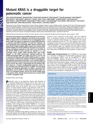
  • 2. We measured accumulated siRNA release from the LODER. LODERs containing 10 μg siG12D were incubated in phosphate buffer solution (PBS), at 37 °C, in a humidified incubator. Fig. 1C shows the cumulative release of siG12D from LODERs in- cubated in PBS for over 2 mo. The release curve reveals that after 60 d in PBS, ∼75% of the encapsulated siG12D was re- leased. Notably, variations in release curves measured for dif- ferent LODERs did not exceed 12%. In parallel, we measured the amount of siRNA retained in the LODERs (Fig. S1D). To assess their ability to protect the siRNA from degradation, LODERs containing 10 μg siG12D were incubated in PBS or placed in a mouse liver tissue ex vivo; siG12D was later extracted from the LODERs and its quantity and integrity were measured using HPLC, gel electrophoresis, and light absorption. Both gel electrophoresis and HPLC show that siG12D remained in its intact form for at least 97 d in PBS and at least 60 d in a liver 3 siG12D LODERs A B C D m97 days PBS 60 days Liver m Fig. 1. LODER characteristics: (A) SEM image of the siG12D LODER removed 45 d after implantation in vivo. (B) LODER actual size picture. (C) Release of siG12D from LODERs containing 10 μg siRNA: siG12D LODERs were incubated in PBS (pH = 7.4), at 37 °C. The siG12D amount was measured in the PBS using NanoDrop (absorption). (D) LODERs protect siG12D from degradation: protection of LODER-embedded siG12D from degradation was assessed by urea-poly- acrylamide gel electrophoresis. siG12D was extracted from LODERs that were incubated at 37 °C in PBS or mouse liver tissue ex vivo (liver). m, siG12D size marker. A B C D 1 2 3 4 1 2 3 4 siGFP siG12D LODERs 1 2 3 4 1 2 3 4 siGFP siG12D transfection 48hrs KRAS -actin KRAS -actin Viability test: XTT 0 0.4 0.8 1.2 1.6 2 48 72 96 120hrs OD siGFP siG12D ** * Cell death test: LDH 0 0.1 0.2 0.3 0.4 0.5 48 72 96 120hrs OD siGFP siG12D * * 0% 20% 40% 60% 80% 100% 10 100 1000 10000 %ofKRSASexpression siRNA pM relative KRAS mRNA levels mock si-scr siG12D 50% β-ac n u/t mock si-scr siG12D 1.5nM KRAS P-Erk P-Akt 0% 50% 100% 150% 200% 250% KRAS P-Erk P-Akt egatnecrep:noisserpxenietorpevitaler fromsi-scrtransfected detected proteins Rela ve protein expression u/t mock si-scr siG12D p=0.01 * p=0.01 * p=0.01 * E F Fig. 2. The silencing effect of the siG12D LODER in human pancreatic cancer cells. (A) Panc1 cells were transiently transfected with siG12D, nontargeting scrambled siRNA (si-scr) or mock transfected using Lipofectamine 2000. A relative level of KRAS mRNA was assessed by real-time PCR. HPRT and UBC were used as endogenous controls. The result is shown as an average of three different samples ± SEM. (B) Panc1-Luc cells were incubated with LODERs con- taining either siGFP or siG12D in the presence of Lipofectamine 2000 or transiently transfected with these siRNAs using Lipofectamine 2000. After 48 h, cells were lysed and the total level of KRAS protein was assessed by Western blot analysis. Shown are representative blots of 48 h of four independent experiments. (C and D) Panc1 cells were left un- treated (u/t) or transiently transfected with siG12D, nontargeting scrambled siRNA (si-scr), or mock transfected using Lipofectamine 2000. At 36 h after the transfection, cells were lysed and relative levels of KRAS, P-Erk, and P-Akt proteins were assessed by Western blot analysis. The results were normalized to the level of β-actin. (C) Representative blots of KRAS, P-Erk, P-Akt, and β-actin. (D) The graph shows the average relative protein level as percentage of mock transfected cells ± SEM. Student t test (p) was calculated compared with mock transfected cells. (E and F) Panc1-Luc cells were incubated with LODERs containing either siGFP or siG12D in the presence of Lipofectamine 2000 for 48, 72, 96, or 120 h. Cell vi- ability (E) and cell death (F) were assessed using XTT and LDH tests, respectively. Representative results of six different samples are shown ± SEM, *P < 0.05; **P < 0.01 according to the Student t test. 20724 | www.pnas.org/cgi/doi/10.1073/pnas.1314307110 Zorde Khvalevsky et al.
  • 3. tissue (Fig. 1D and Fig. S1E). These data substantiate the po- tential of the LODER to function as an efficient and stable delivery device for siRNA in vivo. LODER-Derived siG12D Significantly Inhibits Growth of Pancreatic Cancer Cells in Vitro. To determine the silencing potential of siG12D, a specific siRNA directed against the mutant KRASG12D , we used the Panc1 human pancreatic carcinoma cells that harbor this specific mutation in the KRAS gene. Cultured Panc1 cells were transfected with siG12D, control scrambled siRNA (si-scr) in decreasing concentrations or mock transfected. Following 24 h, mRNA levels of KRAS were assessed by quantitative PCR (qPCR) (Fig. 2A). This analysis revealed that siG12D led to reduced KRAS mRNA levels, with a half maximal inhibitory concentration (IC50) of 67 pM. We confirmed the site-specific, siG12D-directed cleavage of the KRAS message, by rapid am- plification of cDNA ends (RACE) (Fig. S2). Next, we compared the effects of LODER-derived siG12D with directly applied siG12D on KRAS protein levels in vitro. Panc1 cells were incubated with siGFP, siG12D, or with LODERs containing these siRNAs, in the presence of a transfection re- agent. At 48 h posttreatment, the protein levels of KRAS were found to be lower in both siG12D groups compared with the siGFP. This decrease was met whether siG12D was applied di- rectly or through a LODER (Fig. 2B and Fig. S3A). These results show that the LODER-driven siRNA can efficiently silence target gene expression, comparable to nonbound siRNA. To verify the specific effect of siG12D on KRAS signaling pathway, we measured the levels of downstream signaling proteins that are activated by KRAS—including Erk (P-Erk) and Akt (P-Akt). Western blot results revealed that inhibition of KRAS caused a decrease in the levels of P-Erk and P-Akt (Fig. 2 C and D). Previous studies reported that inhibition of KRAS expression led to the inhibition of pancreatic cancer cell growth (12, 16). To examine the potential growth-inhibiting effect of siG12D, we measured both cell viability and cell death of Panc1 cells treated with the siG12D LODER. A time-dependent decrease in cell vi- ability correlating with an increase in cell death was met in the presence of siG12D LODERs compared with the siGFP LODERs (Fig. 2 E and F). To confirm the effects of LODER-derived siG12D on cell growth, Panc1-Luc cells, a subclone of Panc1 cells stably expressing the luciferase reporter gene under the constitutive pro- moter [CMV early enhancer/chicken β actin (CAG) promoter], wereusedforvitalityassays.ThesecellswereincubatedwithsiG12D LODER, siLuc LODER (as a positive technical control), or the siGFP LODER. Fig. S3B shows that in the presence of either siLuc orsiG12DLODERs,asignificantinhibitionofluciferaseexpression is met compared with the siGFP LODER. These results both em- phasize the ability of LODERs to efficiently release functional siRNA and confirm the feasibility of Panc1-Luc cells to serve as indicators of effects on growth for further in vivo assessment. The Effect of the siG12D LODER on Epithelial–Mesenchymal Transition. In the course of tumor development cancer cells may undergo an epithelial–mesenchymal transition (EMT). In this process, the cells acquire mesenchymal characteristics and lose epithelial ones. It was previously reported that KRAS inhibition reduces EMT, as manifested by a decrease in cell migration and re- duction in contact inhibition (16). To study the effects of siG12D on EMT, we conducted a series of experiments in which we examined different aspects of this phenomenon. We analyzed the migration characteristics of Panc1 cells following transfection with siG12D or scrambled siRNA, using the scratch and Trans- well-migration assays (17, 18). TGFβ was used as a positive control. Migration ability was assessed using a modified Boyden chamber assay, which revealed that siG12D inhibited Panc1 migration by more than 30% (Fig. S4A). The scratch assay revealed that after 24 h, the nontreated and the scrambled siRNA transfected cells migrated and narrowed the gap created by the scratch, whereas the siG12D transfected cells barely mi- grated (Fig. S4B). LODER-Derived siRNA Decreases Gene Expression in Vivo. To confirm the functionality of LODER-driven siRNA in vivo, we tested the ability of a LODER containing siRNA targeting the luciferase gene, siLuc LODER, to reduce luciferase expression in normal and tumor tissues constitutively expressing the luciferase gene. We therefore implanted empty or siLuc LODERs into the livers of transgenic mice expressing the luciferase gene (MUP-Luc) in liver cells (19). In vivo imaging results showed that siLuc LODERs led to a significant decrease in luciferase levels compared with empty LODERs (Fig. S5A). Next, LODERs carrying siLuc or siGFP were implanted into CT26 cell-derived s.c. synograft tumors. Three days after implantation, measurements of luciferase activity in vivo revealed that siLuc released from the LODERs inhibited luciferase expression (Fig. S5B). This decrease in luciferase activity was not correlated to nonspecific effects on tumor growth, as tumor weights were similar within both groups (Fig. S5C). Together these results show that siRNA is released in vivo from the LODER and silences the target gene in vivo, thus demonstrating that the LODER can serve as a delivery platform for active siRNA in both normal and tumor tissues in vivo. To study the toxicity of the LODER-driven siRNA, we com- pared the effect of the siG12D LODER (16, 32, and 64 μg siG12D/kg body weight) to that of the empty LODER, sham- operated, or untreated animals. LODERs were implanted into the pancreas of normal mice and rats. Animals of both species, males and females, were tested. In parallel, the same and higher doses of siG12D were intraperitoneally injected into normal mice and rats (16, 160, and 320 μg siG12D/kg body weight). The duration of this study was 2 wk in mice and 8 wk in rats. The results demonstrated that the siG12D LODER had no effect on animal mortality, behavior, or body and liver weight. Hematological and biochemical tests failed to reveal any statistically significant A B Empty LODER siG12D LODER 1mm 1mm 0% 20% 40% 60% 80% 100% Empty LODER siG12D LODER Percentageofnecrocarea Percentage of necro carea PancO2 cells, subcutan model * Fig. 3. Silencing of KRAS inhibited s.c. tumor growth in immune-proficient settings. (A and B) Syngeneic s.c. PancO2 tumors were implanted with empty LODERs or LODERs containing siG12D. (A) Representative pictures of H&E immunostaining are shown 1 wk after the implantation. (B) Calculated percentage of necrotic area. The calculation was done using the ImageJ program. *P < 0.05 according to the Student t test. Zorde Khvalevsky et al. PNAS | December 17, 2013 | vol. 110 | no. 51 | 20725 MEDICALSCIENCES
  • 4. differences between the tested groups. Gross and histopathology analyses revealed that all changes were of minimal severity and typical in untreated mice or rats of the same age and strain. Based on the lack of adverse reaction following siRNA treatment, the maximum dose of 0.32 mg siRNA/kg body weight was considered as not representing an acute toxic risk. LODER-Driven siG12D Inhibits Tumor Growth in Vivo. We next as- sessed the ability of LODERs to inhibit the growth of tumors derived from human pancreatic tumor cell lines in vivo. We used human pancreatic cancer cell lines, Panc1 and Capan1, consti- tutively expressing the luciferase gene. Both these cell lines bear mutations in the KRAS gene, G12D in Panc1 cells and G12V in Capan1 cells. Immune-deficient mice bearing Capan1-Luc– derived s.c. tumors at an average size of 1 cm3 were divided into four groups: intratumorally implanted with siG12D, siG12V, or empty LODERs, and untreated. In Fig. S6A, the immunohisto- chemistry (IH) staining results are shown for the proliferation marker CDC47 at 1 mo after LODER implantation. Measure- ment of the percentage of necrotic area reveals that in both siG12D and siG12V LODER-treated groups, necrotic areas were significantly larger than in the untreated and empty LODER-treated control groups (Fig. S6B). Thus, the siG12D and siG12V LODERs severely hampered the growth of Capan1- Luc–derived tumors. In mice bearing s.c. Panc1-Luc tumors, results revealed that tumors treated with the siG12D LODER had significantly smaller volumes than those treated with the siLuc LODERs (Fig. S6C). Furthermore, histological analysis of LODER-treated Panc1 tumor tissues showed in the siG12D group a case of tumor tissue that was almost totally replaced by an adipose tissue (Fig. S6D). Correlated to these, the survival of mice bearing s.c. Panc1 tumors was significantly increased (P < 0.05) compared with each of the other three groups: untreated, empty LODER, and intraperitoneally (i.p.) injected siG12D (Fig. S6E). Next, we tested the effects of the siG12D LODER treatment on tumor growth in immune proficient settings. Toward this end, we used in a syngeneic mouse model, the Panc02 mouse pan- creatic cell line bearing a G12D mutation. Notably, in vitro, siG12D inhibited Panc02 cell growth in a similar mode as in Panc1 cells (Fig. S7). In vivo, siG12D LODERs implanted in the s.c. Panc02-derived tumors resulted in extensive necrosis 1 wk following implantation (Fig. 3A). Quantification of the necrotic area showed that it exceeds 60% of the total tumor tissue area (Fig. 3B). To determine the long-term potential effect of the siRNA LODERs, we analyzed the remaining siRNA content of LODERs isolated from tumors after implantation. The results revealed that the LODER structure protected the siG12D from degradation for over a 70-d period. Collectively, these results demonstrate that the siG12D LODER effectively inhibited tumor growth and can lead to necrotic destruction of tumor tissue in vivo. siG12D LODERs Impede Orthotopic Pancreatic Tumor Growth. We established an orthotopic pancreatic cancer model in which mice were operated on and Panc1-Luc or Capan1-Luc cells were injected into the tail of the mouse pancreas. Tumor growth was monitored by in vivo luciferase imaging. When tumors were detected, mice were divided into the different treatment groups, keeping a similar average level of luciferase intensity in each group. Mice then underwent laparotomy and two LODERs were stitched to the pancreatic tumor mass. In Panc1-Luc–derived tumors, in vivo luciferase measurements revealed that tumor growth was retarded in the siG12D LODER group throughout the study in comparison with the control groups (Fig. 4 A and B). Importantly, the overall sur- vival of siG12D LODER-treated mice was considerably pro- longed compared with all of the different control groups (Fig. 4C). Similar results were obtained with Capan1-Luc cells (Fig. S8). To show the direct effects of siG12D LODER on tumor tissue, we assessed the effect of the siG12D LODER on KRAS protein expression in xenograft tumor tissues. IH with a KRAS-specific antibody was performed on tumor tissue sections obtained from xenograft tumors, 24 d after treatment with: siG12D LODERs, empty LODERs, or untreated. This analysis shows that KRAS staining in siG12D LODER-treated tumors was weaker compared with the empty LODER or untreated tumors (representative tissue sections in Fig. 5A). Image analysis software was used to calculate the percentage of KRAS positive cells vs. nonstained cells, in 0.1 × 0.1 mm2 squares, in various consecutive distances from the LODER implantation sites (representative micrographs shown in Fig. S9A). The percent of KRAS positive cells was found to be in an anticorrelation with the distance from the LODER, whereas in other groups, untreated and empty LODER implanted, the graph remained linear (Fig. 5B and Fig. S9B). No- tably, the ratio of KRAS positive stained versus nonstained cells (positive/negative ratio) in siG12D LODER-treated tumors was ∼10-fold lower compared with the two other groups. These data attest to the specific silencing effect of siG12D LODER treatment on KRAS expression. A B Empty LODER Day4Day24 siG12D LODERsiG12D injecteduntreated PancreaticPanc1tumors C Fig. 4. siG12D LODERs inhibit growth of orthotopic pancreatic tumors of human origin. (A and B) Representative mouse pictures (A) and luciferase activity graph (B) in mice bearing Panc1-Luc pancreatic tumors from the different treatment groups: untreated, i.p. injected with siG12D (injected) or implanted with empty or siG12D LODERs. (C) Survival curve in mice bearing Panc1 pancreatic tumors from the different treat- ment groups: untreated, i.p. injected with siG12D, or implanted with empty or siG12D LODERs. 20726 | www.pnas.org/cgi/doi/10.1073/pnas.1314307110 Zorde Khvalevsky et al.
  • 5. Having shown the effects of KRAS silencing on pancreatic tumor cell proliferation, we performed IH staining for CDC47, a marker for dividing cells. Quantification of this staining (Fig. 5C and Fig. S9 C and D) reveals that the siG12D LODER inhibited the growth of tumor cells with increased efficiency in the immediate periphery of the LODER. These results confirm that the observed tumor growth inhibition was due to the si- lencing of KRAS by the siG12D LODER. Altogether, our results demonstrate that the LODER-driven siG12D hampered KRAS expression and significantly inhibited the in vivo growth of pancreatic tumors in both s.c. and ortho- topic, xenograft, and synograft mouse models. Discussion In this study, we present a unique platform technology for siRNA delivery. We show that the mutated KRAS, previously considered as an undruggable oncogene, can be targeted by siRNA in vivo in mouse models of pancreatic cancer. Our strategy of siRNA delivery to cancer cells was through em- bedding of the siRNA within a miniature biodegradable polymeric matrix that both protects and enables the release of the siRNA drug for an extended duration of time regionally within tumor tissue. We show the efficacy of this drug and this delivery system as being efficient, specific, and safe. The notion of oncogene addiction lies at the base of the bulk of cancer research performed today (20). KRAS has long been underlined as a beneficial target for cancer therapeutics, because its activation drives many of the tumor-cell–related traits, especially growth and proliferation (21). Specific KRAS targeting in tumors—especially in pancreatic cancer—offers hope for cancer patients by attenuating tumor growth through the deprivation of this oncogene (22). In conclusion, the siG12D LODER overcomes the current siRNA delivery obstacles, including enzymatic degradation of the drug, renal clearance, and the need for active target- ing. Moreover, it improves efficacy by eliminating the need for distal vectors and/or chemical modifications and reduces the required siRNA dose. Importantly, this approach is uni- versal and can be easily applied to target other genes that are pivotal for progression and growth of various solid tumors, using different sets of siRNAs. In addition to being safe and effective, the strategy we propose is applicable, reproducible, cost-effective, and withstands a much lower dose than siRNAs administered systemically. Materials and Methods The LODER. LODER is a biopolymeric cylindrical implant comprising an siRNA drug that is released throughout a period of months into a tumor. Dimensions are optimized for insertion into the pancreatic tumor by a 19- gauge endoscopic ultrasound needle (D ∼ 0.8 mm). The used drug load was from submicrogram levels according to in vivo requirements, up to 375 μg for the optimal dose in clinical use. For siRNA integrity tests, siRNA was separated on 10% (wt/vol) urea- polyacrylamide gels followed by ethidium bromide staining. Band intensity was quantified using TINA software. Cell Lines and Cell Culture Conditions. Panc1 and Capan1 cell lines were obtained from American Type Culture Collection (ATCC). The Panc02 cell line is a gift from Dr. M. Elkin (Sharett Institute, Hadassah-Hebrew University Medical Center, Jerusalem). The three cell lines were cultured in RPMI-1640 medium, supplemented with 10% (vol/vol) heat-inactivated FCS, 2 mM glutamine, 100 units/mL penicillin, and 100 μg/mL streptomycin. The cell cultures were maintained in a humidified atmosphere of 5% (vol/vol) CO2 at 37 °C. Transfections, Stable Transfections, and Gene Silencing Assays. For establish- ing stably transfected Panc1-Luc, Panc02-Luc, and Capan1-Luc cell lines, pCDNA3.1 (Invitrogen) plasmid bearing the neomycin gene was cotrans- fected with the pCAG-Luc plasmid (obtained from E. Zeira, Hebrew Uni- versity, Jerusalem) in a ratio of 1:5, using TransIT-LT1Mirus transfection reagent, according to the manufacturer’s instructions. The selection was performed using G418. Luciferase level was assessed using the Steady- Glo luciferase assay (Promega). siRNA transfections were performed using Lipofectamine 2000 (Invitrogen), according to the manufacturer’s in- structions. Cell viability was assessed using 2,3-bis-(2-methoxy-4-nitro-5- sulfophenyl)-2H-tetrazolium-5-carboxanilide (XTT) (Biological Industries) or the lactate dehydrogenase (LDH) (Roche) kit, according to the manufacturer’s instructions. Synthetic siRNAs. KRAS-targeting siRNA sequences were described previ- ously (12, 16). Control siRNA sequences were 5-CUUACGCUGAGUACU- UCGAdTdT and 5′-UCGAAGUACUCAGCGUAAGdTdT for si-Luc; 5′-GCUGACCCUGAAGUUCAUCdTdT and 5-’GAUGAACUUCAGGGUCAGCdTdT for siGFP; and 5′-GUUGGAGGCGGUAUGUGAGdTdT and CDC47 staining as a function of distance from LODER 0% 20% 40% 60% 80% 100% 0 500 1000 1500 2000 2500 distance from LODER [μm]* posiveCDC47staining siG12D injection siG12D LODER empty LODER untreated siG12DLODER- treated *untreated samples measurements were taken from the tumor edge A G12D LODER-treated tumors Untreated tumors T T TT L B C 40% 50% 60% 70% 80% 90% 100% 0 200 400 600 800 1000posiveKRASstaining distance from LODER [μm]* siG12D LODER empty LODER untreated Linear (siG12D LODER) Linear (empty LODER) Linear (untreated) *untreated samplesmeasurementsweretakenfromthe tumoredge Percentage of positively KRAS – stained cells as a function of distance from LODER Fig. 5. siG12D LODER inhibits KRAS expression and growth of orthotopic pancreatic tumors of human origin. (A) Representative pictures of KRAS immunostaining of Panc1 pancreatic tumor tissues 24 d posttreatment: implanted with siG12D LODERs or untreated. (B) Graph represents per- centage of KRAS positively stained cells in squares 0.1 × 0.1 mm2 as a func- tion of distance from LODER or tumor edge, measured in three groups: untreated, empty LODER, and siG12D LODER treated. (C) Graphs represent percentage of CDC47 positively stained versus unstained cells as a function of distance from LODER or tumor edge, measured in four groups: untreated, siG12D injected, empty LODER, and siG12D LODER treated. Zorde Khvalevsky et al. PNAS | December 17, 2013 | vol. 110 | no. 51 | 20727 MEDICALSCIENCES
  • 6. 5′-CUCACAUACCGCCUCCAACdTdT for scrambled control. All siRNAs were synthesized as ready-to-use duplexes by Sigma-Aldrich or by Biospring. Real-Time PCR. RNA was isolated using TRIzol (Invitrogen; 15596). cDNA was synthesized using qScript cDNA Synthesis kit (Quanta BioSciences; 95047). The primers used were as follows: for KRAS, forward 5′-GAGGCCTGCTGAAA- ATGACTG-3′ and reverse 5-ATTACTACTTGCTTCCTGTAGG-3′; for hypoxan- thine-guanine phosphoribosyltransferase (HPRT), forward 5′-GGTCCTTTTCACCAGCAAGCT-3′ and reverse 5′-TGACACTGGCAAAACAATGCA-3′; and for human ubiquitin C pro- moter (hUBC), forward 5′-ATTTGGGTCGCGGTTCTTG-3′ and reverse 5′-TGCCTTGACATTCTCGA- TGGT-3′. RACE. Panc1 cells were transfected with siG12D using Lipofectamine 2000 (Invitrogen) according to the manufacturer’s instructions. Cells were har- vested at 1, 4, and 8 h posttransfection. RNA was extracted from cells using TRIzol Reagent (Ambion) according to the manufacturer’s instructions. A total of 2 μg RNA was reverse transcribed using KRAS-specific primer (5′- CTGTTCTAGAAGGCAAATCAC-3′) and M-MLV reverse transcriptase (Prom- ega). The 5′ poly-A tails were added using terminal transferase (NEB) and dATP. PCR amplification of the RACE-specific target was done using the 3′ KRAS-specific primer (5′-CTGTTCTAGAAGGCAAATCAC-3′) and an oligo-dT primer containing 4 bp at the 3′ complementary to the 5′ nick site (5′- TTTTTTTTTTTTTTTTTTGATG-3′). Western Blot Analysis. Western blot assays were carried out using routine pro- cedures. Briefly, cells were homogenized in lysis buffer A [0.25 M sucrose, 20 mM Tris pH 7.6, 1.5 mM Mg Cl2, 10% glycerol, 1 mM EDTA, and ‘‘Complete mini’’ protein inhibitor mixture (Roche Diagnostics)], incubated on ice for 10 min and centrifuged at 15,000 × g for 15 min at 4 °C for supernatant collection. Pri- mary Abs are as follows: anti-KRAS (Abcam; ab84573), anti-P-Erk (Sigma; M 8159), anti-P-Akt (Cell Signaling; 4060), and anti–β-actin (ICN/MP Biomedicals; 691001). Secondary Abs are as follows: Dako EnVision system labeled polymer-HRP anti-mouse (Dako; K4001) and anti-rabbit (Dako; K4003). Pro- teins were visualized by the EZ-ECL chemiluminescence detection kit for HRP (Biological Industries; 20-500-120). Results are expressed as a ratio protein of interest/β-actin to correct for loading for each sample. Animals. Female C57B/6 5-wk old mice, nude and SCID/bg female 6-wk-old mice were purchased from Harlan Laboratories. All mice were kept in a specific pathogen-free facility. Mice were handled according to the criteria outlined in the Guide for the Care and Use of Laboratory Animals (23) prepared by the National Academy of Sciences and published by the Na- tional Institutes of Health. All experiments were approved by the Animal Care Committee of Hebrew University. Tumor Models. The mice were allowed to acclimate to the facility for at least 1 wk before manipulation. Mice had free access to water and chow at all times. All animal procedures were performed under general anesthesia with i.p. administered xylazine 10 mg/g body weight (Chanelle Pharmaceuticals Manufacturing) and ketamine, 450 mg/g body weight (Fort Dodge Animal Health). After surgery, mice were allowed food and water ad libitum. S.c. tumors. Tumor xenografts were established by s.c. injection of log-phase growth viable cells, 107 (in 150 μL PBS) in the case of Panc1 cells or 106 (in 100 μL PBS) in the case of PancO2 cells; the cells were injected into the flanks of the mice. When tumors reached an average volume of 80 mm3 , mice were divided into equal groups. LODERs were implanted into tumors under an- esthesia. The tumor volume was calculated according to the following for- mula: V = largest diameter × small diameter2 /2. Intrapancreatic orthotopic tumors. The mice were anesthetized, their abdomens were sterilized with alcohol (70%), and they were positioned laterally. A small, left abdominal flank incision was made, and the pancreas tail with the spleen was carefully exposed under aseptic conditions. The tumor cells (106 cells/30 μL PBS) were injected into the tail of pancreas using a 27-gauge tuberculin syringe. After replacement of the pancreas into the abdominal cavity, the incision was closed in two layers using an absorbable surgical 6- 0 vicryl suture for the peritoneum and a 4-0 vicryl suture for the skin. After surgery, mice were inspected daily. Tumor growth was followed by mea- surement of luciferase level. When the tumors were detected, mice were stratified and divided into treatment groups according to the luciferase levels and treated as noted. For LODER implantation, mice were anes- thetized, pancreas was exposed as described, and LODERs were attached to the tumor using a 7-0 vicryl suture. The abdominal cavities were closed as described. Pancreatic tumor growth was followed by luciferase measure- ment twice a week. Immunohistochemical Staining. Immunohistochemistry was done on 4-μm-thick formalin-fixed paraffin-embedded tissue sections by standard procedure. Deparaffinization and rehydration were followed by antigen retrieval using a pressure cooker with citrate buffer (pH 6) for P-ERK and glycine buffer (pH 9) for KRAS and CDC47. Primary antibodies were diluted 1:100 for KRAS (Abnova; PAB14811) and ERK (M8159; Sigma); 1:50 for CDC47 (Biocare Medical CM137b; Pharmatrade). Secondary antibodies were from Dako. Staining was developed with diamonobenzine using a kit from Zymed. Statistical Analysis. All data were subjected to statistical analysis using the Excel software package (Microsoft). A two-tailed Student t test was used to determine the difference between the groups. Differences were considered significant at P < 0.05. Data are given as mean ± SEM. ACKNOWLEDGMENTS. We thank Ms. Evelyne Zeira for her contribution to establishing the in vivo tumor models, Dr. Daniel Goldenberg for MUP-Luc mice, Prof. Nahum Goldberg for numerous useful comments, and Dr. Eliel Ben-David for CT interpretation. 1. Hidalgo M (2010) Pancreatic cancer. N Engl J Med 362(17):1605–1617. 2. Karamitopoulou E (2012) Tumor budding cells, cancer stem cells and epithelial-mes- enchymal transition-type cells in pancreatic cancer. Front Oncol 2:209. 3. Delpu Y, et al. (2011) Genetic and epigenetic alterations in pancreatic carcinogenesis. Curr Genomics 12(1):15–24. 4. Jones S, et al. (2008) Core signaling pathways in human pancreatic cancers revealed by global genomic analyses. Science 321(5897):1801–1806. 5. Belda-Iniesta C, et al. (2008) Molecular biology of pancreatic cancer. Clin Transl Oncol 10(9):530–537. 6. Maitra A, Hruban RH (2008) Pancreatic cancer. Annu Rev Pathol 3:157–188. 7. Feldmann G, Beaty R, Hruban RH, Maitra A (2007) Molecular genetics of pancreatic intraepithelial neoplasia. J Hepatobiliary Pancreat Surg 14(3):224–232. 8. Quinlan MP, Quatela SE, Philips MR, Settleman J (2008) Activated Kras, but not Hras or Nras, may initiate tumors of endodermal origin via stem cell expansion. Mol Cell Biol 28(8):2659–2674. 9. Sun C, et al. (2001) Characterization of the mutations of the K-ras, p53, p16, and SMAD4 genes in 15 human pancreatic cancer cell lines. Oncol Rep 8(1):89–92. 10. Villanueva A, et al. (1996) Diagnostic utility of K-ras mutations in fine-needle aspi- rates of pancreatic masses. Gastroenterology 110(5):1587–1594. 11. Singh A, et al. (2009) A gene expression signature associated with “K-Ras addiction” reveals regulators of EMT and tumor cell survival. Cancer Cell 15(6):489–500. 12. Réjiba S, Wack S, Aprahamian M, Hajri A (2007) K-ras oncogene silencing strategy reduces tumor growth and enhances gemcitabine chemotherapy efficacy for pan- creatic cancer treatment. Cancer Sci 98(7):1128–1136. 13. Leucuta SE (2012) Drug delivery systems with modified release for systemic and bi- ophase bioavailability. Curr Clin Pharmacol 7(4):282–317. 14. Mohtaram NK, Montgomery A, Willerth SM (2013) Biomaterial-based drug deliv- ery systems for the controlled release of neurotrophic factors. Biomed Mater 8(2): 022001. 15. Hudson D, Margaritis A (2013) Biopolymer nanoparticle production for controlled release of biopharmaceuticals. Crit Rev Biotechnol, 10.3109/07388551.2012.743503. 16. Fleming JB, Shen GL, Holloway SE, Davis M, Brekken RA (2005) Molecular consequences of silencing mutant K-ras in pancreatic cancer cells: Justification for K-ras-directed therapy. Mol Cancer Res 3(7):413–423. 17. Liang CC, Park AY, Guan JL (2007) In vitro scratch assay: A convenient and inexpensive method for analysis of cell migration in vitro. Nat Protoc 2(2):329–333. 18. Kramer N, et al. (2013) In vitro cell migration and invasion assays. Mutat Res 752(1):10–24. 19. Klopstock N, Levy C, Olam D, Galun E, Goldenberg D (2007) Testing transgenic reg- ulatory elements through live mouse imaging. FEBS Lett 581(21):3986–3990. 20. Sharma SV, Settleman J (2007) Oncogene addiction: Setting the stage for molecularly targeted cancer therapy. Genes Dev 21(24):3214–3231. 21. Friday BB, Adjei AA (2005) K-ras as a target for cancer therapy. Biochim Biophys Acta 1756(2):127–144. 22. Zimmermann G, et al. (2013) Small molecule inhibition of the KRAS-PDEδ interaction impairs oncogenic KRAS signalling. Nature 497(7451):638–642. 23. Committee on Care and Use of Laboratory Animals (1985) Guide for the Care and Use of Laboratory Animals (Natl Inst Health, Bethesda), DHHS Publ No (NIH) 85-23. 20728 | www.pnas.org/cgi/doi/10.1073/pnas.1314307110 Zorde Khvalevsky et al.