Download as PDF, PPTX

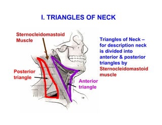
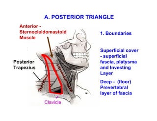


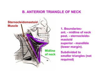


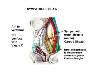


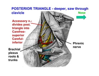


The document provides an outline on the triangles of the neck and deep structures of the neck. It begins by introducing the sternocleidomastoid muscle as the most useful landmark for locating neck structures. It then describes the boundaries and contents of the posterior and anterior triangles of the neck, noting key nerves, vessels, and muscles located in each. The document concludes by detailing structures such as the thyroid gland, parathyroid glands, sympathetic chain, and thoracic duct found deeper in the neck.


