More Related Content Similar to POPULATION DYNAMICS OF MICROFILARIL PARASITES IN HUMAN BEINGS FROM OMARGA TAHSIL DISTRICT OSMANABAD, (M.S.), INDIA (20) 1. www.sciencejournal.in
Volume 4 Issue 3 (2015) ISSN: 2319 – 314X (Print); 2319 – 3158 (Online) © 2015 DAMA International. All rights reserved. 18
POPULATION DYNAMICS OF MICROFILARIL PARASITES IN HUMAN BEINGS FROM OMARGA
TAHSIL DISTRICT OSMANABAD, (M.S.), INDIA
Sandhya Salunkhe* and Dama L.B.**
*Department of Zoology, Shri Shivaji College, Barshi, Dist. Solapur Maharashtra, India.
** Department of Zoology, D.B.F. Dayanand College of Arts and Science, Solapur, Maharashtra, India
ABSTRACT
The population dynamics of microfilaria was examined in human beings from different localities of Omerga tahsil,
District Osmanabad, (M.S.), India, during the two annual cycles June 2013 - March 2015. To determine the effects
of intensity, incidence, density and index of infection. The result shows that the infection was more during summer
followed by rainy seasons. The infection was single or in association with other symptoms like, fevers shaking
chills, sweating, headache, vomiting and joint pains, skin ulcers. The results were analyzed by tabulation and
graphical representation.
KEY WORDS: human being, microfilarial parasite, Omerga, Population dynamics.
INTRODUCTION
Filariasis is one of the major parasitic infections of mankind, which is widely spread throughout the tropical and
subtropical places. Filariasis that are caused by thread like filarial nematode worms and transmitted by mosquitoes.
They occur in the poor in underdeveloped regions of South America, Central Africa, Asia, and the Pacific Islands. The
nematode microfilaria (Wuchereria bancrofti) affects more than 115 million people worldwide. Filariasis or
Elephantisis is a major public health and socio-economic problem in India (Das and Pani , 2000; Das and Pani, 2001;
Ramaiah et al., 1996), approximately 420 million people reside in endemic areas and 48.11 million are infected
(Michael et al., 1996).
The World Health Organization (WHO) has identified filariasis as second leading cause of permanent and long-term
disability next only to mood affecting disorder. In India, filariasis has been recognized as disease of National
importance because of continuous spread of disease and protracted suffering and disability caused in the affected
population. India contributes to 40% cases of bancroftian filariasis in the global scenario. Elephantisis is a more intense
in people who don’t live in this area, because many native people have built up some immunity. Symptoms of
elephantiasis include fever, shaking chills. Sweating, headaches, and vomiting.
Enlarge lymph nodes, swelling of affected area, skin ulcers, bone and joint pain, tiredness, and red streaks along the
arm or leg also may occur. Abscesses can from in lymph nodes or in the lymphatic vessels. They may appear at the
surface of the skin as well as long-term infection with lymphatic filariasis can lead to lymph edema, hydrocele in the
scrotum, and elephantiasis of the legs, scrotum, arms, penis, breasts, and vulvae. The most common site of
elephantiasis is the leg. It typically begins in the ankle and progresses to the foot and leg can resemble an elephant’s
foreleg in size, texture and color.
The swollen leg eventually becomes hard and thick. The skin may appear darkened and may even crack, allowing
bacteria to infect the leg and complicate the disease. True elephantiasis is caused by parasitic infections from kinds
round worms. These worms block the body’s lymphatic system-a network of channels, lymph nodes, and organs that
help maintain proper fluid levels in the body by draining lymph from tissues into the blood stream. This blockage
causes fluids to collect in the tissues, which can lead to great swelling, called “lymphedema.” Elephantiasis is also
called Barbados leg, elephant leg, morbus herculeus, mal de Cayenne and myelymphangioma. The present study
includes epidemiologic survey of Filariasis in Osmanabad District, Maharashtra State, India.
MATERIALS AND METHODS
Microfilaria is a nematode parasite found in the lymphatic system of host (human), diagnosis was based on the
demonstration of parasite which includes the field work, was carried out, and survey at night between 8 pm and12pm,
from a randomly selected population (Rokade, 2012). The blood slide were prepared Conventional Night blood smears
examination. Microscopic examination of 20 mm3
stained blood film was prepared from a finger prick, still the best
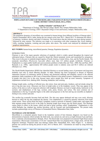
diagnostic technique. Proposed study area Omerga Taluka Dist: Osmanabad, M.S., India (Figure 1). in filariasis
patients was carried out during the period of June, 2013 to March, 2015. Calculations are made by using following
formulae.
2. www.sciencejournal.in
Volume 4 Issue 3 (2015) ISSN: 2319 – 314X (Print); 2319 – 3158 (Online) © 2015 DAMA International. All rights reserved. 19
A B C
Figure-1. A- Map of India B-Map of Maharashtra C- Map of Osmanabad District the geographical area of Filariasis
infection in the framework of this study.
1) Incidence of infection- It is the percentage of host infected by particulate by the following formula.
Infected host
Incidence of infection = ----------------------------- x 100
Total hosts Examined
2) Intensity of infection- It takes in to account the total no of microfilaria of nematode parasites in infected host
population, observations are recorded annually and calculated by the following formula.
Number of Parasites collected in a sample
Intensity of infection = --------------------------------------------------------------
Number of Infected hosts
3) Density of infection- It is the measure of concentration of microfilarial parasites per unit space (single host).
Observations are recorded annually and calculated by the following formula.
Number of parasites collected in a sample
Density of infection ----------------------------------------------------------------
Total hosts Examined
4) Index of infection- It is calculated with the help of the formula Tenoru and Zejde, 1974. Observations are recorded
annually and calculated by the following formula.
No. of hosts infected x No. of parasites collected
Index of infection = -------------------------------------------------------------------------
(T0tal host examined)
RESULTS AND DISCUSSION
The data of population dynamic of microfilaria in human being from Omerga tahsil, Osmanabad district (MS.) India,
during June 2013- May 2015 shown in Table 1 and In Figure 2,3,4 and 5.
3. www.sciencejournal.in
Volume 4 Issue 3 (2015) ISSN: 2319 – 314X (Print); 2319 – 3158 (Online) © 2015 DAMA International. All rights reserved. 20
Table 1. Month wise population dynamics of Microfilaria from Omerga Taluka, Osmanabad District, (M.S.),
India
Name of
Month
No.of host
Examined
No.of
host
infected
Total
no.of
parasite
Incidence
%
Intensity Density Index of
infection
Locality
June. 13 477 06 11 1.25 1.83 0.02 0.0002 Turori
Jully.13 467 00 00 00 00 00 0.0 Guggalgao
n
Aug.13 714 03 10 0.42 3.33 0.01 0.0005 Chichkot
Sept.13 845 00 00 00 00 00 00 Korewadi
Oct.13 219 00 00 00 00 00 00 Trikoli
Nov.13 285 00 00 00 00 00 00 Dhudhanal
Dec.13 206 00 00 00 00 00 00 Trikoli
Jan.14 286 01 1 0.34 1 0.003 0.0001 Trikoli
Feb.14 00 00 00 00 00 00 00 Kunhali
Mar.14 172 01 04 0.58 04 0.023 0.00013 Hundal
Apr.14 214 11 00 00 00 00 00 Hundal
May.14 1086 16 26 0.014 16.25 0.023 0.0003 Dhudhanal
June.14 1243 09 21 0.72 2.33 0.01 0.00012 Karali
Jully.14 710 00 00 00 00 00 00 Hipparga
Aug.14 320 00 00 00 00 00 00 Hundal
Sept.14 523 00 00 00 00 00 00 Jangdalwad
i
Oct.14 500 00 00 00 00 00 00 TalmodTan
da
Nov.14 125 00 00 00 00 00 00 TalmodTan
da
Dec.14 130 00 00 00 00 00 00 Talmod
Jan.15 1184 22 00 00 00 00 00 Talmod
Feb.15 1396 22 06 1.57 0.27 0.004 0.0047 Diggi
Mar.15 1394 00 00 00 00 00 00 Diggi
Figure 2. Shows incidence % of microfilaria parasite from human being during June 2013- March 2015.
4. www.sciencejournal.in
Volume 4 Issue 3 (2015) ISSN: 2319 – 314X (Print); 2319 – 3158 (Online) © 2015 DAMA International. All rights reserved. 21
Figure 3. Shows intensity of microfilaria parasite from human being during June 2013- March 2015.
Figure 4. Shows density of microfilaria parasite from human being during June 2013- March 2015.
Figure 5. Shows index of infection of microfilaria parasite from human being during June 2013- March 2015.
The analysis data showed that the occurrence of microfilaria parasites variable according to seasons. The high
incidence, intensity, density and index of infection of all the nematode parasites (microfilaria) occurred in summer
season followed by rainy seasons whereas lower infection in winter seasons.
5. www.sciencejournal.in
Volume 4 Issue 3 (2015) ISSN: 2319 – 314X (Print); 2319 – 3158 (Online) © 2015 DAMA International. All rights reserved. 22
According to the Kennedy (1971, 1975 and 1977) and Rodhe (1993) the temp, humidity and rainfall, feeding habits of
host, availability of infective host and parasite maturation, and such factors are responsible for influencing the parasitic
infection. Experimental studies by Kennedy (1971) have shown that the nematode parasites bancroftian filariasis can
establish in humans and survive for longer period at low temperature. Hence he explained the temperature was major
controlling seasonal periodicity of microfilarial infection. Rodhe, 1993 explained the temperature controls
parasitization. He explained the infections are more in warm seas than in cold ones. Jadhav (1976, 2005 and 2006)
explained the development of parasites should be needed high temperature, low rainfall and sufficient moisture. Hence
the high prevalence occurs in summer followed by other season.
CONCLUSION
After the analysis of data the present study can be concluded that the high infection of microfilarial
parasites (incidence, intensity, density and index of infection) was occurred in summer season followed by winter
where as low in monsoon season. This type of results indicated that environmental factors were influencing the
seasonality of parasitic infection either directly or indirectly. The influence of environmental factors and climatic
factors as they affect the dynamics of population growth of the brancroftian filariasis vector in the Omerga taluka of
Osmanabad district, Maharashtra state, India.
REFERENCE
Das P.K. and Pani S.P. (2000). Towards elimination of lymphatic filariasis in India: Problems, challenges,
opportunities and new initiatives. Journal of International Medical Sciences Academy (Special Issue: emerging and re-
emerging parasitic infestations in India). 13:18.
Das P.K. and Pani S.P. (2001). "Filariasis" Epidemiology and control in Helminthology in India; Ed M. L. Sood.
Jadhav (2006). Population dynamics of helmith parasite in freshwater fishes from Marathwada region, (MS)
India.Flora and fauna Vol-12, N-2, pp.143-148.
Kennedy C.R. (1971). Population biology of cestode proteocephalus torulisus (Bat Sch) indace Leucisus leucisus (L)
of the river Avon. J. fish Biol. 1(3):209-219.
Michael E., Bunday, DAP and Grenfell B.T. (1996). Reassessment the global Prevalence and distribution of
lymphatic filariasis. Parasitology. 112: 409 – 28.
Ramaiah K. D., Ramu Vijaya., Kumar K. N. and Guyatt H. (1996). Epidemiology of acute filarial episodes caused
by Wuchereria bancrofti infection in two rural villages in Tamil-Nadu, South-India. Trans. Roy. Soc. Trop. Med. Hyg.;
90:639.
Rohde K. (1993). Ecology of Marine Parasites 2nd Edition. CAB International, Oxford.
URL: http:// www.filariasis .org/
Rokade et al., (2012). Prevalence of Filariasis in Solapur District, Maharashtra, State, India. Trends Parasitology Res.
47-52.
World Health Organization (1994). Lymphatic filariasis infection and disease control strategies Report of
WHO/CTD/TDR consultative meeting 1.
World Health Organization (1997). Lymphatic filariasis Reasons for hope. Division of control of tropical diseases.
WHO/CTD/FIL/97.4 World Health Organization 1997; 1
WHO (2002). World Health Organization. Geneva. Defining the roles of vector control and xenomonitoring in the
global programme to eliminate lymphatic filariasis. 1.
World Health Organization (2014). Global programme to eliminate lymphatic filariasis: progress report 2000–2009
and strategic plan 2010 – 2020:
halfway towards eliminating filariasis. Geneva: WHO, 2010. Document No. WHO/HTM/NTD/PCT/2010.6.
http://whqlibdoc.who.int/