
Recommended
More Related Content
Similar to Posterior Tibial By Dr. Chaman Lal (CK).ppt
Similar to Posterior Tibial By Dr. Chaman Lal (CK).ppt (20)
Recently uploaded
Recently uploaded (20)
Posterior Tibial By Dr. Chaman Lal (CK).ppt
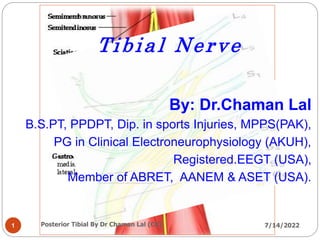
- 1. Tibial Nerve By: Dr.Chaman Lal B.S.PT, PPDPT, Dip. in sports Injuries, MPPS(PAK), PG in Clinical Electroneurophysiology (AKUH), Registered.EEGT (USA), Member of ABRET, AANEM & ASET (USA). 1 7/14/2022 Posterior Tibial By Dr Chaman Lal (CK)
- 2. Posterior Tibial The tibial nerve also known as posterior tibial nerve is the largest nerve branch of sciatic division. The anterior divisions of L4 ,L5 ,S1,S2 & S3 unites to form Tibial Nerve. 7/14/2022 2 Posterior Tibial By Dr Chaman Lal (CK)
- 3. Tibial Nerve Course: Descends through popliteal fossa to posterior compartment of leg, accompanied with posterior tibial vessels. Passes deep to flexor retinaculum (through the tarsal tunnel, behind medial malleolus) to reach the sole of foot where it divides into 2 terminal 7/14/2022 3 Posterior Tibial By Dr Chaman Lal (CK)
- 4. 7/14/2022 4 Posterior Tibial By Dr Chaman Lal (CK)
- 5. The popliteus (L4,L5,S1) , muscle in the leg is used to unlock the knee by laterally rotating the femur on the tibia during a closed chain movement (such as one with the foot in contact with the 7/14/2022 5 Posterior Tibial By Dr Chaman Lal (CK)
- 6. 7/14/2022 6 Posterior Tibial By Dr Chaman Lal (CK)
- 7. In the popliteal fossa the nerve gives off branches to GASTROCNEMI US , is involved in standing, walking, running and jumping. Along with the soleus muscle it forms the calf 7/14/2022 7 Posterior Tibial By Dr Chaman Lal (CK)
- 8. The soleus is a powerful muscle in the back part of the lower leg (the calf). It runs from just below the knee to the heel, and is involved in 7/14/2022 8 Posterior Tibial By Dr Chaman Lal (CK)
- 9. Cont’d Below the soleus muscle the tibial nerve lies close to the tibia bone and supplies the tibialis posterior, the flexor digitorum longus and the flexor hallucis longus. 7/14/2022 9 Posterior Tibial By Dr Chaman Lal (CK)
- 10. Below the soleus muscle the nerve lies close to the tibia and supplies the Tibialis posterior. The Tibialis posterior is the most center of all the leg muscles. It is the key stabilizing muscle of the lower leg. 7/14/2022 10 Posterior Tibial By Dr Chaman Lal (CK)
- 11. Muscular Branches 1. Muscles of posterior compartment of leg (Planter flexors of ankle, Flexors of toes 2. Intrinsic muscles of sole. 3. ONE Invertor of foot (tibialis posterior). 7/14/2022 11 Posterior Tibial By Dr Chaman Lal (CK)
- 12. Flexor digitorum longus is situated on the tibial side of the leg. At its origin it is thin and pointed, but it gradually increases in size as it descends. This muscle serves to curl the second, third, fourth, and 7/14/2022 12 Posterior Tibial By Dr Chaman Lal (CK)
- 13. Flexor hallucis longus. (L5,S1,S2) The Flexor hallucis longus is situated on the fibular side of the leg. It arises from the inferior two-thirds of the posterior surface of the body of the 7/14/2022 13 Posterior Tibial By Dr Chaman Lal (CK)
- 14. Tibial nerve cont’d . . . The nerve passes into the foot running posterior to the medial malleolus. Here it is bound down by the flexor retinaculum in company with the posterior tibial artery The tibial nerve, a major artery, veins, and tendons travel in a bundle along this pathway, through the tarsal tunnel. In the tunnel, the nerve splits into three different paths. One nerve (calcaneal) continues to the heel, the other two (medial plantar nerve and 7/14/2022 14 Posterior Tibial By Dr Chaman Lal (CK)
- 15. Cutaneous innervation The sural nerve is joined by fibres from tibial and common peroneal nerve and runs down the calf to supply the lateral and posterior skin of the distal third of the leg . The skin overlapping over the lateral melleolus and lateral aspect of the foot and little toe. 7/14/2022 15 Posterior Tibial By Dr Chaman Lal (CK)
- 16. 7/14/2022 16 Posterior Tibial By Dr Chaman Lal (CK)
- 17. Medial plantar nerve The medial planter nerve also gives off articular, cutaneous and muscular branches. Cutaneous branches supplies skin of the sole, foot, including the digit branches of the hallux, the seconds, third and half of the fourth toe. Muscular branches supply the Abductor hallucis, Flexor digitorum brevis, Flexor brevis and First lumbrical. 7/14/2022 17 Posterior Tibial By Dr Chaman Lal (CK)
- 18. Sensory innervations Motor innervations 7/14/2022 18 Posterior Tibial By Dr Chaman Lal (CK)
- 19. Lateral Planter Nerve:- The lateral plantar nerve (external plantar nerve) is a branch of the tibial nerve, in turn a branch of the sciatic nerve and supplies the skin of the fifth toe and lateral half of the fourth, as well as most of the deep muscles, its distribution being similar to that of the ulnar nerve in the hand. Muscular branches supply most deep muscles of the foot including Flexor digitorium accessories Abductor digiti minimi( quinti) Flexor digiti minimi brevis Second to fourth lumbricals and adductor hallucis. 7/14/2022 19 Posterior Tibial By Dr Chaman Lal (CK)
- 20. 7/14/2022 20 Posterior Tibial By Dr Chaman Lal (CK)
- 21. 7/14/2022 21 Posterior Tibial By Dr Chaman Lal (CK)
- 22. Entrapment of tibial nerve:- Tarsal tunnel syndrome is caused by compression of the tibial nerve or its b ranches in the tarsal tunnel. The tibial nerve entrapment and injury in the foot and ankle may be a source of great pain and functional impairment. Tibial nerve problem In the foot and ankle may cause difficulties with weight bearing and non weight bearing activities, sleep and footwear. 7/14/2022 22 Posterior Tibial By Dr Chaman Lal (CK)
- 23. Motor Nerve Study:- It is recorded from the abductor hallucis muscle. It is stimulated from the ankle and knee. The normative values are 5.8msec for distal latencies, 41msec for the conduction velocity, 4mV for the amplitudes and 56msec for the F- wave latencies. 7/14/2022 23 Posterior Tibial By Dr Chaman Lal (CK)
- 24. 7/14/2022 24 Posterior Tibial By Dr Chaman Lal (CK)
- 25. 7/14/2022 25 Posterior Tibial By Dr Chaman Lal (CK)
- 26. Anatomical Path of Mixed Planter Nerves 7/14/2022 26 Posterior Tibial By Dr Chaman Lal (CK)
- 27. Mixed Planter recording Technique. Medial planter Mixed Nerve- Recording Electrodes are placed at medial ankle. Site: G1 placed above and posterior to the medial malleolus. G2 placed 3-4cm proximally. G 3 in between active recording and stimulating electrodes. Distance:- Distance between recording and stimulating electrode should be 14 cm. Stimulation: Stimulate while placing cathode at the base of big toe. 7/14/2022 27 Posterior Tibial By Dr Chaman Lal (CK)
- 28. Medial planter sensory stimulation 7/14/2022 28 Posterior Tibial By Dr Chaman Lal (CK)
- 29. Lateral planter Mixed Nerve study Lateral planter Mixed study :- Recording Electrodes are placed at medial ankle. Site: G1 placed above and posterior to the medial malleolus. G2 placed 3-4cm proximally. G 3 in between active recording and stimulating electrodes. Distance:- Distance between recording and stimulating electrode should be 14 cm. Stimulation: Stimulate while placing cathode at the base of little toe. 7/14/2022 29 Posterior Tibial By Dr Chaman Lal (CK)
- 30. Lateral planter sensory stimulation 7/14/2022 30 Posterior Tibial By Dr Chaman Lal (CK)
- 31. A smile is an inexpensive way to improve your looks. Thanks 7/14/2022 31 Posterior Tibial By Dr Chaman Lal (CK)