More Related Content
Similar to A Rationale for Postsurgical Laser Use to Effectively Treat Dental Implants_Froum_2020 (2).pdf
Similar to A Rationale for Postsurgical Laser Use to Effectively Treat Dental Implants_Froum_2020 (2).pdf (20)
More from DrCarlosIICapitan
More from DrCarlosIICapitan (20)
A Rationale for Postsurgical Laser Use to Effectively Treat Dental Implants_Froum_2020 (2).pdf
- 1. The International Journal of Periodontics & Restorative Dentistry
© 2020 BY QUINTESSENCE PUBLISHING CO, INC. PRINTING OF THIS DOCUMENT IS RESTRICTED TO PERSONAL USE ONLY.
NO PART MAY BE REPRODUCED OR TRANSMITTED IN ANY FORM WITHOUT WRITTEN PERMISSION FROM THE PUBLISHER.
- 2. Volume 40, Number 4, 2020
561
Submitted April 19, 2019; accepted June 25, 2019.
©2020 by Quintessence Publishing Co Inc.
1
Department of Periodontology, Baltimore College of Dental Surgery, University of
Maryland Dental School, Baltimore, Maryland, USA; Dental Implantology, James Cook
University Dental School, Cairns, Australia; Private practice, Yardley, Pennsylvania;
Private practice, New York, New York, USA.
2
State University of New York, Stony Brook Department of Periodontology;
Private practice, New York, New York, USA.
3
Ashman Department of Periodontology and Implant Dentistry, New York University
College of Dentistry, New York, New York, USA; Private practice, New York, New York, USA.
Correspondence to: Dr Paul S. Rosen, 27 West 54th Street, Suite 1C/D, New York, NY, USA.
Email: psr907@gmail.com
A Rationale for Postsurgical Laser Use to
Effectively Treat Dental Implants
Affected by Peri-implantitis:
Two Case Reports
Peri-implantitis is a biologic complication that can affect the survival of a dental
implant. Most surgical and nonsurgical treatments have been relatively ineffective
even when using targeted antimicrobial approaches. A growing number of reports
are documenting the presence of titanium granules and/or cement in the soft
tissues surrounding peri-implantitis–affected dental implants. Two case reports are
presented demonstrating how the Nd:YAG or a carbon dioxide (CO2) laser used
following regenerative surgeries changed failures into successes as measured by
radiographic bone fill and improved clinical parameters. These cases suggest that
successful peri-implantitis treatment may need to incorporate decontamination of
the soft tissues in addition to the implant’s surface. Further studies are warranted to
determine if each of these lasers would be successful over a larger patient cohort.
Int J Periodontics Restorative Dent 2020;40:561–568. doi: 10.11607/prd.4428
The use of dental implants has
been a reliable treatment option
for replacing missing or soon-to-
be-missing teeth. While dental im-
plants have demonstrated great
reliability, their long-term use has
uncovered problems that were not
necessarily apparent or reported in
the early years when they were first
implemented.1–3
Biologic problems
represent one particular category
of dental implant complications that
have recently received a great deal
of attention.4,5
Peri-implantitis has been com-
monly described in the literature as
a pathologic condition occurring
in tissues around dental implants,
characterized by inflammation in
the peri-implant mucosa and pro-
gressive loss of supporting bone.5,6
The recent World Workshop held
by the American Academy of Peri-
odontology (AAP) and the European
Federation of Periodontists (EFP)
on the classifications of periodontal
and peri-implant diseases identi-
fied strong evidence that there is an
increased risk of developing peri-
implantitis in patients who have a
history of chronic periodontitis, poor
plaque control skills, and no regular
maintenance care after implant ther-
apy.5,6
The data analyzed by this con-
ference regarding both smoking and
diabetes as potential risk factors/
indicators for peri-implantitis was
found to be inconclusive.5
Moreover,
Paul S. Rosen, DMD, MS1
Scott H. Froum, DDS2
Stuart J. Froum, DDS3
© 2020 BY QUINTESSENCE PUBLISHING CO, INC. PRINTING OF THIS DOCUMENT IS RESTRICTED TO PERSONAL USE ONLY.
NO PART MAY BE REPRODUCED OR TRANSMITTED IN ANY FORM WITHOUT WRITTEN PERMISSION FROM THE PUBLISHER.
- 3. The International Journal of Periodontics Restorative Dentistry
562
the literature evidence that the peri-
implantitis group reviewed5
pointed
to there being some limited evi-
dence linking peri-implantitis to oth-
er factors, such as postrestorative
presence of submucosal cement,
lack of peri-implant keratinized mu-
cosa, and positioning of implants
that make it difficult to perform oral
hygiene and maintenance.
When reviewing the literature
regarding treatment approaches
for peri-implantitis, most systematic
reviews affirm that there is no best
approach for effective treatment.7,8
Nonsurgical approaches have been
consistently ineffective9,10
and sur-
gery has been disappointing in its
ability to arrest the disease, even
when targeted antimicrobial ther-
apy has been employed.11,12
It has
been speculated that the reason
targeted antimicrobial therapy may
be ineffective is that no one agent
kills all the bacteria and that a dual
antibiotic approach, which has been
absent in many of the historic ap-
proaches, may be necessary.13
There have been a number of
reports in the literature regarding
foreign materials being found in
biopsy material obtained around
peri-implantitis–affected implants.14
These materials have included tita-
nium particles and/or residual ce-
ment.15
These two foreign materials
may not have been eliminated by
the locally or systemically adminis-
tered antimicrobials or even by sur-
gical approaches that concentrate
on mechanical/chemotherapeutic
implant-surface decontamination.16
One possible method for ablating
these particles is the use of a laser,
which will affect the surrounding
soft tissues.17
This article presents
two case reports demonstrating
how the use of either the Nd:YAG or
CO2
laser in the first several months
following surgery changed an in-
complete treatment into very suc-
cessful outcomes.
Case 1
A 63-year-old Caucasian man was
referred for evaluation and treat-
ment of several dental implants that
were failing due to peri-implantitis.
The lesion had reached an advanced
level of disease as evidenced by
greater than 50% bone loss.18
The
gravest concern was the failing den-
tal implant at the mandibular left
first-molar site (Fig 1a). His medical
history included hypertension for
which enalapril was being taken.
The implant under examination had
probing depths that reached 9 to
10 mm circumferentially with puru-
lence and bleeding elicited upon
both probing and pressure to the
tissues. The adjacent teeth and a
dental implant had probing depths
that ranged from 3 to 4 mm, and the
patient was being seen every 3 to 4
months for his periodontal mainte-
nance. Due to the severe bone loss
and probing that suggested the
lesion was contained, it was deter-
mined that a surgical regenerative
approach would be performed.
The protocol for this treatment
has been previously described.19,20
In summary, informed consent was
first obtained. Local anesthesia
was administered using Septocaine
(Septodont) 4% with 1:100,000 epi-
nephrine. Full-thickness flaps were
elevated with periosteal release to
allow for adequate flap mobilization
for visualization, implant surface ac-
cess, and coronal advancement at
time of closure. Surface debride-
ment was performed using air-
borne-particle abrasion with glycine
(Fig 1b) followed by citric acid (50%
solution) applied for approximately
1 minute with cotton pellets. Each
step was separated by a vigorous
rinse of sterile water. Recombinant
human platelet-derived growth
factor-BB (rhPDGF-BB) (Gem 21,
Lynch Biologics) was then placed on
the decontaminated surface of the
implant, and the circumferential le-
sion received a composite graft of
freeze-dried bone/demineralized
freeze-dried bone allografts in a
70:30 ratio (Creos Allo.Gain, Nobel
Biocare) (Fig 1c) hydrated by the rh-
PDGF-BB and layered/contained by
a collagen membrane (Creos Xeno-
protect, Nobel Biocare) (Fig 1d). The
graft was hydrated with rhPDGF-BB
together with enamel matrix deriva-
tive (Emdogain, Straumann). The
site was sutured with 5-0 polytetra
fluoroethylene sutures (Omnia)
(Fig 1e). Postoperative pain man-
agement was achieved with 600 mg
ibuprofen, and infection control in-
cluded systemic use of amoxicillin
for 7 days (875 mg twice daily) and
an anti-inflammatory triple botani-
cal rinse (PeriActive, Izun Oral Care).
The patient was seen every 2 weeks
for follow-up.
At 5 months, the outcome of
treatment appeared to be subop-
timal, as evidenced by highly in-
flamed soft tissues (Fig 1f) and poor
osseous profile/bone fill (Fig 1g). The
decision was made to treat the area
© 2020 BY QUINTESSENCE PUBLISHING CO, INC. PRINTING OF THIS DOCUMENT IS RESTRICTED TO PERSONAL USE ONLY.
NO PART MAY BE REPRODUCED OR TRANSMITTED IN ANY FORM WITHOUT WRITTEN PERMISSION FROM THE PUBLISHER.
- 4. Volume 40, Number 4, 2020
563
Fig 1 Case 1. (a) Pretreatment radiograph
suggests severe bone loss on the anterior
implant in the mandibular left first-molar
site. (b) The lesion seen is a circumfer-
ential moat around this anterior implant
at the first molar. There is some residual
glycine powder on the dental implant
following airborne-particle abrasion.
(c) Following implant surface decontamina-
tion, the lesion is filled with an allograft of
mineralized:demineralized bone in a 70:30
ratio. The graft has been hydrated with
rhPDGF-BB. (d) A collagen membrane has
been adapted over the composite graft in
an effort to contain it. (e) The flaps have
been advanced to completely cover the
graft membranes and were secured with
5-0 polytetrafluoroethylene sutures using
an interrupted technique. (f) The clinical
view at 5 months demonstrates that the
soft tissues are still fairly inflamed. When
lightly cleaning and probing the area, there
is bleeding with this provocation. (g) The
5-month postoperative radiograph sug-
gests some improvement with incomplete
healing. (h) The site has been treated using
an Nd:YAG laser with two passes. Interven-
ing these two passes, the site received
curettage of the soft tissue along with
postoperative use of amoxicillin. (i) Clinical
view 1 year after the initial treatment. The
soft tissues surrounding the dental implant
are greatly improved. Probing depths were
consistent with good health and there was
an absence of bleeding. (j) The radiograph
taken at the 1-year follow-up suggests a
substantial improvement in the hard tissues
surrounding the dental implant, which
starkly contrasts both the pretreatment and
5-month radiographs.
a
e
c
g
i
b
f
d
h
j
© 2020 BY QUINTESSENCE PUBLISHING CO, INC. PRINTING OF THIS DOCUMENT IS RESTRICTED TO PERSONAL USE ONLY.
NO PART MAY BE REPRODUCED OR TRANSMITTED IN ANY FORM WITHOUT WRITTEN PERMISSION FROM THE PUBLISHER.
- 5. The International Journal of Periodontics Restorative Dentistry
564
with the Nd:YAG laser (PerioLase,
Millennium Dental Technologies).
A first pass was performed with a
setting of 3.6 watts, 100 millisec-
onds, and 20 Hz delivering 65 J,
followed by curettage of the tissue
and then a second pass with a set-
ting of 3.6 watts, 650 milliseconds,
and 20 Hz for 77 J (Fig 1h). Amoxi-
cillin (875 mg, twice daily) was again
given for 7 days as a postoperative
antibiotic. The patient was seen
at 1 week, 4 weeks, and 3 months
postoperative and every 3 months
thereafter. Probing depth measure-
ments were performed at 6 months
and were reduced to 4 mm with an
absence of bleeding. Two years af-
ter the initial surgery, there was ra-
diographic evidence of a favorable
gain in bone, and the soft tissue pa-
rameters were consistent with good
health (Figs 1i and 1j).
Case 2
A 67-year-old woman, with a non-
contributory medical history taking
a multivitamin with no stated food
or drug allergies, was referred for
evaluation and treatment of a can-
tilevered implant partial denture in
the mandibular left quadrant that
was failing due to peri-implantitis.
She had hydroxyapatite-coated
dental implants (Calcitek, Zimmer
Biomet) placed over 20 years ago
at the mandibular left first- and
second-molar sites that were splint-
ed with a cantilever distal pon-
tic at the third-molar site (Fig 2a).
These implants were affected by
severe peri-implantitis18
with prob-
ing depths greater than 7 mm, over
50% bone loss (Figs 2b and 2c), and
keratinized tissue measuring less
than 1 mm in width and thickness.
In addition, she had a rough-surface
implant (Osseotite, Zimmer Biomet)
placed at the second-premolar site
approximately 10 years earlier with
evidence of early to moderate peri-
implantitis.18
The dentition of this
Figs 2a to 2c Case 2. (a) Preoperative clinical
view of an implant-supported fixed partial denture
showing recession around dental implants with lack
of gingival tissue. (b) Probing depth is 7 mm with
bleeding and suppuration. (c) The preoperative
radiograph suggests severe peri-implantitis with
greater than 50% bone loss around the implants.
a
c
b
© 2020 BY QUINTESSENCE PUBLISHING CO, INC. PRINTING OF THIS DOCUMENT IS RESTRICTED TO PERSONAL USE ONLY.
NO PART MAY BE REPRODUCED OR TRANSMITTED IN ANY FORM WITHOUT WRITTEN PERMISSION FROM THE PUBLISHER.
- 6. Volume 40, Number 4, 2020
565
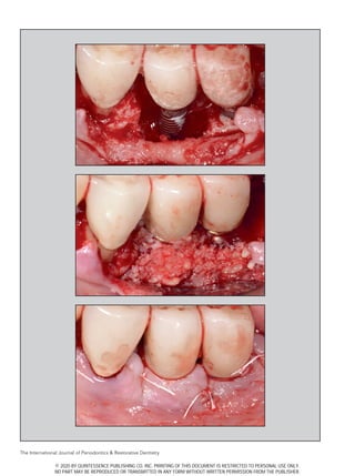
Figs 2d to 2i Case 2. (d) Regenerative graft material (anorganic bovine bone collagen hydrated with rhPDGF-BB) was
placed around the implants at the time of surgery. (e) A xenograft collagen soft tissue graft was placed over the bone-
replacement graft material mixed with enamel matrix derivative. (f) Soft tissue healing at 3 months after the initial surgery
appears poor as ongoing suppuration is present. (g) Surgical reentry procedure allowing for soft tissue laser ablation
of the flap using a 9.3-micron CO2 laser. (h) Favorable soft tissue healing is seen at 3 years after the initial procedure,
demonstrated by both an increase in the gingival tissue and the lack of bleeding on probing. (i) The radiograph taken
at the 3-year follow-up shows bone fill of the hard tissue defect, suggesting improved health of the implants.
e
g
i
f
d
h
© 2020 BY QUINTESSENCE PUBLISHING CO, INC. PRINTING OF THIS DOCUMENT IS RESTRICTED TO PERSONAL USE ONLY.
NO PART MAY BE REPRODUCED OR TRANSMITTED IN ANY FORM WITHOUT WRITTEN PERMISSION FROM THE PUBLISHER.
- 7. The International Journal of Periodontics Restorative Dentistry
566
patient was periodontally stable,
with probing depths ranging from
2 to 4 mm. She was seen at 3-month
intervals for maintenance visits. Due
to the severe bone loss and lack of
keratinized tissue, it was determined
that a surgical hard and soft tissue
regenerative approach would be
performed.
The protocol for treatment has
been previously described.19,20
In
summary, informed consent was
first obtained. Local anesthesia
was administered using Septocaine
(Septodont) 4% with 1:100,000 epi-
nephrine. Full-thickness flaps were
elevated with periosteal release to
allow for adequate flap mobiliza-
tion ensuring visualization, implant
surface access, and coronal ad-
vancement at the time of closure.
Implant-surface debridement was
performed using airborne-particle
abrasion with glycine followed by
mechanical debridement with a
titanium brush (RotoBrush, Salvin
Dental) followed by citric acid (60%
solution) applied for approximately
1 minute with cotton pellets. Each
step was separated by a vigorous
rinse with sterile water. rhPDGF-BB
(Gem 21) was then applied to the
decontaminated surface of the im-
plant and layered with anorganic
bovine bone with 10% collagen
(Bio-Oss, Geistlich) (Fig 2d). Be-
cause of the limited keratinized tis-
sue, a bovine collagen soft tissue
matrix (Mucograft, Geistlich) (Fig 2e)
was placed over the bone replace-
ment graft. The graft was hydrated
with rhPDGF-BB along with enamel
matrix derivative (Emdogain). The
tissue was coronally advanced and
secured using a sling suture tech-
nique with 5-0 polygalactin sutures
(Vicryl, Ethicon). Postoperative pain
was managed with 800 mg ibupro-
fen, and infection control included
systemic use of amoxicillin (500 mg,
tid) for 10 days and a homeopathic
antibacterial/anti-inflammatory rinse
(VEGA Oral Rinse, StellaLife). The
patient was seen every 2 weeks for
follow-up.
At 3 months, healing appeared
suboptimal as the soft tissues
showed high inflammation with
suppuration (Fig 2f). The decision
was made to treat the area with the
9.3-micron-CO2
Solea laser (Con-
vergent Dental). A mucoperiosoteal
full-thickness flap was reflected, and
both the implant surface and the
soft tissue flap were ablated using
a setting of 1.0 watts, lower power
mode, 1.2-mm spot site, 30% cut-
ting speed, 100% mist (13 mL/
minute), with a contra-angle hand-
piece (Fig 2g). Amoxicillin (500 mg,
tid) was again given for 10 days as a
postoperative antibiotic. The patient
was seen at 1 week, 3 weeks, and 3
months postoperative and every 3
months thereafter. Probing-depth
measurements were performed at
1 year and were reduced to 3 mm
with an absence of bleeding. Three
years after the initial surgery, there
was a favorable gain in both hard
and soft tissues, indicative of good
health (Figs 2h and 2i).
Discussion
Dental implants differ from teeth in
their ability to handle inflammation,
which stems from their differences in
the interfacial soft tissue interfaces.
Teeth have both connective tissue
fibers inserting into their root surface
cementum in addition to a hemides-
mosomal adherence of the sulcular
epithelium. Implants, on the other
hand, have only a hemidesmosomal
adhesion of the supracrestal epithe-
lium to act as a barrier to the oral/
sulcularenvironment,sincethesupra-
crestal connective tissue fibers run
parallel to the surface of the implant.
Thus, anything that might impact oral
sulcular epithelial homeostasis would
be of great concern.
The most recent World Work-
shop held by the AAP and EFP
reaffirmed plaque as the primary
etiology to peri-implantitis.6
At the
time of the conference, they also
felt that the available evidence did
not allow an evaluation of the role
that titanium or metal particles play
in the pathogenesis of peri-implant
diseases. Hence, the concept of
implant tribocorrosion as an inflam-
matory initiator is a departure from
the long-held bacterial pathogen-
esis model.14
This newer perspective
may help explain why many of the
treatment efforts have failed, and
it may well be the reason clinicians
have had to continually explore
other treatment methods to better
manage this challenging problem.
One possible solution has been
the additive use of a dental laser to
the regenerative treatment for peri-
implantitis. Both the Nd:YAG and
CO2
lasers have an impact on the
soft tissues. Their ability to ablate re-
sidual titanium and/or cement may
help enable the sites to successfully
heal. In both case reports, a modi-
fication of a regenerative surgical
algorithm was used and met with
© 2020 BY QUINTESSENCE PUBLISHING CO, INC. PRINTING OF THIS DOCUMENT IS RESTRICTED TO PERSONAL USE ONLY.
NO PART MAY BE REPRODUCED OR TRANSMITTED IN ANY FORM WITHOUT WRITTEN PERMISSION FROM THE PUBLISHER.
- 8. Volume 40, Number 4, 2020
567
high success.19,20
This begins with
the algorithm’s ability to decontami-
nate the infected implant surface21
but may in some instances fall short
if soft tissue decontamination is not
achieved.
The cause(s) of these titanium
particles to be present in the sur-
rounding soft tissues is currently un-
known. There are several possible
scenarios, with several having some
supporting evidence from the litera-
ture. First, titanium particles might
be produced during the insertion of
the dental implant into the bone.22
Second, there may be dissolution
of titanium from the dental implant
into the submucosal plaque as a re-
sult of the peri-implantitis disease it-
self.23
Third, titanium particles might
come from the micromotion pres-
ent at the abutment-implant inter-
face.24,25
Fourth, titanium particles
might be produced when perform-
ing certain professionally adminis-
tered hygiene procedures around
the dental implant.26
The ability of
both the Nd:YAG and CO2
lasers to
ablate the soft tissue of this particu-
late material while killing some of
the residual translocated/invasive
bacteria may facilitate soft and hard
tissue healing.
Suárez-López Del Amo et al22
evaluated as part of their study the
impact of the titanium debris that
may be created. They took titanium
debris that they created from den-
tal implants and cultured the ma-
terial with normal oral keratinocyte
spontaneously immortalized cells.
The authors determined that the
particles/debris may contribute to
the disruption of epithelial homeo-
stasis and potentially compromise
the oral epithelial barrier by dam-
aging the cellular DNA. They also
speculated that the particles could
be the result of corrosive forces trig-
gered through surface degradation
and leaching of metal ions and de-
bris. Nevertheless, further investiga-
tive work needs to be performed on
this growing area of concern.
The results achieved in these
two case reports are just a sample
of a growing number of patients
treated by these authors. To date,
there are no controls to determine
how the lasers are truly impacting
the treatment outcomes. However,
it is the authors’ collective observa-
tion from a number of their patients
treated for peri-implantitis that it is
a complex disease entity with many
factors associated with its initiation
and progression. The merit of add-
ing laser treatment in those patients
who have been appropriately treat-
ed with surgery and are not com-
pletely responding to care should
be evaluated in controlled trials.
Conclusions
These two cases are, to the authors’
understanding, the first patient case
reports in which the use of a laser
following regenerative surgery pro-
vided a successful solution for resid-
ual pathology. Further case reports
and controlled trials are needed to
determine if each of these lasers
would be successful with a larger
patient cohort who were treated by
a greater number of clinicians.
Acknowledgments
The authors declare no conflicts of interest.
References
1. Goodacre CJ, Bernal G, Rungcharas-
saeng K, Kan JY. Clinical complications
with implants and implant prostheses. J
Prosthet Dent 2003;90:121–132.
2. Froum SJ. Dental Implant Complica-
tions: Etiology, Prevention, and Treat-
ment, ed 2. Singapore: Wiley-Blackwell,
2016.
3. American Academy of Periodontology.
Peri-implant mucositis and peri-implan-
titis: A current understanding of their
diagnosis and clinical implications. J
Periodontol 2013;84:436–443.
4. Froum SJ, González de la Torre E, Rosen
PS. Peri-implant mucositis. Int J Peri-
odontics Rest Dent 2019;39:e46–e57.
5. Schwarz F, Derks J, Monje A, Wang
HL. Peri-implantitis. J Periodontol
2018;89(suppl 1):s267–s290.
6. Berglundh T, Armitage G, Araujo MG, et
al. Peri-implant diseases and conditions:
Consensus report of workgroup 4 of the
2017 World Workshop on the Classifi-
cation of Periodontal and Peri-Implant
Diseases and Conditions. J Periodontol
2018;89(suppl 1):s313–s318.
7. Esposito M, Grusovin MG, Tzanetea
E, Piattelli A, Worthington HV. Inter-
ventions for replacing missing teeth:
treatment of perimplantitis. Cochrane
Database Syst Rev 2010:CD004970.
8. Heitz-Mayfield LJA, Mombelli A. The
therapy of peri-implantitis: A systematic
review. Int J Oral Maxillofac Implants
2014;29(suppl):s325–s345.
9. Renvert S, Roos-Jansåker AM, Claffey
N. Non-surgical treatment of peri-
implant mucositis and peri-implantitis:
A literature review. J Clin Periodontol
2008;35(suppl):s305–s315.
10. Renvert S, Samuelsson E, Lindahl C,
Persson GR. Mechanical non-surgical
treatment of peri-implantitis: A double-
blind randomized longitudinal clinical
study. I: Clinical results. J Clin Periodon-
tol 2009;36:604–609.
11. Leonhardt A, Dahlén G, Renvert S. Five-
year clinical, microbiological, and radio-
logical outcome following treatment of
peri-implantitis in man. J Periodontol
2003;74:1415–1422.
© 2020 BY QUINTESSENCE PUBLISHING CO, INC. PRINTING OF THIS DOCUMENT IS RESTRICTED TO PERSONAL USE ONLY.
NO PART MAY BE REPRODUCED OR TRANSMITTED IN ANY FORM WITHOUT WRITTEN PERMISSION FROM THE PUBLISHER.
- 9. The International Journal of Periodontics Restorative Dentistry
568
12. Charalampakis G, Leonhardt Å, Rabe P,
Dahlén G. Clinical and microbiological
characteristics of peri-implantitis cases:
A retrospective multi-centre study. Clin
Oral Implants Res 2012;23:1045–1054.
13. Rams T, Degener JE, van Winkelhoff AJ.
Antibiotic resistance in human peri-im-
plantitis microbiota. Clin Oral Implants
Res 2014;25:82–90.
14. Mombelli A, Hashim D, Cionca N. What
is the impact of titanium particles and
biocorrosion on implant survival and
complications? A critical review. Clin
Oral Implants Res 2018 Oct;29(suppl
18):37–53.
15. Wilson TG Jr, Valderrama P, Burbano
M, et al. Foreign bodies associated with
peri-implantitis human biopsies. J Peri-
odontol 2015;86:9–15.
16. Froum SJ, Dagba AS, Shi Y, Perez-Asenjo
A, Rosen PS, Wang WCW. Successful
surgical protocols in the treatment of
peri-implantitis: A narrative review of
the literature. Implant Dent 2016;25:
416–426.
17. Aoki A, Mizutani K, Schwarz F, et al. Peri-
odontal and peri-implant wound heal-
ing following laser therapy. Periodontol
2000 2015;68:217–269.
18. Froum SJ, Rosen PS. A proposed clas-
sification for peri-implantitis. Int J Peri-
odontics Restorative Dent 2012;32:
533–540.
19. Froum SJ, Froum SH, Rosen PS. Suc-
cessful management of peri-implantitis
with a regenerative approach: A con-
secutive series of 51 treated implants
with 3 to 7.5 year follow-up. Int J Peri-
odontics Rest Dent 2012;32:11–20.
20. Froum SJ, Froum SH, Rosen PS. A regen-
erative approach to the successful treat-
ment of peri-implantitis: A consecutive
series of 170 implants in 100 patients
with 2- 10-year follow-up. Int J Periodon-
tics Rest Dent 2015;35:857–863.
21. Rosen PS, Qari M, Froum SJ, Dibart S,
Chou LL. A pilot study on the efficacy of
a treatment algorithm to detoxify den-
tal implant surfaces affected by peri-
implantitis. Int J Periodontics Rest Dent
2018;38:261–268.
22. Suárez-López Del Amo F, Rudek I,
Wagner VP, et al. Titanium activates the
DNA damage response pathway in oral
epithelial cells: A pilot study. Int J Oral
Maxillofac Implants 2017;32:1413–1420.
23. Safioti LM, Kotsakis GA, Pozhitkov AE,
Chung WO, Daubert DM. Increased lev-
els of dissolved titanium are associated
with peri-implantitis—A cross-sectional
study. J Periodontol 2017;88:436–442.
24. Fretwurst T, Buzanich G, Nahles S, et
al. Metal elements in tissue with dental
peri-implantitis: A pilot study. Clin Oral
Implants Res 2016;27:1178–1186.
25. Fretwurst T, Nelson K, Tarnow DP, Wang
HL, Giannobile WV. Is metal particle re-
lease associated with peri-implant bone
destruction? An emerging concept.
J Dent Res 2018;97:259–265.
26. Harrel SK, Wilson TG Jr, Pandya M,
Diekwisch TGH. Titanium particles gen-
erated during ultrasonic scaling of im-
plants. J Periodontol 2019;90:241–246.
© 2020 BY QUINTESSENCE PUBLISHING CO, INC. PRINTING OF THIS DOCUMENT IS RESTRICTED TO PERSONAL USE ONLY.
NO PART MAY BE REPRODUCED OR TRANSMITTED IN ANY FORM WITHOUT WRITTEN PERMISSION FROM THE PUBLISHER.